Medical Terminology Daily (MTD) is a blog sponsored by Clinical Anatomy Associates, Inc. as a service to the medical community. We post anatomical, medical or surgical terms, their meaning and usage, as well as biographical notes on anatomists, surgeons, and researchers through the ages. Be warned that some of the images used depict human anatomical specimens.

You are welcome to submit questions and suggestions using our "Contact Us" form. The information on this blog follows the terms on our "Privacy and Security Statement" and cannot be construed as medical guidance or instructions for treatment.


We have 134 guests online


A Moment in History

Larsen

William J. Larsen, PhD
(1942-2000)

An American scientist, Dr. Larsen was a gifted scientist, consistently producing research at the forefront of cell, developmental, and reproductive biology. Early in his career he published a landmark paper that conclusively established mitochondrial fission as the mechanism of mitochondrial biogenesis. He went on to become the first to demonstrate the endocytosis of gap junctions. Moreover, his work on the hormonal regulation of gap junction formation and growth culminated in an authoritative review article in Tissue and Cell, “Structural Diversity of Gap Junctions (1988)”, which became a citation classic.

Throughout his 25 year teaching career, his sixty-seven peer reviewed publications—not to mention numerous invited reviews, abstracts, and book chapters—covered a wide range of research areas including adrenal cortical tumor cells, human ovarian carcinomas, preterm labor, cumulus expansion, oocyte maturation, ovulation, folliculogenesis, and in-vitro fertilization.

In addition to his many contributions to basic research, Dr. Larsen loved to teach and was much appreciated by his students. His exceptional ability was reflected in the four teaching awards he received as a professor at the University of Cincinnati.

Notably, he was the author of Human Embryology, a textbook for medical students that was the first to incorporate modern experimental research into a subject that had traditionally been taught in a strictly descriptive style. On its initial publication in 1998 it was hailed as, “a magnificent book…” by the European Medical Journal. With the release of the fourth edition in 2008, the book was renamed “Larsen’s Human Embryology” in recognition of Dr. Larsen's place as the originator of this revolutionary text. This book is today in it's 6th Edition.

His stellar scientific career would be enough for most people, but Dr. Larsen pursued his numerous and varied interests with such extraordinary passion, energy, and skill that he seemed to have more hours in a day than the ordinary person. He was fascinated with the American Southwest and studied and collected traditional arts and crafts of the Hopi, Zuni, and Navajo peoples. He was a woodworker who built three harpsichords and a fortepiano for his wife, and, with his two children, over 100 pieces of gallery-quality furniture. In addition, he loved to regale his friends, colleagues, and students with jokes and stories, and to share his love for gourmet cooking.

The William J. Larsen Distinguished Lecture Series

An annual lecture series was created for the Department of Cancer & Cell Biology at the University of Cincinnati to honor Dr. Larsen's research which was at the forefront of cell developmental and reproductive biology. This series recognizes forward-thinking research scientists in the field of developmental biology and asks that they share their research and findings with students and faculty of the University of Cincinnati, College of Medicine.

Personal note: I had the opportunity to meet and attend Dr. Larsen’s embryology lectures as he and I worked in the Anatomy, Embryology, and Histology program at the University of Cincinnati Medical College. Unfortunately, I never had the opportunity to have Dr. Larsen sign my personal copy of his book. He is sorely missed, Dr. Miranda

Sources:

1. "The William J. Larsen Distinguished Lecture Series" University of Cincinnati, College of Medicine.
2. https://www.larsenbooks.com
3. 2022 Larsen Lecture Series brochure (download here)
4. Dr. Larsen's family personal communications


 "Clinical Anatomy Associates, Inc., and the contributors of "Medical Terminology Daily" wish to thank all individuals who donate their bodies and tissues for the advancement of education and research”.

Click here for more information


abebooks banner

bookplateink.com


Rib

The ribs form the lateral bony wall of the thorax.  The ribs are paired and there are 12 pairs of ribs in the human thorax, most of them connecting the thoracic spine with the sternum. They are numbered from superior to inferior.

Ribs have different anatomical characteristics depending on their level. A typical rib has the following characteristics from posterior to anterior:

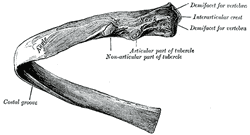
• Head: The head of each rib articulates with one or two ribs depending on their level. Typically ribs number 1,2, 10, 11, and 12 articulate with one vertebra, while the rest articulate with two vertebrae.

Thoracic rib, Posteroinferior view

Thoracic rib - Posteroinferior view.
Click on the image for a larger version.

Facets: These are the articular surfaces found in the head of each rib. They are covered by hyaline cartilage and form part of the costovertebral synovial joints. In the case of ribs 3 to 9, since they articulate with two vertebrae, they have two facets, each one called a demifacet, with an interarticular crest between them.

• Neck: A short, somewhat narrower portion of the rib that projects straight posterolaterally.

• Costal tubercle: A bony protuberance, usually with two components, one articular and one non-articular. The articular part of the costal tubercle presents with a facet that articulates with the transverse process of a thoracic vertebra.

• Costal angle: A sharp posterior curvature of the rib. The body when supine rests of these costal angles which deflect pressure from the thoracic spine.

• Costal body: The area of the rib anterior to the costal angle. In most ribs this oval-shaped region of the rib presents with an inferior and internal groove. This is the costal groove or costal sulcus. The corresponding level intercostal artery, vein, and nerve are found in the costal sulcus.

• Costal cartilage: All ribs have an anterior fibrocartilaginous component. Some of them attach directly to the sternum (chondrosternal joints), while some of them attach only to other costal cartilages (chondrochondral joints).

The 12 pairs of ribs are divided as follows:

• True ribs: Ribs 1-7, which attach by way of their costal cartilage directly to the sternum

• False ribs: Ribs 8-10, whose costal cartilage attach only to the cartilage of the superior rib, creating a lower border for the thoracic cage known as the costal margin.

• Free or "floating" ribs: Ribs 11 and 12. Their anterior cartilaginous end does not attach to sternum or other cartilage, so the end is free. The term "floating" although used, is a misnomer as these ribs do attach posteriorly to the thoracic spine.

There can be anatomical variations to the ribs, including the existence of extra cervical or lumbar ribs.

Images courtesy of Bartleby.com.For more information: click here
 
Back to MTD Main Page Subscribe to MTD