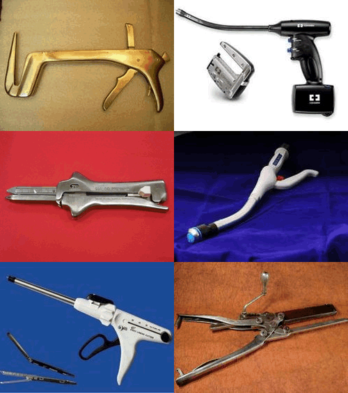
This article is part of the series "A Moment in History" where we honor those who have contributed to the growth of medical knowledge in the areas of anatomy, medicine, surgery, and medical research.

Surgical staplers through history
In the mid 1900’s the Soviet Ministry of Health established in Moscow the Scientific Research Institute for Experimental Surgical Apparatus and Instruments. This was forced by the need to train surgeons to perform complex operations at long distances from the capital cities. The Institute developed an incredible array of instruments including single line, linear cutter, and circular staplers which had applications such as gastrointestinal stapling, bone staplers, skin staplers, cornea and vascular staplers. etc. One of the problems of these instruments was that since they were hand-made, the parts from different staplers were not necessarily interchangeable.
The Moscow Thoracic Surgical Institute had very good results with the bronchial stapler, and it was here that in 1958, almost by chance, Dr. Mark M. Ravitch (1910 – 1989) and three other American physicians had the opportunity to evaluate patients that had been operated with this instrument, as well as seeing it in use.
Again, totally by chance, Dr. Ravitch happened to find a store that carried this instrument and as he tells “…quite unnecessarily I am sure, I identified myself plainly as an American. The only instrument they had in stock that day was the bronchial stapler…” He brought the instrument to the Thoracic Institute where the personnel calibrated it. This instrument came back to the USA with him to start a revolution in surgery.
Back in the USA, Dr. Ravitch started research with this and other instruments he procured in later trips. He recruited Dr. Felicien Steichen (1926 – 2011) to work with him, starting a friendship and collaboration that would last until his death. Both Drs. Ravitch and Steichen helped perfect and develop the modern instruments we use today: The linear stapler, the linear cutter, and the circular stapler.
Once these instruments were introduced, the development and advancement of the technology was pioneered by medical industry in the USA. First with Mr. Leon Hirsch, founder of the U.S. Surgical Corporation (today Covidien Surgical Devices), and later by Johnson and Johnson’s Ethicon Endo-Surgery (today Ethicon). These companies developed first the reloadable reusable surgical staplers and later the disposable reloadable surgical staplers.
Minimally invasive surgery (MIS) was common with gynecologist, but not used by general surgeons. Dr. Erich Muhe (1938 – 2005) was the first to perform a laparoscopic cholecystectomy in 1985, followed by many others. With the advent of MIS, these companies launched the development of laparoscopic surgical staplers, quite common today.
What about the future? First is the development of newer stapling technologies that take into account the viscoelastic behavior of tissues under rapid compression, multiple height staple lines, microstaplers, etc. Then, the advent of NOTES (Natural Orifice Transluminal Endoscopic Surgery) needs the development of smaller and smaller surgical staplers that can be used through a natural orifice and delivered through a flexible endoscope. That is, for now, the new frontier of surgical stapling.
The history of surgical stapling [1] ; [2]; [3]; [Video]
Sources
1. "Surgical stapling" Mallina, R F 1962 Scientific American 207, 48
2. “Science of Stapling: Urban Legend and Fact” Pfiedler & Ethicon EndoSurgery
3. “Cholecystointestinal, gastrointestinal, enterintestinal anastomosis, and approximation without sutures” Murphy JB. Med Rec (1892) 42: 665
4. “Study of Tissue Compression Processes in Suturing Devices” Astafiev, G. (1967 (USSR Ministry of Health, Ed.)
5. “Rese?as Hist?ricas: John Benjamin Murphy” Parquet, R.A. Acta Gastroenterol Latinoam 2010;40:97br />6. “The Science of Stapling and Leaks” Baker, R. S., & et al. (2004) Obesity Surgery, 14, 1290-1298.
7. “John Benjamin Murphy – Pioneer of gastrointestinal anastomosis”Bhattacharya, K., & Bhattacharya, N. (2008). Indian J. Surg., 70, 330-333.
8. “The Story of Surgery” Graham, H. (1939) New York: Doubleday, Doran & Co.. Inc.
9. “Compression Anastomosis: History and Clinical Considerations”Kaidar-Person, O, et al, e. (2008) Am J Surg, 818-826.
10. “Current Practice of Surgical Stapling”Ravitch, M. M., Steichen, F. M., & Welter, R. (1991) Philadelphia: Lea& Febiger.
11. “Aladar Petz (1888-1956) and his world-renowned invention: The gastric stapler” Olah, A. Dig Surg 2002: 19; 393-399
NOTE: The copyright notice for the images in this article can be found in the series "The History of Surgical Stapling" in this website


