

Medical Terminology Daily - Est. 2012

Medical Terminology Daily (MTD) is a blog sponsored by Clinical Anatomy Associates, Inc. as a service to the medical community. We post anatomical, medical or surgical terms, their meaning and usage, as well as biographical notes on anatomists, surgeons, and researchers through the ages. Be warned that some of the images used depict human anatomical specimens.

You are welcome to submit questions and suggestions using our "Contact Us" form. The information on this blog follows the terms on our "Privacy and Security Statement" and cannot be construed as medical guidance or instructions for treatment.


We have 780 guests online


A Moment in History

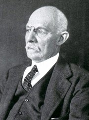
William S. Halsted, MD

William S. Halsted, MD
(1852 – 1922)

American anatomist, teacher, and surgeon, William Stewart Halsted was born in New York City, USA to a wealthy family of English origin. His father was involved in charitable work and Governor and trustee to a city hospital. Not a brilliant student initially, Halsted took an undergraduate in Liberal Arts in Yale, CT., after which he entered the Medical College of Physicians at the Columbia College, where he excelled.

As a second-year medical student Halsted applied and obtained a position in surgery at a local hospital. In here he learned about Lister’s antiseptic technique and became an adamant proponent of it to reduce infection. In 1877 Halsted obtained his MD. After a short time as House Physician at the New York Hospital, Halsted traveled to Europe to further his education studying for two years at the Universities of Vienna, Leipzig, and W?rzburg.

Besides being at the forefront of surgical and antiseptic techniques (introducing the use of rubber gloves in surgery), Halsted was extremely concerned with the way medical students were taught in the US. He pioneered bedside clinical round discussions with the medical students after two years of basic sciences studies. Halsted developed the idea of a patient chart; he also developed the residency program for medical students in use today.

Halsted is probably the most influential researcher and surgeon at the turn of the century. He dedicated time to the study of intestinal anastomoses and the use of silk as a suture material. His experimental work in 1887 proved that the inclusion of the submucosa layer in an anastomosis was mandatory, as well that a single layered anastomosis was enough to attain closure. Perhaps Halsted’s most important contribution was the application and use of the scientific method to surgical questions. Halsted’s principles set the standards used today in surgical suturing and surgical stapling.

He also pioneered the development and surgical techniques for radical mastectomy as a treatment for breast cancer.

As a side effect of this studied in anesthesia and the use of cocaine for anesthesia, Halsted became addicted to this substance, a problem that followed him through the years. Without impairing his capacity as a researcher and a surgeon, Halsted eventually recovered. He died in Baltimore in 1922 as a complication to surgery.

Sources:
1. Dubay, A. D., & Franz, G. M. (2003). Acute Wound Healing: The Biology of Acute Wound Failure. Surg Clin NA, 83, 463-481.
2. Halsted, W. S. (1887). Circular Suture of the Intestine - An Experimental Study. Am J Med Sci, 436-461.
3. “William Stewart Halsted: his life and contributions to surgery” Osborne, P. Lancet Oncol 2007; 8: 256–65
4. “William Stewart Halsted: Surgical pioneer” Burress, P Endoc Today (2010), 8: (2) 22
5. “William Stewart Halsted (1852–1922) Neurological stamp” Haas, LF J Neurol Neurosurg Psych 2000;69:641
Original image courtesy of "Images from the History of Medicine" at  www.nih.gov


 "Clinical Anatomy Associates, Inc., and the contributors of "Medical Terminology Daily" wish to thank all individuals who donate their bodies and tissues for the advancement of education and research”.

Click here for more information


bookplateink.com


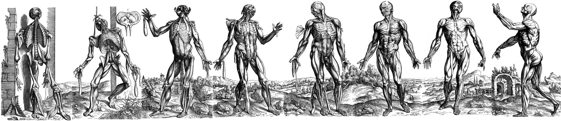
 Eight-series landscape panorama of Vesalius' muscle men plates. Paintings attributed to Jan Stephan Van Calcar

UPDATED: There is no doubt that the book “De Humani Corporis Fabrica Libri Septem” written by Andreas Vesalius and published by Johannes Oporinus in May, 1543 is one of the most important scientific anatomical and medical books ever written. Much has been said about the place this book has in medical history as part of the discarding of dogmas and the establishment of scientific observation and thinking.

Some of the most intriguing images published in the book are fourteen woodcuts in the second book. These amazing and detailed images show the muscles in a whole body as it is dissected. The text details the structures and the procedure of how the dissection is performed.

It is believed that these images were done by Jan Stephan Van Calcar, an artist from the Titian’s studio, although there are indications that these images may have been authored by somebody else, or even that they were the effort of more than one artist working under the close supervision of Andreas Vesalius.

Today’s anatomical images are very descriptive and the artistry is relegated to the technique used for the depiction of the image. In Vesalius’ muscle men plates each image has a background showing a landscape. It was not until 1903 that it was discovered that the landscapes of the different images were part of a complete landscape. This is evident if these images are placed side by side. There have been several books and articles published on these images. Interestingly, some of the images had to be reversed to be placed in the panorama. This is due to the process of cutting the woodblocks.

The image in this article is one of two identified and composed by Harvey Cushing in 1943 (see sources). He calls it the "eight-series". The "six-series" can be seen here. To see Cushing's original template click here. The panorama of the six-series was identified by Cushing to be an area of the Euganean hills near Venice and Padua, Italy. There landscape formed by the eight-series is an actual region near Padua, Italy showing the ruins of old Roman baths. Cavanagh (1938) adds more information on these images, for his .pdf article, click here.

The image shown at the top of this article was created using original images from Vesalius’ Fabrica and composed using Adobe Fireworks CS5. Click on the image for a larger depiction. The large image is 1800px wide.

NOTE: In June 2023, I was able to purchase a single leaf of the 1543 Fabrica, page 187, which depicts one of the "muscle men". In the panorama image in this article it is figure number 3, from left to right. For more information on this image click here. This image is now properly framed in my office. Dr. Miranda.

This article continues here: "The landscape panorama of Vesalius' Muscle Men (2)"

Sources:
1. “A New View of the Vesalian Landscape” Cavanagh,  GST Med Hist 1983, 27: 77-79
2. “The Panorama of Vesalius: A 'Lost' Design From Titian's Studio” Skandalakis, JE JAMA May 28, 1997, Vol 277, No. 20
3. “A Drawing for the Fabrica; and some Thoughts Upon the Vesalius Muscle-Men” Kemp. M. Med Hist. Jul 1970; 14(3): 277–288
4. “Andreas Vesalius: The Making, the Madman and the Myth” Joffe, SN. Persona Publishing 2009
5. "A Bio-Bibliography of Andreas Vesalius" Cushing, H. (1943) Schumann's