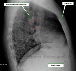
Anatomy: The term [aortopulmonary window] is a radiological term that refers to a small space in the left mediastinal region. It is bounded anteriorly by the ascending aorta, posteriorly by the descending aorta, superiorly by the aortic arch, inferiorly by the left pulmonary artery, medially by the arterial ligament [ligamentum arteriosum] and left main bronchus, and laterally by the pleura and left lung. It contents the recurrent laryngeal nerve, lymph nodes, and adipose tissue.
Clinical: The aortopulmonary window is commonplace for lymphadenopathy in various inflammatory and neoplastic diseases.
Radiology: In a frontal projection, it corresponds to a focal concavity on the left border of the mediastinum, inferior to the aorta and superior to the left pulmonary artery. In the lateral projection (which is the proper image to identify this area), it is seen as a radiolucency inferior to the aortic arch and superior to the left pulmonary artery. Its appearance can be modified by tortuosity of the aorta.
Congenital heart disease: This term also refers to a congenital heart disease similar in appearance to patent ductus arteriosus, (or truncus arteriosus) with the difference that this involves septal defect. It is described as a communication between the ascending aorta and pulmonary trunk portion or right pulmonary artery. It is a rare anomaly that represents 0.2% -0.6% of congenital cardiac abnormalities.
Sources:
1. Heitzman ER. The infraaortic area. In: The mediastinum: With correlations radiologic anatomy and pathology. Berlin, Germany: Springer-Verlag, 1988; 151: -168.
2. Blank N, Castellino RA. Patterns of pleural reflections of the left upper mediastinum: Normal anatomy and distortions produced by adenopathy. Radiology 1972; 102: 585-589.
3. Marc Dewey, Donna Magid, Paul S. Wheeler and Bernd Hamm aortopulmonary Angle Window or on the Chest Radiograph? American Journal of Roentgenology. 2004; 182: 1085-1086.
4. SY Ho, Gerlis LM, Anderson C, Devine WA, Smith A. The morphology of aortopulmonary windows With regard to their classification and morphogenesis. Young Cardiol 1994; 4: 146-55.
5. Kutsche LM, Van Mierop LHS. Anatomy and pathogenesis of aorticopulmonary septal defect. Am J Cardiol 1987; 59: 443-7.
6. Stevenson, Roger E .; Hall, Judith G. (2006). Human malformations and related anomalies. Oxford University Press US. pp. 119-. ISBN 978-0-19-516568-5
7. Donoghue, Veronica B .; Bj?rnstad, Per G. Radiological Imaging of the Neonatal Chest. Springer. pp. 330-. ISBN 978-3-540-33748-5.


