Medical Terminology Daily (MTD) is a blog sponsored by Clinical Anatomy Associates, Inc. as a service to the medical community. We post anatomical, medical or surgical terms, their meaning and usage, as well as biographical notes on anatomists, surgeons, and researchers through the ages. Be warned that some of the images used depict human anatomical specimens.

You are welcome to submit questions and suggestions using our "Contact Us" form. The information on this blog follows the terms on our "Privacy and Security Statement" and cannot be construed as medical guidance or instructions for treatment.


We have 103 guests online


A Moment in History

Larsen

William J. Larsen, PhD
(1942-2000)

An American scientist, Dr. Larsen was a gifted scientist, consistently producing research at the forefront of cell, developmental, and reproductive biology. Early in his career he published a landmark paper that conclusively established mitochondrial fission as the mechanism of mitochondrial biogenesis. He went on to become the first to demonstrate the endocytosis of gap junctions. Moreover, his work on the hormonal regulation of gap junction formation and growth culminated in an authoritative review article in Tissue and Cell, “Structural Diversity of Gap Junctions (1988)”, which became a citation classic.

Throughout his 25 year teaching career, his sixty-seven peer reviewed publications—not to mention numerous invited reviews, abstracts, and book chapters—covered a wide range of research areas including adrenal cortical tumor cells, human ovarian carcinomas, preterm labor, cumulus expansion, oocyte maturation, ovulation, folliculogenesis, and in-vitro fertilization.

In addition to his many contributions to basic research, Dr. Larsen loved to teach and was much appreciated by his students. His exceptional ability was reflected in the four teaching awards he received as a professor at the University of Cincinnati.

Notably, he was the author of Human Embryology, a textbook for medical students that was the first to incorporate modern experimental research into a subject that had traditionally been taught in a strictly descriptive style. On its initial publication in 1998 it was hailed as, “a magnificent book…” by the European Medical Journal. With the release of the fourth edition in 2008, the book was renamed “Larsen’s Human Embryology” in recognition of Dr. Larsen's place as the originator of this revolutionary text. This book is today in it's 6th Edition.

His stellar scientific career would be enough for most people, but Dr. Larsen pursued his numerous and varied interests with such extraordinary passion, energy, and skill that he seemed to have more hours in a day than the ordinary person. He was fascinated with the American Southwest and studied and collected traditional arts and crafts of the Hopi, Zuni, and Navajo peoples. He was a woodworker who built three harpsichords and a fortepiano for his wife, and, with his two children, over 100 pieces of gallery-quality furniture. In addition, he loved to regale his friends, colleagues, and students with jokes and stories, and to share his love for gourmet cooking.

The William J. Larsen Distinguished Lecture Series

An annual lecture series was created for the Department of Cancer & Cell Biology at the University of Cincinnati to honor Dr. Larsen's research which was at the forefront of cell developmental and reproductive biology. This series recognizes forward-thinking research scientists in the field of developmental biology and asks that they share their research and findings with students and faculty of the University of Cincinnati, College of Medicine.

Personal note: I had the opportunity to meet and attend Dr. Larsen’s embryology lectures as he and I worked in the Anatomy, Embryology, and Histology program at the University of Cincinnati Medical College. Unfortunately, I never had the opportunity to have Dr. Larsen sign my personal copy of his book. He is sorely missed, Dr. Miranda

Sources:

1. "The William J. Larsen Distinguished Lecture Series" University of Cincinnati, College of Medicine.
2. https://www.larsenbooks.com
3. 2022 Larsen Lecture Series brochure (download here)
4. Dr. Larsen's family personal communications


 "Clinical Anatomy Associates, Inc., and the contributors of "Medical Terminology Daily" wish to thank all individuals who donate their bodies and tissues for the advancement of education and research”.

Click here for more information


abebooks banner

bookplateink.com


Canal of Nuck

The "Canal of Nuck" is the patent embryological remnant of the processus vaginalis in the female. The processus vaginalis is an extension of the peritoneum that forms to the side of the gubernaculum, a small fibrous cord that is attached to the lower pole of the gonad in the embryo. On the other end, the gubernaculum attaches to the inner aspect of the labioscrotal fold, an embryonic structure that will become the scrotum in the male and the labia majora in the female.

In the male, the processus vaginalis accompanies the gubernaculum and the testicle, on its descent towards the scrotum. In the female, the gonad (ovary) stays in the pelvis and the embryological remnants of the gubernaculum become the proper ovarian ligament (uteroovarian ligament) and the round ligament of the uterus which enters the inguinal canal, splits into multiple small fibers that disappear in the tissues of the labium majus.

In the male (and female) the walls of the processus vaginalis normally fuse, closing the communication between the scrotum (and the labia majora) and the main peritoneal cavity. If they remain open, the name is different, although the pathological consequences are similar (hernia, cysts or hydrocele). In the male, it is called a “patent processus vaginalis”, and in the female it is called the “Canal of Nuck”, which is found patent in 10-20% of the cases, although its presence does not per se imply the presence of pathology.

It was first described by Anton Nuck, a Dutch surgeon and anatomist (1650-1692) in his book "Adenographia & uteri anatome nova" published in 1722. In this book he questions why do some females present with inguinal hernias: "Haecce , praeter alias herniarium species , in utroque sexu obvias auditoribus meis anno fuperiori demonftrandi , difficile vifum fuit explicare , qui Hernia foeminarum inguinales orirentur?" Why when it is easy to see (the canal) in other species it is so difficult to explain to those listening why only some women have inguinal hernias?

In figure XL of the same book he proceeds to show the open processus vaginalis which was from then on known as the eponymic "Canal of Nuck"

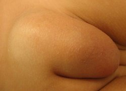
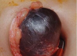
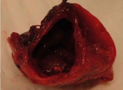
The images in this article are from “Case Report: Infected Hydrocele of the Canal of Nuck” by Mandahan, P and Batthi, K. (see sources) Figure 1 shows the superficial hydrocele herniation; figure 2 shows the infected hydrocele; and figure 3 shows the excised opened hydrocele. Read the full article here. 

http://dx.doi.org/10.1155/2013/275257

My personal thanks to Dr. Sanford Osher who suggested this article. Dr. Miranda

Infected Canal of Nuck Hydrocele, Mandhan, P; Bhatti, K. Infected Canal of Nuck Hydrocele, Mandhan, P; Bhatti, K. 
Click on the image for a larger depiction
 Infected Canal of Nuck Hydrocele, Mandhan, P; Bhatti, K. Infected Canal of Nuck Hydrocele, Mandhan, P; Bhatti, K. 
Click on the image for a larger depiction
 Infected Canal of Nuck Hydrocele, Mandhan, P; Bhatti, K. Infected Canal of Nuck Hydrocele, Mandhan, P; Bhatti, K. 
Click on the image for a larger depiction

Sources:
1. 
Adenographia curiosa et uteri foeminei anatome nova” Classic pages in Obstetrics and Gynecology Lawrence Longo, M.D. Volume 123, Issue 1, 1 September 1975, Page 66
2. Nuck, Antonio “Adenographia curiosa et uteri foeminei anatome nova” (Latin) Apud Samuellem Luchtmans   https://archive.org/details/bub_gb_tQZJYL7qyssC 
3. Poghosyanm T, et al “Hydrocele of canal of Nuck” Applied Radiology; Scotch Plains43.12 (Dec 2014): 37-38.
4. Bagley, J. “Cyst of Canal of Nuck.” Journal of Diagnostic Medical Sonography Vol 31, Issue 2, pp. 111 - 114
5. Bagu, A. et al Endometriosis in the canal of Nuck hydrocele: An unusual presentation
6. Mandham, P; Bhatti, K “Case Report: Infected Hydrocele of the Canal of Nuck” Case Reports in Urology Volume 2013  http://dx.doi.org/10.1155/2013/275257
 
MTD Main Page Subscribe to MTD