Medical Terminology Daily (MTD) is a blog sponsored by Clinical Anatomy Associates, Inc. as a service to the medical community. We post anatomical, medical or surgical terms, their meaning and usage, as well as biographical notes on anatomists, surgeons, and researchers through the ages. Be warned that some of the images used depict human anatomical specimens.

You are welcome to submit questions and suggestions using our "Contact Us" form. The information on this blog follows the terms on our "Privacy and Security Statement" and cannot be construed as medical guidance or instructions for treatment.


We have 189 guests and no members online


A Moment in History

Georg Eduard Von Rindfleisch

Georg Eduard Von Rindfleisch
(1836 – 1908)

German pathologist and histologist of Bavarian nobility ancestry. Rindfleisch studied medicine in Würzburg, Berlin, and Heidelberg, earning his MD in 1859 with the thesis “De Vasorum Genesi” (on the generation of vessels) under the tutelage of Rudolf Virchow (1821 - 1902). He then continued as a assistant to Virchow in a newly founded institute in Berlin. He then moved to Breslau in 1861 as an assistant to Rudolf Heidenhain (1834–1897), becoming a professor of pathological anatomy. In 1865 he became full professor in Bonn and in 1874 in Würzburg, where a new pathological institute was built according to his design (completed in 1878), where he worked until his retirement in 1906.

He was the first to describe the inflammatory background of multiple sclerosis in 1863, when he noted that demyelinated lesions have in their center small vessels that are surrounded by a leukocyte inflammatory infiltrate.

After extensive investigations, he suspected an infectious origin of tuberculosis - even before Robert Koch's detection of the tuberculosis bacillus in 1892. Rindfleisch 's special achievement is the description of the morphologically conspicuous macrophages in typhoid inflammation. His distinction between myocardial infarction and myocarditis in 1890 is also of lasting importance.

Associated eponyms

"Rindfleisch's folds": Usually a single semilunar fold of the serous surface of the pericardium around the origin of the aorta. Also known as the plica semilunaris aortæ.

"Rindfleisch's cells": Historical (and obsolete) name for eosinophilic leukocytes.

Personal note: G. Rindfleisch’s book “Traité D' Histologie Pathologique” 2nd edition (1873) is now part of my library. This book was translated from German to French by Dr. Frédéric Gross (1844-1927) , Associate Professor of the Medicine Faculty in Nancy, France. The book is dedicated to Dr. Theodore Billroth (1829-1894), an important surgeon whose pioneering work on subtotal gastrectomies paved the way for today’s robotic bariatric surgery. Dr. Miranda.

Sources:
1. "Stedmans Medical Eponyms" Forbis, P.; Bartolucci, SL; 1998 Williams and Wilkins
2. "Rindfleisch, Georg Eduard von (bayerischer Adel?)" Deutsche Biographie
3. "The pathology of multiple sclerosis and its evolution" Lassmann H. (1999)  Philos Trans R Soc Lond B Biol Sci. 354 (1390): 1635–40.
4. “Traité D' Histologie Pathologique” G.E.
Rindfleisch 2nd Ed (1873) Ballieres et Fils. Paris, Translated by F Gross


 "Clinical Anatomy Associates, Inc., and the contributors of "Medical Terminology Daily" wish to thank all individuals who donate their bodies and tissues for the advancement of education and research”.

Click here for more information


abebooks banner

bookplateink.com


Atrium

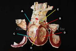
Heart model - LAO cranial view [UPDATED] The term [atrium] is Latin, its plural form is [atria]. The atrium was the center hall of a Roman home, around which the rest of the rooms opened. Since the atrium was the first area of the house that was entered once passing through the front door, the term [atrium] has been used to describe the "entrance hall', such as the atrium of a hotel. The atria are the two superior chambers of the heart. (see image, items "A=right atrium" and "B=left atrium")

An interesting question is why are the atria called so, since they are part of the heart, and not just the entrance?. The reason is that early anatomists considered the heart to be composed only by the ventricles. The atria were then chambers where blood would wait before entering the "heart proper", ergo [atria].

Each atrium has a smooth wall (sinus venarum) and a muscular extension akin to a closed-end bag. These are the atrial appendages or auricles. Anatomically they are quite different. The right atrial appendage communication or opening to the right atrium is wide and allows blood to easily flow from and to the atrium. On the contrary, the left atrial appendage has a very small opening (ostium) and its morphology is convoluted with lobulations and a complicated mesh of atrial muscle wall.

The very structure of the left atrial appendage is quite conducive to the formation of clots in atrial fibrillation (AFib). These anchored clots (thrombus/thrombi) can detach and become free clots (embulus/emboli) that will enter the blood stream, pass into the left ventricle, then though the aortic valve, and then pass into the ascending aorta and main circulation. Unfortunately, two of the first arteries that arise from the aorta are the common carotid arteries that take blood to the brain and these thrombi can cause a brain stroke.

Personal note: On November 7, 2023 Dr. Randall K. Wolf invited me to a seminar where we reviewed the anatomy of the left atrial appendage, the problems it can cause in atrial fibrillation as a cause for stroke, and the reasons for its exclusion in AFib surgery. 

Image property of:CAA.Inc.Photographer:D.M. Klein