Medical Terminology Daily (MTD) is a blog sponsored by Clinical Anatomy Associates, Inc. as a service to the medical community. We post anatomical, medical or surgical terms, their meaning and usage, as well as biographical notes on anatomists, surgeons, and researchers through the ages. Be warned that some of the images used depict human anatomical specimens.

You are welcome to submit questions and suggestions using our "Contact Us" form. The information on this blog follows the terms on our "Privacy and Security Statement" and cannot be construed as medical guidance or instructions for treatment.


We have 142 guests and no members online


A Moment in History

Andreas Vesalius Bruxellensis (1514- 1564)

A Flemish anatomist and surgeon, Andreas Vesalius was born on December 31, 1514 in Brussels, Belgium. He is considered to be the father of the science of Anatomy. Up until his studies and publications human anatomy studies consisted only on the confirmation of the old doctrines of Galen of Pergamon (129AD - 200AD). Anatomy professors would read to the students from Galen's work and a demonstrator would point in a body to the area being described, if a body was used at all. The reasoning was that there was no need to dissect since all that was needed to know was already written in Galen's books. Vesalius, Fallopius, and others started the change by describing what they actually saw in a dissection as opposed to what was supposed to be there. 

Vesalius had a notorious career, both as an anatomist and as a surgeon. His revolutionary book "De Humani Corporis Fabrica: Libri Septem" was published in May 26, 1543. One of the most famous anatomical images is his plate 22 of the book, called sometimes "The Hamlet". You can see this image if you hover over Vesalius' only known portrait which accompanies this article. Sir William Osler said of this book "... it is the greatest book ever printed, from which modern medicine dates" 

After the original 1543 printing, the Fabrica was reprinted in 1555. It was re-reprinted and translated in many languages, although many of these printings were low-quality copies with no respect for copyright or authorship.

The story of the wood blocks with the carved images used for the original printing extends into the 20th century. In 1934 these original wood blocks were used to print 617 copies of the book "Iconaes Anatomica". This book is rare and no more can be printed because, sadly, during a 1943 WWII bombing raid over Munich all the wood blocks were burnt.

One interesting aspect of the book was the landscape panorama in some of his most famous woodcuts which was only "discovered" until 1903.

Vesalius was controversial in life and he still is in death. We know that he died on his way back from a pilgrimage to Jerusalem, but how he died, and exactly where he died is lost in controversy. We do know he was alive when he set foot on the port of Zakynthos in the island of the same name in Greece. He is said to have suddenly collapsed and die at the gates of the city, presumably as a consequence of scurvy. Records show that he was interred in the cemetery of the Church of Santa Maria delle Grazie, but the city and the church were destroyed by an earthquake and Vesalius' grave lost to history. Modern researchers are looking into finding the lost grave and have identified the location of the cemetery. This story has not ended yet.

For a detailed biography of Andreas Vesalius CLICK HERE.

Personal note: To commemorate Andrea Vesalius' 500th birthday in 2014, there were many scientific meetings throughout the world, one of them was the "Vesalius Continuum" anatomical meeting on the island of Zakynthos, Greece on September 4-8, 2014. This is the island where Vesalius died in 1564. I had the opportunity to attend and there are several articles in this website on the presence of Andreas Vesalius on Zakynthos island. During 2015 I also attended a symposium on "Vesalius and the Invention of the Modern Body" at the St. Louis University. At this symposium I had the honor of meeting of Drs. Garrison and Hast, authors of the "New Fabrica". Dr. Miranda


 "Clinical Anatomy Associates, Inc., and the contributors of "Medical Terminology Daily" wish to thank all individuals who donate their bodies and tissues for the advancement of education and research”.

Click here for more information


Rare & Collectible Books at AbeBooks.com 

bookplateink.com

Incisura angularis

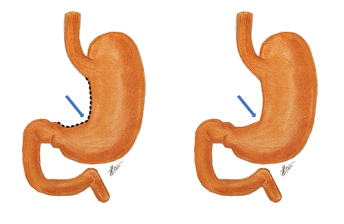
Anterior view of the stomach


The term "incisura" is Latin, derived from the verb [incidere]* meaning "to cut" or [incisura]” meaning a "notch" or indentation in a structure, suggesting a distinctive incision, or cut. The second component, "angularis," is also Latin, derived from "angulus," which translates to "angle." The term "incisura angularis" can be translated as the "angular notch", a term that is also use for this gastric anatomical landmark.

The incisura angularis is a notch located along the lesser curvature of the stomach. Externally, it marks the transition between the body (corpus) and antrum of the stomach, an abdominal viscus. It is related to the gastrohepatic portion of the lesser omentum superolaterally. It should be mentioned that the lesser curvature vascular arcade runs within the lesser omentum, closely related to the gastric lesser curvature.

Found approximately midway between the esophagogastric junction and the pylorus, this external anatomical feature is easily identifiable internally during gastric endoscopy.

Structurally, the incisura angularis is formed by a fold of mucous membrane on the inner surface of the stomach, creating a small recess along the lesser curvature.

The stomach, although it has the same layers as the rest of the GI tract, presents an extra muscular layer in the area of the lesser curvature, which renders this area less distensible forming a muscular channel called the magenstrasse.

The mucosa layer is the deepest of the stomach layer. Within it, three areas of gastric mucosa are usually described: pyloric, transitional, and fundic. The incisura angularis corresponds mostly to the transitional zone. When there are mucosal changes that shown an invasion of another type of mucosa, it can mean preneoplastic changes. For this reason, the incisura angularis is an area that, when biopsied, can show early cancerous changes, as well as muscular atrophy, intestinal metaplasia, and dysplasia.

Preservation of the anatomy of the incisura angularis is critical during a sleeve gastrectomy, the most common bariatric procedure worldwide. The objective of a sleeve gastrectomy is to reduce the size of the stomach by placing a curved staple line along the left border of the magenstrasse, a lesser known gastric anatomy term.

Because of the location of the incisura angularis, improper placement of a straight gastric stapler could cause stenosis or stricture at this level. Another potential postoperative problem in this procedure is gastroesophageal reflux disease (GERD) where some authors have proposed an omentopexy as a way to modify the angle of the incisura angularis.

Sources:
1. "The Origin of Medical Terms" Skinner, HA 1970 Hafner Publishing Co.
2. "Medical Meanings - A Glossary of Word Origins" Haubrich, WD. ACP Philadelphia
3 "Tratado de Anatomia Humana" Testut et Latarjet 8 Ed. 1931 Salvat Editores, Spain
4. “The Rarely Sampled Incisura Angularis Is Useful for the Detection of Gastric Preneoplastic Lesions” Singhal, A. , Saboorian, H. , Turner, K. , Rugge, M. & Genta, R. (2023). The American Journal of Gastroenterology, 118 (10S), S1407-S1407.
5. “Incisura angularis belongs to fundic or transitional gland regions in Helicobacter pylori-naive normal stomach: Sub-analysis of the prospective multi-center study” Nakajima, S at al Digestive Endoscopy 2021; 33: 125–132
6. “Increasing the angle at the incisura angularis using omentopexy reduces/prevents GERD symptoms five years after laparoscopic sleeve gastrectomy?” Presidential Grand Rounds. Surgery for Obesity and Related Diseases, Volume 18, Issue 8, Supplement, 2022
7. “Gastric POEM to treat incisura angularis torsion after sleeve gastrectomy” Baptista, A; Davila, M; Guzman, M. Endoscopy 2019; 51(04)
8. “Obstruction after Sleeve Gastrectomy, Prevalence, and Interventions: a Cohort Study of 9,726 Patients with Data from the SOReg” Sillen, L; Andersson, E, Edholm, D. OBES SURG 31, 4701–4707 (2021)

Note: Google Translate includes the symbol (?). Clicking on it will allow you to hear the pronunciation of the word.