Sponsor   

Medical Terminology Daily (MTD) is a blog sponsored by Clinical Anatomy Associates, Inc. as a service to the medical community, medical students, and the medical industry. We post anatomical, medical or surgical terms, their meaning and usage, as well as biographical notes on anatomists, surgeons, and researchers through the ages. Be warned that some of the images used depict human anatomical specimens.

Click on the link below to subscribe to the MTD newsletter. If you think an article could be interesting to somebody else, feel free to forward the link of the article. Should you want to use the information on the article, please follow the CAA, Inc Privacy and Security Statement found at the bottom of this page. 

You are welcome to submit questions and suggestions using our "Contact Us" form. The information on this blog follows the terms on our "Privacy and Security Statement"  and cannot be construed as medical guidance or instructions for treatment. 


We have 180 guests and no members online


A Moment in History

Self-portrait, Henry Vandyke Carter, MD (Public Domain)

Henry Vandyke Carter, MD
(1831 – 1897)

English physician, surgeon, medical artist, and a pioneer in leprosy and mycetoma studies.  HV Carter was born in Yorkshire in 1831. He was the son of Henry Barlow Carter, a well-known artist and it is possible that he honed his natural talents with his father. His mother picked his middle name after a famous painter, Anthony Van Dyck. This is probably why his name is sometimes shown as Henry Van Dyke Carter, although the most common presentation of his middle name is Vandyke.

Having problems to finance his medical studies, HV Carter trained as an apothecary and later as an anatomical demonstrator at St. George’s Hospital in London, where he met Henry Gray (1872-1861), who was at the time the anatomical lecturer. Having seen the quality of HV Carter’s drawings, Henry Gray teamed with him to produce one of the most popular and longer-lived anatomy books in history: “Gray’s Anatomy”, which was first published in late 1857.  The book itself, about which many papers have been written, was immediately accepted and praised because of the clarity of the text as well as the incredible drawings of Henry Vandyke Carter.

While working on the book’s drawings, HV Carter continued his studies and received his MD in 1856.

In spite of initially being offered a co-authorship of the book, Dr. Carter was relegated to the position of illustrator by Henry Gray and never saw the royalties that the book could have generated for him. For all his work and dedication, Dr. Carter only received a one-time payment of 150 pounds. Dr.  Carter never worked again with Gray, who died of smallpox only a few years later.

Frustrated, Dr. Carter took the exams for the India Medical Service.  In 1858 he joined as an Assistant Surgeon and later became a professor of anatomy and physiology. Even later he served as a Civil Surgeon. During his tenure with the India Medical Service he attained the ranks of Surgeon, Surgeon-Major, Surgeon-Lieutenant-Colonel, and Brigade-Surgeon.

Dr. Carter dedicated the rest of his life to the study of leprosy, and other ailments typical of India at that time. He held several important offices, including that of Dean of the Medical School of the University of Bombay. In 1890, after his retirement, he was appointed Honorary Physician to the Queen.

Dr. Henry Vandyke Carter died of tuberculosis in 1897.

Personal note: Had history been different, this famous book would have been called “Gray and Carter’s Anatomy” and Dr. Carter never gone to India. His legacy is still seen in the images of the thousands of copies of “Gray’s Anatomy” throughout the world and the many reproductions of his work available on the Internet. We are proud to use some of his images in this blog. The image accompanying this article is a self-portrait of Dr. Carter. Click on the image for a larger depiction. Dr. Miranda

Sources:
1. “Obituary: Henry Vandyke Carter” Br Med J (1897);1:1256-7
2. “The Anatomist: A True Story of ‘Gray’s Anatomy” Hayes W. (2007) USA: Ballantine
3. “A Glimpse of Our Past: Henry Gray’s Anatomy” Pearce, JMS. J Clin Anat (2009) 22:291–295
4. “Henry Gray and Henry Vandyke Carter: Creators of a famous textbook” Roberts S. J Med Biogr (2000) 8:206–212.
5. “Henry Vandyke Carter and his meritorious works in India” Tappa, DM et al. Indian J Dermatol Venereol Leprol (2011) 77:101-3


"Clinical Anatomy Associates, Inc., and the contributors of "Medical Terminology Daily" wish to thank all individuals who donate their bodies and tissues for the advancement of education and research”.

Click here for more information


Rare & Collectible Books at AbeBooks.com 

bookplateink.com


Sinotubular junction (STJ)

The sinotubular junction (STJ) is a well-defined circular ridge found between the superior or tubular segment of the ascending aorta and the inferior, bulbous segment of the ascending aorta, known as the aortic root.

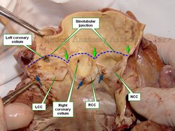
The STJ follows the superior contour of the three sinuses of Valsalva that form the bulbs of the aortic root and intersects the commissures of the aortic valve. In the accompanying image the STJ is depicted by a blue dotted line and the commissures are depicted by green arrows.

The ostia of the coronary arteries are usually found inferior to the STJ, although they can be on it or superior to the STJ. There seems to be a correlation with anomalous origins of the coronary ostia and sudden-death syndrome.

Aortic root of the ascending aorta open by dissection
Aortic root open. Click on the image for a larger version.
The image also shows the three cusps of the aortic valve: the non-coronary cusp (NCC), the right coronary cusp (RCC) and the left coronary cusp (LCC). The blue arrows indicate the location of the nodules of Arantius.

Medical terminology notes: There are many scholarly texts that hyphenate the word as [sino-tubular]. This is incorrect, as the root term is [-sin-], meaning "sinus". The addition of the [-o-] to the root term creates the combining form [-sino-] which is then used to connect to the root term [-tubul-]. Since it is a vowel and a consonant combining and they are euphonic, there is no need to add the hyphen. In fact, a word should use as a combining element either a hyphen or an [-o-], but not both. The word with the hyphen then should be [sin-tubular] which is definitely not euphonic. For more information on combining root terms, click here.

Others write [sinutubular]. The paragraph above clearly explains why this is a mistake. The root term is [-sin-] meaning "sinus" and the addition of the [-o-] creates the combining form [-sino-] and not [-sinu-].

Sources:
1. “Clinical Anatomy of the Aortic Root” Anderson, RH Heart 200; 84: 670–673
2. “The Anatomy of the Aortic Root” Loukas, E et al. Clin Anat 2014; 27:748-756
3: "Tratado de Anatomia Humana" Testut et Latarjet 8th Ed. 1931 Salvat Editores, Spain
Image property of: CAA.Inc. Photographer: D.M. Klein 

MTD Main Page Back to MTD Main Page