
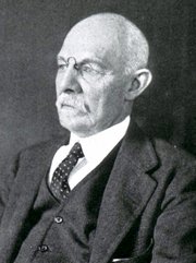




Dr. José Manuel Revuelta
Personal Note: This is article originally published in Spanish by Dr. Jose Manuel Revuelta, a a Professor of Surgery and Professor Emeritus at the University of Cantabria. Former Head of Cardiovascular Surgery at Valdecilla Hospital in Santander, Spain. Dr. Revuelta has contributed several articles to this blog
The article's title (in Spanish) is "La Libertad del Corazón" (The Freedom of the Heart), a discussion of the heart's independence from the nervous system.
He has graciously granted us permission to translate and publish his article in “Medical Terminology Daily”. Dr. Miranda.
The Freedom of the Heart
There is a common belief that if the brain stops functioning, the body collapses instantly and the heart stops. The heart is not merely an executor of the brain's commands, but a central organ with astonishing self-governance. In fact, if we were to isolate a human heart in a suitable, oxygenated, and nutrient-rich environment, it would continue to beat autonomously, as we observe every day in donor organs for transplantation.
This capacity for self-excitation resides in its automaticity, a property of a group of specialized cells (the sinoatrial or sinus node) that acts as a biological pacemaker. Located in the upper wall of the right atrium, this peculiar cluster of cells spontaneously generates its own electrical activity rhythmically, triggering heart contractions without needing any impulse from the cerebral cortex or brainstem.
After reading this, we wonder about the true role of the brain in directing and coordinating the human body. More than a conductor creating the music, the brain acts like a sound engineer adjusting the volume and rhythm according to the needs of the environment. Through the nervous system, the brain operates pedals to accelerate (sympathetic nervous system, driven by norepinephrine) or decelerate the heart (parasympathetic nervous system, guided by the vagus nerve and acetylcholine).
When we run or feel fear, the brain commands the heart rate to accelerate; when we sleep, it presses the brakes, so the heart functions more calmly. In a healthy person, the moment-to-moment interaction between the sympathetic and parasympathetic nervous systems generates micro-oscillations in the interval between heartbeats (R-R interval), a variability that reflects the system's adaptability.
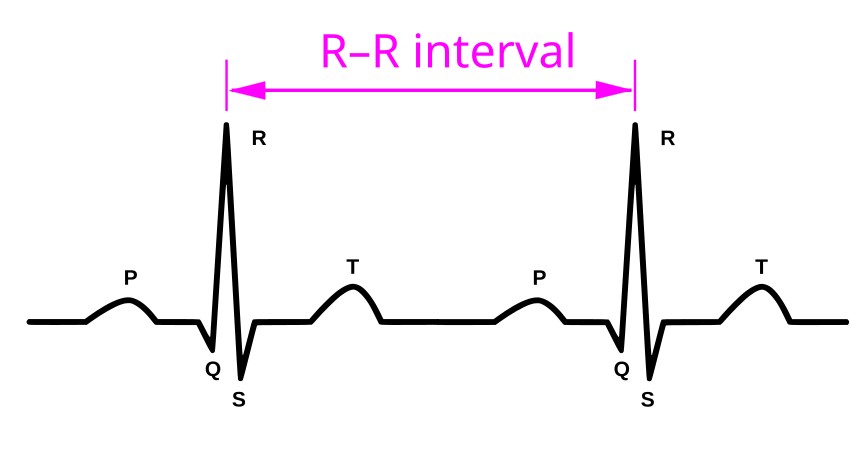
R-R Interval in an ECG
Generally, the heart and brain form a harmonious "marriage"; both need each other and collaborate in life's challenges. However, there are physiological, pathological, or surgical situations in which this good communication breaks down, and the accelerator or brake pedals cease to function properly. At this point, the heart takes control of the biological machinery, disobeying central commands. At this threshold of independence, we find the most surprising mysteries of our organism and its extraordinary constitutional complexity.
The Independence of the Heart
To understand this temporary or permanent disconnection, it is helpful to remember that the first organ to form in the human fetus is the heart. It begins to beat, seemingly without order, to provide oxygen and nutrients to the millions of specialized cells that will form the different organs and tissues, including the brain. We know that it begins beating around the sixth week of gestation, when its development is complete, a crucial moment that allows it to pump blood throughout the body.
From day one, fulfilling its vital function, the heart strives to be part of the ordered microcosm of human biology, while also possessing the powerful capacity to become independent at any moment in the face of certain extraordinary situations, whether internal or external. In fact, modern neurocardiology has highlighted a fundamental finding: "a heart that is too obedient is a vulnerable heart."
When the dialogue breaks down: The arrival of brain signals that are incomprehensible to the heart, which could damage it or create a dangerous scenario for the body due to an excessive response from the autonomic sympathetic nervous system, triggers its aforementioned disobedience and disconnection. An excessive surge of cortisol, adrenaline, and noradrenaline causes a very rapid tachycardia that could lead to ventricular fibrillation or cardiac arrest, forcing the heart to slow down these exaggerated impulses from the nervous system.
On the other hand, the brain can miscalculate, sending disproportionate bursts of vagal tone, known as vasovagal syncope or neurocardiogenic syndrome, in response to certain triggers such as acute pain, significant emotional stress, or prolonged standing. The sudden release of an illogical amount of acetylcholine induces extreme bradycardia or temporary asystole, combined with severe peripheral vasodilation, causing cerebral blood flow to stop and the person to lose consciousness and fall to the ground (fainting, also known as a syncope). At that moment, the heart takes over, regulating heart rate and blood pressure to restore consciousness, since the brain has temporarily ceased functioning.
Sometimes, the brain sends the correct signal, but the heart's internal wiring fails, or ectopic foci of the myocardium decide to act independently. When the specific cardiac electrical conduction system fails to respond, impulses from the sinoatrial node reach the atrioventricular node, but cannot pass through the bundle of His to the ventricles (partial or complete atrioventricular block). This forces the Purkinje network or the ventricular myocardium itself to take control, activating an escape rhythm. The heart then beats at a very slow intrinsic rate (20–40 beats per minute, bpm), completely dissociated from atrial activity and brain control. In situations of cardiac ischemia or significant myocardial damage, multiple ectopic foci generate impulses at extremely high and disorganized heart rates, potentially causing death from ventricular fibrillation or cardiac arrest.
A Definitive Separation
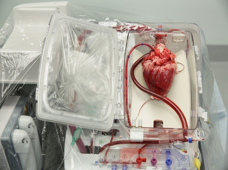
Heart transplant transport system
The most fascinating scenario of forced physiological insubordination occurs after a heart transplant. During the surgical procedure, the parasympathetic (vagal) and postganglionic sympathetic nerve fibers connecting the brain to the donor's heart are irreversibly severed.
In the transplant recipient, lacking the "brake" of acetylcholine, the donor's sinoatrial node operates with an uninhibited intrinsic rhythm, registering a higher resting heart rate (between 90 and 100 bpm). Without the physiological "beat-to-beat adjustment" coordinated by the vagus nerve and the brain, the electrocardiographic tracing of a transplanted heart shows extremely rigid and constant R-R intervals.
During physical exercise, the transplant recipient's brain sends the signal to increase cardiac output, but since there are no direct nerve fibers to the heart, the acceleration is not instantaneous. The heart rate will increase, but gradually, and will take longer to decrease during the recovery phase, as it depends exclusively on the arrival of circulating catecholamines (adrenaline and noradrenaline) secreted by the adrenal glands of the transplant recipient. Furthermore, because the sensory afferent pathways that transmit chest pain to the brain of the transplant recipient are also severed, any episodes of insufficient blood flow to the transplanted heart do not cause the classic angina pectoris, requiring very close and frequent clinical monitoring through echocardiography and specialized tests. It should be noted that "a myocardial infarction in a transplant recipient never causes pain."
The heart's automaticity and its frequent disobedience to the brain's intelligent commands demonstrate that the heart is not a submissive vassal, but a strategic partner endowed with self-governance. Evolution has endowed this marvelous organ with the capacity to sustain itself, through a fascinating biological pacemaker and its own system of neurons. The brain provides the necessary flexibility to adapt to the changing environment, but when the physical or functional connection is interrupted, whether by self-defense, pathology, or the prodigious surgery of transplantation, the heart demonstrates its biological tenacity and justified freedom in defense of life.
“The heart is an organ endowed with such tenacious automatism that it continues beating long after the mind has fallen silent.”
Claude Bernard (1813-1878), French physician, founder of Experimental Medicine.
Personal note: Dr. Revuelta presents in this article something that had been mentioned in “The little brain of the heart”, a prior article he authored, and it resonates with Dr. Randall K. Wolf’s theory that the heart independency (or freedom) exists because of the presence of a larger superficial and smaller deep interconnected network of neurons which are independent of the “classic” nervous system. These are the ganglionated plexi of the heart.
For more information, here are links to videos and articles on this topic:
Article: The Rhythm Control System of the Heart
Article: Conduction System of the Heart
Article: The Heart's Invisible Engineering that Keeps us Alive
Video: Atrial Fibrillation: A Deep Dive into the Autonomic Control of Heart Rhythm
Notes:
1. R-R interval image attribution: ECG-P+QRSkomplex+T.svg: *ECG-PQRST+popis.svg: *SinusRhythmLabels.svg: Created by Agateller (Anthony Atkielski), converted to svg by atom. derivative work: Kychot (talk) derivative work: Kychot (talk) derivative work: Kychot, Copyrighted free use, via Wikimedia Commons. Public domain
2. Heart transplant image attribution: Korozia45, CC BY-SA 4.0, via Wikimedia Commons. Public Domain.

