Medical Terminology Daily (MTD) is a blog sponsored by Clinical Anatomy Associates, Inc. as a service to the medical community. We post anatomical, medical or surgical terms, their meaning and usage, as well as biographical notes on anatomists, surgeons, and researchers through the ages. Be warned that some of the images used depict human anatomical specimens.
You are welcome to submit questions and suggestions using our "Contact Us" form. The information on this blog follows the terms on our "Privacy and Security Statement" and cannot be construed as medical guidance or instructions for treatment.
We have 1110 guests online

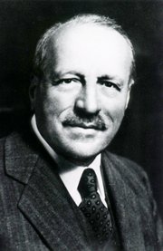
Jean George Bachmann
(1877 – 1959)
French physician–physiologist whose experimental work in the early twentieth century provided the first clear functional description of a preferential interatrial conduction pathway. This structure, eponymically named “Bachmann’s bundle”, plays a central role in normal atrial activation and in the pathophysiology of interatrial block and atrial arrhythmias.
As a young man, Bachmann served as a merchant sailor, crossing the Atlantic multiple times. He emigrated to the United States in 1902 and earned his medical degree at the top of his class from Jefferson Medical College in Philadelphia in 1907. He stayed at this Medical College as a demonstrator and physiologist. In 1910, he joined Emory University in Atlanta. Between 1917 -1918 he served as a medical officer in the US Army. He retired from Emory in 1947 and continued his private medical practice until his death in 1959.
On the personal side, Bachmann was a man of many talents: a polyglot, he was fluent in German, French, Spanish and English. He was a chef in his own right and occasionally worked as a chef in international hotels. In fact, he paid his tuition at Jefferson Medical College, working both as a chef and as a language tutor.
The intrinsic cardiac conduction system was a major focus of cardiovascular research in the late nineteenth and early twentieth centuries. The atrioventricular (AV) node was discovered and described by Sunao Tawara and Karl Albert Aschoff in 1906, and the sinoatrial node by Arthur Keith and Martin Flack in 1907.
While the connections that distribute the electrical impulse from the AV node to the ventricles were known through the works of Wilhelm His Jr, in 1893 and Jan Evangelista Purkinje in 1839, the mechanism by which electrical impulses spread between the atria remained uncertain.
In 1916 Bachmann published a paper titled “The Inter-Auricular Time Interval” in the American Journal of Physiology. Bachmann measured activation times between the right and left atria and demonstrated that interruption of a distinct anterior interatrial muscular band resulted in delayed left atrial activation. He concluded that this band constituted the principal route for rapid interatrial conduction.
Subsequent anatomical and electrophysiological studies confirmed the importance of the structure described by Bachmann, which came to bear his name. Bachmann’s bundle is now recognized as a key determinant of atrial activation patterns, and its dysfunction is associated with interatrial block, atrial fibrillation, and abnormal P-wave morphology. His work remains foundational in both basic cardiac anatomy and clinical electrophysiology.
Sources and references
1. Bachmann G. “The inter-auricular time interval”. Am J Physiol. 1916;41:309–320.
2. Hurst JW. “Profiles in Cardiology: Jean George Bachmann (1877–1959)”. Clin Cardiol. 1987;10:185–187.
3. Lemery R, Guiraudon G, Veinot JP. “Anatomic description of Bachmann’s bundle and its relation to the atrial septum”. Am J Cardiol. 2003;91:148–152.
4. "Remembering the canonical discoverers of the core components of the mammalian cardiac conduction system: Keith and Flack, Aschoff and Tawara, His, and Purkinje" Icilio Cavero and Henry Holzgrefe Advances in Physiology Education 2022 46:4, 549-579.
5. Knol WG, de Vos CB, Crijns HJGM, et al. “The Bachmann bundle and interatrial conduction” Heart Rhythm. 2019;16:127–133.
6. “Iatrogenic biatrial flutter. The role of the Bachmann’s bundle” Constán E.; García F., Linde, A.. Complejo Hospitalario de Jaén, Jaén. Spain
7. Keith A, Flack M. The form and nature of the muscular connections between the primary divisions of the vertebrate heart. J Anat Physiol 41: 172–189, 1907.
"Clinical Anatomy Associates, Inc., and the contributors of "Medical Terminology Daily" wish to thank all individuals who donate their bodies and tissues for the advancement of education and research”.
Click here for more information
- Details
This term arises from the Latin [lumbus] meaning "loin". The "loins" region of the body is the area lateral to the umbilicus, wrapping around posteriorly. In fact, there are two abdominal regions known as the "lumbar regions".
Galen of Pergamon(129AD - 200AD) named and described the lumbar vertebrae.
- Details
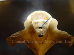
[UPDATED] The basivertebral foramen is an opening in the posterior aspect of each vertebral body (click on the picture for a larger image) which allows the exit of the basivertebral veins. These foramina can be single (as seen in the image) or multiple. The basivertebral veins represent a communication that allows drainage of the vertebral body venous sinuses into the extensive complex venous network of the internal venous plexuses that surrounds the spinal cord.
The plexus of veins around the spinal cord are known as Batson's plexus. this plexus is named after Oscar Vivian Batson (1894 - 1979), an American anatomist and ENT.
Image property of: CAA, Inc. Photographer: David M. Klein
- Details
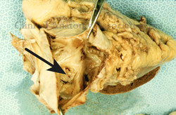
[UPDATED] Latin words meaning "oval fossa" or "oval depression". The fossa ovalis is, as it names implies, an oval-shaped depression in the interatrial wall of the right ventricle. (see image, pointer "A"). The fossa ovalis represents in the adult the fetal communication between the right and left atrium allowing for fetal oxygenated blood to bypass the pulmonary circulation and enter the systemic circulation directly. The fossa ovalis is closed upon birth by two opposing membranes, and the higher pressure on the left side of the heart.
The persistence of the communication between the right and left atrium is known as an Atrial Septal Defect (ASD) and will need surgical correction. Some anatomists refer to this depression as the "foramen ovale" and it is surrounded by a well-defined muscular border known as the "limbus fossa ovalis", also known by the eponym "ring or anulus of Vieussens"
The interatrial opening in the fetus, and the persistent ASD in the adult is referred by the eponym "foramen of Botallus, remembering Leonardo Botallus.
The image shows a human heart with the right atrium opened. The black arrow points to the depression of the foramen ovale in the adult.
For more information:
• On fetal circulation
• On the fossa ovalis (Gray's Anatomy)
Image property of: CAA, Inc. Photographer: David M. Klein
- Details
This root term is Greek and means "diaphragm", referring specifically to the respiratory diaphragm. Because of the importance to life of the region that includes the respiratory diaphragm, liver, and heart, Greek and Roman physicians saw this area as the seat of emotions, a concept that later evolved to include mental instability, and mind. So, for them the seat or location of the emotional processes was not in the brain, but in the area of conjunction of liver, heart, and respiratory diaphragm.
In the late 1700's, when Franz J. Gall developed a pseudoscience that looked at protuberances in the cranium as predictors of mental capabilities and mental pathologies, he named it [phrenology], or "study of the mind". In fact, look at the word "frenzy" and you will see a variation of the root [-phren-] in it!
Today we use the root term [-phren-] mostly to denote "respiratory diaphragm", although it can be found in medical words such as [schizophrenia] meaning "mind".
- Details
This article is part of the series "A Moment in History" where we honor those who have contributed to the growth of medical knowledge in the areas of anatomy, medicine, surgery, and medical research.

George Papanicolaou, MD, PhD
George Papanicolau, MD, PhD (1883 - 1962) Greek physician, cytologyst, physyiologist, and zoologist, Papanicolaou was born on the island of Euboea, Greece. His original Greek name is Georgios Nikolaou Papanikolaou. He initially attended the University of Athens, earning his MD in 1904.
After two years as a conscript in the Army, Papanicolaou continued additional studies in Zoology earning his PhD in 1910 at the University of Munich, later going back to the army as a physician.
Papanicolaou immigrated to the USA in 1913 with his wife. He took a position at the anatomy department of the Cornell University where over the next 45 years established the field of diagnostic cytology, becoming a Professor Emeritus of Clinical Anatomy.
His studies took him to study the cytological patterns in the internal genitalia of guinea pigs, establishing a “cytological calendar”. This study he continued in humans, where he discovered malignant changes in some of the cells obtained. His study on vaginal smears was published in 1943, starting what today is known as a “Pap smear”, a simple annual study that saves thousands of women from cervical and uterine cancer.
Dr. Papanicolaou died in 1960. His personal motto was “I live to serve life”
Sources:
1. “George Nicholas Papanicolaou, 1883-1962” Owens, C. Endoc T 2008:6(5) 28
2. "George N Papanicolaou (1883-1962) MD" Frangos, CC. J Me Biog 17 (3) 134
3. "George N. Papanicolaou, MD, PhD" Elgert, P A; Lab Med (2009) 40;4 245
4. "The diagnostic value of vaginal smears in carcinoma of the uterus" Papanicolaou, GN; Traut HF; Amm J Obst Gyn 1941; 42:193-206.
- Details
This compound term is Latin, from [bifurcus] meaning double-pronged. The prefix [bi-] means "two" or "double", and the root [-furcat- ], derived from the Latin [furca] meaning "a fork". Bifurcation means "to fork into two".
In anatomy the term is used to denote the splitting of a structure into two, such as the "bifurcation of the aorta". A variation of the term is "bifid".


