Medical Terminology Daily (MTD) is a blog sponsored by Clinical Anatomy Associates, Inc. as a service to the medical community. We post anatomical, medical or surgical terms, their meaning and usage, as well as biographical notes on anatomists, surgeons, and researchers through the ages. Be warned that some of the images used depict human anatomical specimens.
You are welcome to submit questions and suggestions using our "Contact Us" form. The information on this blog follows the terms on our "Privacy and Security Statement" and cannot be construed as medical guidance or instructions for treatment.
We have 1113 guests online

Jean George Bachmann
(1877 – 1959)
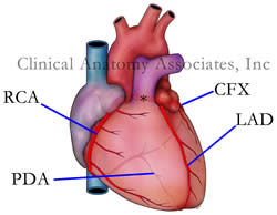
French physician–physiologist whose experimental work in the early twentieth century provided the first clear functional description of a preferential interatrial conduction pathway. This structure, eponymically named “Bachmann’s bundle”, plays a central role in normal atrial activation and in the pathophysiology of interatrial block and atrial arrhythmias.
As a young man, Bachmann served as a merchant sailor, crossing the Atlantic multiple times. He emigrated to the United States in 1902 and earned his medical degree at the top of his class from Jefferson Medical College in Philadelphia in 1907. He stayed at this Medical College as a demonstrator and physiologist. In 1910, he joined Emory University in Atlanta. Between 1917 -1918 he served as a medical officer in the US Army. He retired from Emory in 1947 and continued his private medical practice until his death in 1959.
On the personal side, Bachmann was a man of many talents: a polyglot, he was fluent in German, French, Spanish and English. He was a chef in his own right and occasionally worked as a chef in international hotels. In fact, he paid his tuition at Jefferson Medical College, working both as a chef and as a language tutor.
The intrinsic cardiac conduction system was a major focus of cardiovascular research in the late nineteenth and early twentieth centuries. The atrioventricular (AV) node was discovered and described by Sunao Tawara and Karl Albert Aschoff in 1906, and the sinoatrial node by Arthur Keith and Martin Flack in 1907.
While the connections that distribute the electrical impulse from the AV node to the ventricles were known through the works of Wilhelm His Jr, in 1893 and Jan Evangelista Purkinje in 1839, the mechanism by which electrical impulses spread between the atria remained uncertain.
In 1916 Bachmann published a paper titled “The Inter-Auricular Time Interval” in the American Journal of Physiology. Bachmann measured activation times between the right and left atria and demonstrated that interruption of a distinct anterior interatrial muscular band resulted in delayed left atrial activation. He concluded that this band constituted the principal route for rapid interatrial conduction.
Subsequent anatomical and electrophysiological studies confirmed the importance of the structure described by Bachmann, which came to bear his name. Bachmann’s bundle is now recognized as a key determinant of atrial activation patterns, and its dysfunction is associated with interatrial block, atrial fibrillation, and abnormal P-wave morphology. His work remains foundational in both basic cardiac anatomy and clinical electrophysiology.
Sources and references
1. Bachmann G. “The inter-auricular time interval”. Am J Physiol. 1916;41:309–320.
2. Hurst JW. “Profiles in Cardiology: Jean George Bachmann (1877–1959)”. Clin Cardiol. 1987;10:185–187.
3. Lemery R, Guiraudon G, Veinot JP. “Anatomic description of Bachmann’s bundle and its relation to the atrial septum”. Am J Cardiol. 2003;91:148–152.
4. "Remembering the canonical discoverers of the core components of the mammalian cardiac conduction system: Keith and Flack, Aschoff and Tawara, His, and Purkinje" Icilio Cavero and Henry Holzgrefe Advances in Physiology Education 2022 46:4, 549-579.
5. Knol WG, de Vos CB, Crijns HJGM, et al. “The Bachmann bundle and interatrial conduction” Heart Rhythm. 2019;16:127–133.
6. “Iatrogenic biatrial flutter. The role of the Bachmann’s bundle” Constán E.; García F., Linde, A.. Complejo Hospitalario de Jaén, Jaén. Spain
7. Keith A, Flack M. The form and nature of the muscular connections between the primary divisions of the vertebrate heart. J Anat Physiol 41: 172–189, 1907.
"Clinical Anatomy Associates, Inc., and the contributors of "Medical Terminology Daily" wish to thank all individuals who donate their bodies and tissues for the advancement of education and research”.
Click here for more information
- Details
This is a word of Greek origin. The prefix [epi] means "outer" or "above"; the root term [-thel-] means "nipple" or "female", and the suffix [-ium] means "layer" or "membrane. The reason for the origin of this words is that in c.1700 Ryusch used this term to refer to the surface layer of cells in the nipple and areola. It was later used to denote any covering superficial layer of cells. The plural form for epithelium is epithelia.
There are many types of epithelia in the body, and they are described by their histology, or how they look under the microscope: single-layer, multilayered, cuboidal, columnar, etc.
- Although the above is the standard accepted etymology for this word, I have a different interpretation, as the Latin term [tela], meaning "fabric" or "cover" could have been used. Thus explained, the word means "outer cover layer". Who knows?. Dr. Miranda -
"The origin of Medical Terms" Skinner, AH, 1970
- Details
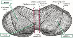
The word [cerebellum] is Latin and means "little brain". The cerebellum is one of the three main gross components of the brain (encephalon), the other two being the cerebral hemispheres and the brain stem.
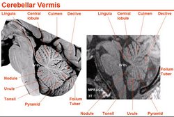
The cerebellum is characterized by a tightly folded external cortex where the gyri are long and parallel to each other and the sulci are not very deep. Upon gross examination, the cerebellum presents with two lateral lobes (left and right) and a median lobe known as the vermis. Other authors divide the cerebellum into an anterior and posterior lobe separated by a primary fissure or sulcus, also known as the preclival sulcus.
The cerebellum is located posterior to the brain stem and posteroinferior to the cerebral hemispheres. It is separated from the occipital lobes of the brain by an extension of dura mater called the tentorium cerebelli. Because of its location, the cerebellum serves as a roof for the 4th ventricle, a component of the ventricular system of the brain. Click here to see a median section, of the cerebellum where you can observe its location and relation to the brain stem, 4th ventricle, and occipital lobe.
The cerebellum is part of the motor control of the brain and is involved in motor coordination, precision, balance and accurate motor timing. Cerebellar dysfunction does not cause paralysis, but produces fine motor control disorders.
Median section image link courtesy of UCLA Radiology.
Sources:
1 "Tratado de Anatomia Humana" Testut et Latarjet 8 Ed. 1931 Salvat Editores, Spain
2. "Anatomy of the Human Body" Henry Gray 1918. Philadelphia: Lea & Febiger
Image modified by CAA, Inc. Original image courtesy of bartleby.com
- Details
This article is part of the series "A Moment in History" where we honor those who have contributed to the growth of medical knowledge in the areas of anatomy, medicine, surgery, and medical research.
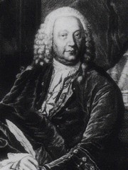
Abraham Vater
Abraham Vater (1684 - 1751) German anatomist and physician, Abraham Vater was the son of a distinguished physician and was born on the city of Wittenberg. He obtained a Doctorate in Philosophy (PhD) in 1706 and his medical degree in 1710 at the University of Leipzig. He became a professor “extraordinarius” of Anatomy and Botany in 1719, continuing his career in anatomy until he obtained the highest professorial degree at the University.
After the death of his father in 1733 Abraham Vater was appointed to the chair of Anatomy. In 1720 he published the discovery of a “biliary diverticulum” , the hepatopancreatic ampulla, known today as the “Ampulla of Vater” or duodenal papilla. His original article was entitled “Dissertatio anatomica qua novum bilis diverticulum circa orificium ductus choledochi”. He also described a large subcutaneous sensory nerve terminal known as the “Vater–Pacinian corpuscle”.
Known as an anatomist, Vater also wrote on surgery, gynecology, pharmacology, pathology, chemistry, and botany, making him a complete scientist. In a twist of fate, Vater died in 1751 after being afflicted with jaundice, probably a consequence of biliary blockage of his eponymic ampulla.
Sources:
1. "Abraham Vater (1684-1751)" Brit Med J; 2,(4) (1951), 1214
2. "Abraham Vater of the Ampulla (Papilla) of Vater" Lerch, MM; Domschke, W. Gastroenterology (2000) 118: (2) 379
- Details
The word [epicardium] is composed by the prefix [epi-], meaning "outer" or "above"; the root term [-card-], meaning "heart"; and the suffix [-ium], meaning "layer" or "membrane". Thus, the word means "outer layer of the heart".
The epicardium is part of a larger structure called the pericardium, in fact, since the pericardium is in contact with a viscus (the heart), it can also be called "visceral pericardium". It must be noted that these two terms are synonyms: "epicardium" and "visceral pericardium".
The epicardium is a type of serosa composed of an outer layer formed by mesothelial cells and an inner layer formed by loose connective tissue containing varying amounts of fat. This inner layer known as the "subepicardial layer" (a misnomer) also contains elastic fibers.
The main arterial and venous components of the coronary circulation are found in the subepicardial layer. When performing a Coronary Artery Bypass Graft (CABG), a surgeon has to open the outer layer of the epicardium, and enter the subepicardial layer to perform the graft anastomosis.
As a serosa, the epicardium is involved in the production and absorption of a clear fluid, the "pericardial fluid". This fluid acts as a lubricant for the pericardium, allowing for the effortless movement of the heart within the pericardial sac.
- Details
The suffix [-iasis] has an original Greek root (such as in [iatros] meaning "healer" or "hospital"). It later became a Latin root meaning "condition, pathology, or disease".
This suffix can be found in many medical terms such as:
- Choledocolithiasis - a condition of stones in the bile duct.
- Elephantiasis - A condiction where a body part grows unusually large, usually lower extremities and/ or scrotum
- Phthiriasis- A condition or infestarion with public or crab lice
- Helminthiasis- A bodily infestation with a type of parasitic worms called helminths (tapeworms)
- Cystolithiasis - Bladder stones
- Details
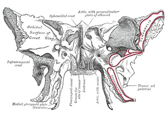
Anterior view of the sphenoid bone
Both these root terms have their origin from the Greek [πτέρυγα] (ptéryga) and mean "wing".
In human anatomy the most common use of this root term is in the word [pterygoid]. Since the suffix [-oid] means "similar to", the word pterygoid means "similar to a wing", or "wing-like".
On the inferior aspect of the sphenoid bone (os sphenoidale) there are two very thin bat-wing-like bony appendages that are called the lateral and medial pterygoid plates. The medial pterygoid plate has a hook-like bony appendage called the hamulus (Latin: little hook).
Related to the pterygoid plates are the lateral and medial pterygoid muscles, both these muscles aid inthe process of mastication.
The root term [pter-] can be found in words such as [pterodactyl] meaning "winged finger", it refers to a phrehistoric winged animal; [pteranodon], and [pterosaurus].
Sources:
1 "Tratado de Anatomia Humana" Testut et Latarjet 8 Ed. 1931 Salvat Editores, Spain
2. "Anatomy of the Human Body" Henry Gray 1918. Philadelphia: Lea & Febiger
Image in the public domain modified by CAA, Inc. Original image courtesy of bartleby.com
Note: Google Translate includes the symbol (?). Clicking on it will allow you to hear the pronunciation of the word.


