![]()
Medical Terminology Daily (MTD) is a blog sponsored by Clinical Anatomy Associates, Inc. as a service to the medical community. We post anatomical, medical or surgical terms, their meaning and usage, as well as biographical notes on anatomists, surgeons, and researchers through the ages. Be warned that some of the images used depict human anatomical specimens.
You are welcome to submit questions and suggestions using our "Contact Us" form. The information on this blog follows the terms on our "Privacy and Security Statement" and cannot be construed as medical guidance or instructions for treatment.
We have 172 guests and no members online
")
Marcia Crocker Noyes
(1869 – 1946)
Further to my comment on old books and research that started with an interesting bookplate (Ex-Libris). I continued my research and found that the person in charge of the Osler library bookplate was a fascinating individual that today maybe a ghost in the MedChi library and building in Baltimore... This is certainly an article that can be called "A Moment in History"
Marcia Crocker Noyes was the librarian at The Maryland State Medical Society from 1896 to 1946 and was a founding member of the Medical Library Association.[1][2][3]
Sir William Osler, MD. a famous Johns Hopkins surgeon was a noted bibliophile and had a large personal collection of books on various topics. When he became the President of MedChi in 1896, he was dismayed at the condition of the library and knew that with the right person and some stewardship, it could become a significant collection. Sir William asked his friend, Dr. Bernard Steiner, a physician and President of the Enoch Pratt Free Library in Baltimore for suggestions of a librarian, and Dr. Steiner recommended Marcia Crocker Noyes. A native of New York, and a graduate of Hunter College, Marcia had moved to Baltimore for a lengthy visit with her sister, and took a “temporary” position at the Pratt Library, which turned into three years. Although she had no medical experience or background, she was enthusiastic, and most importantly, she was willing to move into the apartment provided for the librarian, who needed to be available 24 hours a day.
The image in this article is Ms. Noyes on her first year on the job. Marcia developed a book classification system for medical books, based on the Index Medicus, and called it the Classification for Medical Literature. The system uses the alphabet with capital letters for the major divisions of medicine and lower-case ones for the sub-sections. The system was used for many years, but it's now dated and the Faculty's original shelving scheme was never changed. The card catalogs still reflect her classification and many of the cards are written in Marcia's back-slanting handwriting.
Marcia knew enough to ask the Faculty's members about medical questions, terminology and literature. She gradually won over the predominantly male membership and they became her greatest allies; Sir William at the start, and then for nearly 40 years, Dr. John Ruhräh, a wealthy pediatrician with no immediate family of his own. She made a point of attending almost every Faculty function, and in 1904, under guidelines from the American Medical Association, Marcia was made the Faculty Secretary. For much of her first 10 years, she was the Faculty's only full-time employee, only being assisted by Mr. Caution, the Faculty's janitor. Later in life Marcia would say that she hired him because of his name!
Within ten years, the library had outgrown its space, and plans, spearheaded by Marcia and Sir William before his move to Oxford, were made to build a headquarters building, mainly to house the library's growing collection of medical books and journals.
Marcia was instrumental in the design and building of the new headquarters. She travelled to Philadelphia, New York and Boston to look at their medical society buildings, and eventually, the Philadelphia architectural firm, Ellicott & Emmart was selected to design and build the new Faculty building. Every detail of the building held her imprimatur, from the graceful staircase, to the light-filled reading room, and all of the myriad details of the millwork, marble tesserae, and most of all, the four-story cast iron stacks. She was on-site, climbing up unfinished staircases, checking out the progress of the building, which was built in less than one year at a cost of $90,000.
Among the features of the new building was a fourth-floor apartment for her. She referred to it as the "first penthouse in Baltimore" and it had a garden and rooftop terrace. The library collection eventually grew to more than 65,000 volumes from medical and specialty societies around the world. Journals were traded back and forth, and physicians eagerly anticipated the arrival of each new issue. At the same time, Marcia was involved in the Medical Library Association as one of eight founding members. The MLA promotes medical libraries and the exchange of information. One of the earliest mandates of the MLA was the Exchange, a distribution and trade service for those who had duplicates or little-used books in their collections. Initially, the Exchange was run out of the Philadelphia medical society, but in 1900 it was moved to Baltimore and Marcia oversaw it. Several hundred periodicals and journals were received and sent each month, a huge amount of work for a tiny staff. In 1904, the Faculty had run out of room to manage the Exchange, so it was moved to the Medical Society of the Kings County (Brooklyn). But without Marcia's excellent administrative skills, it floundered and in 1908, the MLA asked Marcia to take charge once again.
In 1909, when the new Faculty building opened, there was enough room to run the Exchange and with the help of MLA Treasurer, noted bibliophile and close friend, Dr. John Ruhräh, it once again became successful. Additionally, Marcia and Dr. Ruhräh combined forces to revive the MLA's bulletin, which had all but ceased publication in 1908, taking the Exchange with it. This duo maintained editorial control from 1911 until 1926. In 1934, around the time of Dr. Ruhräh's death, Marcia became the first “unmedicated” professional to head the MLA. During her tenure, the MLA incorporated, the first seal was adopted, and the annual meeting was held in Baltimore. Marcia wanted to write the history of the MLA once she retired from full-time work at the Faculty, but her health was beginning to fail. She had back problems and had suffered a serious burn on her shoulder as a young woman, possibly from her time running a summer camp, Camp Seyon, for young ladies in the Adirondack Mountains. In 1946, a celebration was planned to honor Marcia's 50 years at the Faculty. But she was adamant that the physicians wait until November, the actual date of her 50 years. However, they knew she was gravely ill, and might not make it until then, so a huge party was held in April. More than 250 physicians attended the celebration, but the ones she was closest to in the early years, were long gone. She was presented with a suitcase, a sum of money to use for travelling, and her favorite painting of Dr. John Philip Smith, a founder of the Medical College in Winchester, Virginia. It was painted by Edward Caledon Smith, a Virginia painter who had been a student of the painter Thomas Sully.[4] She adored this painting and vowed, jokingly, to take it with her wherever she went.
The painting was not to stay with her for very long, for she died in November 1946, and left it to the Faculty in her will. Her funeral was held in the Faculty's Osler Hall, named for her dear friend. More than 60 physicians served as her pallbearers, and she was buried at Baltimore's Green Mount Cemetery. In 1948, the MLA decided to establish an award in the name of Marcia Crocker Noyes. It was for outstanding achievement in medical library field and was to be awarded every two years, or when a truly worthy candidate was submitted. In 2014, the Faculty began giving a bouquet of flowers to the winner of the award in Marcia's name, and in honor of her work. Much evidence exists for this tradition, as we know that the physicians, especially Drs. Osler and Ruhräh, frequently gave her bouquets of flowers. Marcia also cultivated flower gardens at the Faculty and decorated the rooms with her work.
Today, the MedChi building is open for tours and if the rumors are to be believed Ms. Marcia Crocker Noyes is still at work in her beloved library as the "resident ghost" [1][5]

NOTE: This article has been modified from the original Wikipedia article on Marcia Crocker Noyes. The article itself is well-written with interesting images of the subject. I would encourage you to visit it. The second insert is from book 00736 in my personal library and shows in pencil, the incredibly small handwriting of Marsha C. Noyes.
Sources:
1. "Marcia, Marcia, Marcia" MedChi Archives blog.
2. "Marcia C. Noyes, Medical Librarian" (PDF). Bulletin of the Medical Library Association. 35 (1): 108–109. 1947. PMC 194645
3. Smith, Bernie Todd (1974). "Marcia Crocker Noyes, Medical Librarian: The Shaping of a Career" (PDF). Bulletin of the Medical Library Association. 62 (3): 314–324. PMC 198800Freely accessible. PMID 4619344.
4. Edward Caledon BRUCE (1825-1901)"
5. Behind the scenes tour MedChiBuilding
"Clinical Anatomy Associates, Inc., and the contributors of "Medical Terminology Daily" wish to thank all individuals who donate their bodies and tissues for the advancement of education and research”.
Click here for more information


- Details

Click for a larger image
The [sphincter of Oddi] is a complex system of smooth muscles that controls flow of bile and pancreatic juice into the duodenum. Although known by its eponym, this structure has the anatomical name of "sphincter of the hepatopancreatic ampulla". Although described previously by others, it was Ruggero Oddi (1864-1913) who described not only its structure, but also its function.
The hepatopancreatic ampulla or "ampulla of Vater" is a dilation found at the conjunction and end of the common bile duct and pancreatic duct. The presence of the hepatopancreatic ampulla creates a nipple-like elevation of the duodenal mucosa called the "duodenal papilla".
The sphincter of Oddi has several components:
- Sphincter papillae: This portion of the sphincter surrounds the papillary and intramural portion of the hepatopancreatic ampulla
- Sphincter choledochus: This portion of the sphincter surrounds the most distal portion of the common bile duct. It must be noted that this is the narrowest portion of the common bile duct, allowing for potential lodging of bile stones, cause for choledocholithiasis
- Sphincter pancreaticus: This portion of the sphincter surrounds the most distal portion of the pancreatic duct and prevents reflux of bile from the hepatopancreatic ampulla to the pancreatic duct.
The duodenal muscular layer parts to allow passage of the complex formed by the hepatopancreatic ampulla and the sphincter of Oddi, creating a window called the "choledochal window". Longitudinal fibers from the duodenal muscularis externa pass and join to the sphincter of Oddi.
- Details
This is a root term of Greek origin. In both presentations [-chol-] or [-chole-] it means "bile" or "gall". The English word "gall" is of Anglosaxon origin and means "bile", referring to its yellowish-green color. The word [bile] is of Latin origin, from [bilis].
These root terms are used in many medical words, such as:
- cholecystitis: [cyst] means "sac" or "bladder", [itis] means "inflammation" or "infection". Gallbladder inflammation
- cholecystectomy: [cyst] means "sac" or "bladder", [ectomy] means "removal". Gallbladder removal
- cholangiogram: [angi] means "vessel", [(o)gram] means "examination". Examination of a bile vessel
- choledocholithiasis: Condition of stones in the bile duct. Click on the link for more information
- cholera: The suffix [-era] is "flow" or "discharge". The term refers to the constant vomiting of bile in patients afflicted with this disease. This term has been heatedly discussed and this is but one of the theories as to the etymology of the word.
In the early days of physiology, yellow bile was considered one of the "four humors" that made up human personality, temperament, and health. A person with an excess of yellow bile would be considered bad tempered, angry, or "choleric". Observe how the root term [chole] also is found in the word "choleric".
Sources:
1. "The Language of Medicine" John H. Dirckx Pub: Harper & Row 1976
2. "Medical Meanings" Haubrich, William S. Am Coll Phys Philadelphia 1997
3. "The origin of Medical Terms" Skinner, AH, 1970
- Details
This article is part of the series "A Moment in History" where we honor those who have contributed to the growth of medical knowledge in the areas of anatomy, medicine, surgery, and medical research.

Ruggero Oddi
Ruggero Oddi (1864-1913). Anatomist and physician, his complete name was Ruggero Ferdinando Antonio Giuseppe Vincenzo Oddi Pampaglini, born on July 20, 1814 in the city of Perugia, Italy. He studied medicine in the University of Perugia, where he had a keen interest in anatomy and physiology, graduation with a medical degree in 1889. In 1887, as a fourth year medical student Oddi published a paper that would make his name eponymically tied to the sphincter found around the hepatopancreatic ampulla; what today is known as the "sphincter of Oddi". His paper was entitled "Di una Speciale Disposizione a Sfintere allo Sbocco del Coledoco" (On a Special Sphincteric Arrangement at the Outlet of the Common Bile Duct).
Although the circular muscle of the sphincter of Oddi had already been described by Glisson in 1681, Oddi was the one who did a complete anatomical and physiological study of this structure uncovering the fact that it was indeed a sphincter. He continued his studies on the hepatobiliary sphincter until 1894, when he moved to Congo and later back to Belgium.
Because of his inclination towards metaphysical studies, Oddi started experimenting with drugs on himself and became addicted.
His later life was surrounded by scandal and controversy, because of drug abuse and fiscal mismanagement of University funds. Oddi died in poverty in 1913 and his site of burial is unknown.
Sources:
1. "Ruggero Ferdinando Antonio Guiseppe Vincenzo Oddi" Lukas, M, et al. World J Surg (2007) 31:2260–2265
2. "Ruggero Oddi; To commemorate the centennial of his original article--"Di unaspeciale disposizione a sfintere allo sbocco del coledoco" Ono, K; Hada, R. Jap J Surg, VOL. 18, No. 4 pp. 373-375, 1988
3. "Ruggero Oddi: 120 years after the description of the eponymous sphincter: A story to be remembered" Capodicasa, E. J Gastroent Hepat 23 (2008) 1200–1203
4. "Oddi: The Paradox of the Man and the Sphincter" Modlin, IM; Ahlman, H. Arch Surg 129 (1994) May 550-557
Original image in the public domain, courtesy of the National Library of Medicine.
- Details
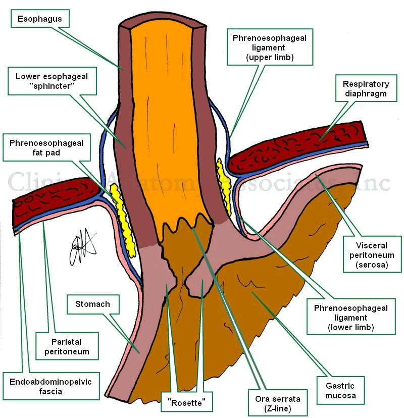
Esophagogastric junction
The "Angle of His" refers to the normally acute angle between the abdominal esophagus and the fundus of the stomach at the esophagogastric junction.
This angle is one of the elements that are important in the prevention of gastroesophageal reflux disease (GERD). When the fundus of the stomach gets expanded by air, because of the normal anatomy and relations of the esophageal hiatus, the esophagogastric junction structures are "pushed" from left to right, pushing close the gastroesophageal flap valve or "rosette". There are other mechanisms that add to the sphincter-like action of the esophagogastric junction structures.
The eponym "angle of His" remembers Dr. Wilhem His Jr. (1864 -1934), a German physician and anatomist, who also described the atrioventricular bundle or "Bundle of His", one of the components of the conduction system of the heart.
Thanks to Debbie Donovan for suggesting this post.
Images property of:CAA.Inc. Artist:Dr. E. Miranda
- Details
![Coronary Arteries. The [*] indicates the left coronary artery](/images/MTD/LargeImages/coronaryarterieslabels_lg.jpg "Coronary Arteries.")
Click for a larger image
The term [coronary dominance] is the answer to the following question: From which coronary artery does the posterior interventricular artery (PDA) arise?
In most of the human species the PDA arises from the right coronary artery, (see accompanying image), therefore most humans (70%) are right dominant. The rest are either left dominant (10%) or have balanced dominance (20%). These statistics have significant variation in different studies.
In the case of balanced dominance, there is either a double posterior interventricular artery, where one is a branch of the right coronary artery and the other a branch of the left coronary artery, or a single PDA receiving blood supply from both coronary arteries.
Coronary dominance is important because the interventricular septum receives blood supply from the PDA in its posterior 1/3rd. If the heart is left dominant, all the blood supply of the interventricular septum is dependant on the left coronary artery. In this case, blockage of the left coronary artery can be catastrophic!
There can be interesting anatomical variations in the coronary arteries of the heart. For a detail on these anatomical variations, click here. Heart and coronary artery anatomy is one of the many lecture topics presented by CAA, Inc
Image property of:CAA.Inc.. Artist:Victoria G. Ratcliffe
- Details
This is a word of Greek origin. The prefix [a-) means "absence of", or "without". The root term [-phon-] means "sound" or "voice". Aphonia is a pathological absence of voice, and was used by both Hippocrates and Galen.
Do not confuse [aphonia] with [dysphonia], where the prefix [dys-] means "abnormal". In aphonia there is total absence of voice, whereas in dysphonia there is an abnormal voice or "hoarseness"
As a side note, the word [phonograph] arises from the combination of the root terms [-phon-] and [-graph-], which means "to write". The word [phonograph] does not relate to the playing of a record, but rather to the process of creating one, transforming sound into a wavy line etched on a rotating wax model that is later cast into records. The modern production of sound CD's is similar, where the sound waves are act upon a laser that "burns" the track into a master CD. It is a similar process, but I guess calling creating a CD a type of "phonography" is too old fashion for modern marketing!