Medical Terminology Daily (MTD) is a blog sponsored by Clinical Anatomy Associates, Inc. as a service to the medical community. We post anatomical, medical or surgical terms, their meaning and usage, as well as biographical notes on anatomists, surgeons, and researchers through the ages. Be warned that some of the images used depict human anatomical specimens.
You are welcome to submit questions and suggestions using our "Contact Us" form. The information on this blog follows the terms on our "Privacy and Security Statement" and cannot be construed as medical guidance or instructions for treatment.
We have 535 guests online

Georg Eduard Von Rindfleisch
(1836 – 1908)
German pathologist and histologist of Bavarian nobility ancestry. Rindfleisch studied medicine in Würzburg, Berlin, and Heidelberg, earning his MD in 1859 with the thesis “De Vasorum Genesi” (on the generation of vessels) under the tutelage of Rudolf Virchow (1821 - 1902). He then continued as a assistant to Virchow in a newly founded institute in Berlin. He then moved to Breslau in 1861 as an assistant to Rudolf Heidenhain (1834–1897), becoming a professor of pathological anatomy. In 1865 he became full professor in Bonn and in 1874 in Würzburg, where a new pathological institute was built according to his design (completed in 1878), where he worked until his retirement in 1906.
He was the first to describe the inflammatory background of multiple sclerosis in 1863, when he noted that demyelinated lesions have in their center small vessels that are surrounded by a leukocyte inflammatory infiltrate.
After extensive investigations, he suspected an infectious origin of tuberculosis - even before Robert Koch's detection of the tuberculosis bacillus in 1892. Rindfleisch 's special achievement is the description of the morphologically conspicuous macrophages in typhoid inflammation. His distinction between myocardial infarction and myocarditis in 1890 is also of lasting importance.
Associated eponyms
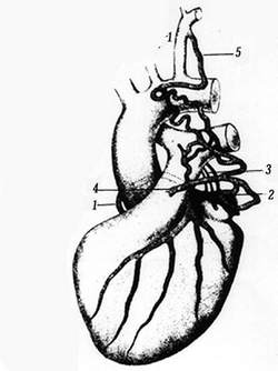
"Rindfleisch's folds": Usually a single semilunar fold of the serous surface of the pericardium around the origin of the aorta. Also known as the plica semilunaris aortæ.
"Rindfleisch's cells": Historical (and obsolete) name for eosinophilic leukocytes.
Personal note: G. Rindfleisch’s book “Traité D' Histologie Pathologique” 2nd edition (1873) is now part of my library. This book was translated from German to French by Dr. Frédéric Gross (1844-1927) , Associate Professor of the Medicine Faculty in Nancy, France. The book is dedicated to Dr. Theodore Billroth (1829-1894), an important surgeon whose pioneering work on subtotal gastrectomies paved the way for today’s robotic bariatric surgery. Dr. Miranda.
Sources:
1. "Stedmans Medical Eponyms" Forbis, P.; Bartolucci, SL; 1998 Williams and Wilkins
2. "Rindfleisch, Georg Eduard von (bayerischer Adel?)" Deutsche Biographie
3. "The pathology of multiple sclerosis and its evolution" Lassmann H. (1999) Philos Trans R Soc Lond B Biol Sci. 354 (1390): 1635–40.
4. “Traité D' Histologie Pathologique” G.E.
Rindfleisch 2nd Ed (1873) Ballieres et Fils. Paris, Translated by F Gross
"Clinical Anatomy Associates, Inc., and the contributors of "Medical Terminology Daily" wish to thank all individuals who donate their bodies and tissues for the advancement of education and research”.
Click here for more information
- Details
"Nothing in the human body is colored, or labeled"
"The Chirurgeon must knowe the Anatomie". Thus states Thomas Vicary (1460 -1561) on the knowledge of Anatomy. He continues: "...for all authors write against those surgeons who work in a man's body not knowing the Anatomie"1. There is no doubt that knowledge must include the awareness of the possibility of anatomical variations. Some anatomical variations, like the "Corona Mortis" can be critical, and in some surgical cases, be the cause for exsanguination!
It is interesting that several medical schools are reducing the total number of hours working on, or moving away from cadaver disection in first year medical school and using computer simulations instead. No computer simulation will give the medical student the detail, variations, and feel of the tissues as actual hands-on experience. I am sure no one wants a surgeon whose first view of the internal aspect of a human body is a living patient...on the surgical table.
It is a fact that "Nothing in the human body is really colored... or labeled" or as someone else said "nothing looks exactly like the anatomy book", unless it is photography, and then each photo is taken after hours of laboring to "Netterize" the organ or area that one is trying to detail. Nothing gives the future professional the exact idea of what to expect in the future patient than the hours and hours of laborious work in the anatomy laboratory.
The same is true with anatomical variations, one "standard" digital cadaver,even with built-in anatomical variations does not give the student the sense of awe and discovery when an anatomical variation is found, interpreted, and analyzed with a group of peers, contributing to the learning process and the formation of future health care professionals.When questioning what is normal or abnormal, Dr. Elizabeth Murray says it most elegantly: "The cadaver is always right"
The image depicts a case of a coronary artery arising from the pulmonary trunk!
Sources:
1. "The Chirurgeon must knowe the Anatomie" R. Shane Tubbs Clin Anat 26:417 (2013)
2. "Two cases of an abnormal coronary artery of the heart arising from the pulmonary artery"Brooks, H; J. Anat. Physiol. 20:26-29, 1886 (anatomyatlases.org)
THIS ARTICLE IS THE THIRD IN A SERIES. TO READ THE FIRST ARTICLE CLICK HERE
- Details
"No anatomical structure has the moral obligation to be where they are supposed to be"
Not only may an anatomical structure be absent, such as in the case of renal aplasia or agenesis, or in the case of a non-existent circumflex coronary artery, but sometimes extra structures can be found. Such is the case where a kidney can present two or even three ureters, all functional. Double inferior vena cavae, cervical ribs, lumbar ribs, the list goes on and on!
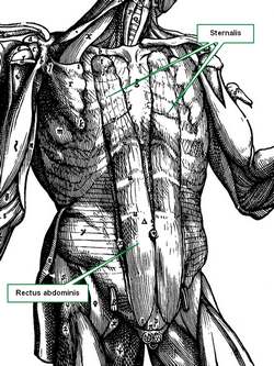
Muscles can be added to this list, again, with absence of a muscle, or with new and completely unexpected attachments. An example of this is the presence of a continuation of the rectus abdominis muscle into the chest region, a variation called a sternalis muscle.
The accompanying image shows the sternalis muscle in one of the "muscle plates" of De Humani Corporis Fabrica Libri Septem, published in 1543 by Andreas Vesalius. This image was criticized by showing a muscle that does not exist, although Vesalius clearly stated in the text of his book that this was an anatomical variation that he had seen.
For many decades surgeons had to operate and "see what they could find". There were the days of the exploratory laparotomy. After the discovery of the application of X-rays by Wilhem Konrad Roentgen (1845 - 1923) and the incredible advances in imaging techniques including CT-scan, MRI, PET, etc, the surgeon is now not usually surprised by anatomical variations.
There are areas in the body that have an high rate of anatomical variation, such as the hepatobiliary region, which includes the "Triangle of Calot". In this area, the standard anatomy is found only in 64% of the cases! In the rest, expect the unexpected. Lahey (1948) states "...the fact that cholecystectomy is a dangerous operation. It is dangerous unless one realizes.... that anomalous anatomy is very common". Today the dangers are less, because of better visualization and technology, but anatomical variations are still there.
Another area where anatomical variations are extremely important is the heart's coronary circulation. Anatomical variations can cause different cardiac dominance. Normal anatomy states that there are two coronary arteries, yet, up to five separate coronary arteries arising directly from the ascending aorta have been described! There is one variation where the left coronary arises from the right coronary artery, effectively having only one artery arise from the aorta and being in charge of all the arterial supply to the heart. What happens if this single artery stenoses? Bear in mind that this is not an "anomalous" vessel, it is just an anatomical variation.
Sources:
1. Lahey DH, discussing the paper "Partial Hepatectomy with Intrahepatic Cholangiojejunostomy" by Wilson H, and Gillespie CE, Ann Surg. 1949 June; 129(6): 756–765
2. "Renal aplasia is the predominant cause of congenital solitary kidneys" Hiraoka, M et al Kidney Int. 2002 May;61(5):1840-4.
This article is the second in a series of three; Click here for the first article.
TO CONTINUE READING: CLICK HERE
- Details
"The only constant in anatomy is variation"
This dictum is incredibly powerful and true. Even the so-called "anatomical constants" are subject to it.
One common misconception is that "we are all the same". This could not be further from the truth. Every body is different from every else's body. Anatomical variations range from the minimal to the incredible. One of the most interesting anatomical variations is the one called "situs inversus". In this case the individual is a mirror image of a human. The apex of the heart points to the right side of the body; the duodenum circles to the right, the liver "hangs" from the left side of the respiratory diaphragm, etc. This particular anatomical variation presents in different degrees and can sometimes coexist with some cardiovascular congenital abnormalities.
Of course there are minor anatomical variations that have no effect on daily life at all and are only discovered by accident, or upon autopsy or dissection. One of the most complete resources on this topic is the Illustrated Encyclopedia of Human Anatomic Variations. An excerpt from this site states: "It is clear that textbook writers and teachers over the centuries, even until today, fail to understand or to transmit to their students the crucial concept that anatomical and physiological diversity and variation is a canon of living organisms. This failure leads to the belief that textbooks are conveying immutable facts with only few anomalous exceptions".
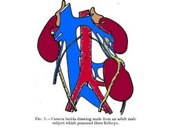
Shown here is an extremely rare case of a third kidney. Dixon (1911) describes in his research paper that as of that date, only 10 cases were known, of these only eight were recorded, with 87% of them found on the left side of the body. Click on the image for a larger depiction.
Source and primary image: "Supernumerary kidney: The occurrence of three kidneys in an adult male subject" Dixon, A.F. J. Anat. Physiol. 45:117-121, 1911.
THIS POST IS CONTINUED, CLICK HERE
- Details
The prefix [para-] has a Greek origin and means "beside" or "alongside". Today we add the meaning of "parallel to".
We see the daily application of this prefix in words such as [paramedic], [parajournalism], [paralogism], and [paranormal]. Medical applications of the term include:
- parasternal: alongside the sternum, such as the internal thoracic vessels
- paramedian: alongside the median plane
- parasagittal: parallel to a sagittal plane (synonym with paramedian)
- paraumbilical: alongside the umbilicus, such as paraumbilical visceral extrusion in a gastroschisis
- parathyroid glands: glands that are found besides the thyroid gland, etc.
- Details
This article is part of the series "A Moment in History" where we honor those who have contributed to the growth of medical knowledge in the areas of anatomy, medicine, surgery, and medical research.
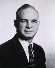
Dr. Otto C. Brantigan
Otto C. Brantigan, MD. (1904-1981) An American surgeon and anatomist, Otto Charles Brantigan was born in Chattanooga, TN in 1904. Having dropped out of high school to help his family and working as a first class machinist, he decided to continue with graduate school. He studied at the Northwestern University in Chicago, where he graduated from the Medical School in 1933. In 1948 he became Chief of Surgery, and eventually became Professor of Surgery, Professor of Thoracic Surgery, and Professor of Anatomy at the Maryland School of Medicine. He retired in 1976 having earned many accolades for his profuse surgical work and publications.
As a surgeon of the times, Dr. Brantigan had a wide area of interest. His over 110 publications and surgical work range from thoracoscopy to vascular, plastic, cardiac, and orthopedic surgery. He is most remembered for the pioneer work he did on chronic obstructive pulmonary disease (COPD), emphysema and lung volume reduction surgery (LVRS), which he presented in 1958. The procedure had (at the time) a very high mortality rate (16 -20%) and Brantigan's work was not readily accepted.
It was not until J. Cooper and his team, revisited the operation proposed by Brantigan that the operation was accepted, now with new surgical stapling and staple line buttressing technology. Dr. Brantigan's name was recognized as a pioneer in lung emphysema surgery, unfortunately 14 years after his death. In 1994 his son, Dr Charles O. Brantigan delivered a beautiful biography of Dr. Otto Brantigan in the same meeting where Cooper presented his results with LVRS.
Personal note: I am proud to own one of the copies of Dr. O.C. Brantigan;s "Clinical Anatomy", a book that I use quite frequently. It is listed in my library catalog. Dr. Miranda.
Sources:
1. "Biography of Otto C Brantigan" C.O. Brantigan 1994 Meeting of the American Association for Thoracic Surgery
2. "LVRS in chronic obstructive pulmonary disease" Davies, L; Calverley, P. Thorax 1996;51(Suppl 2):S29-S34
3. ""Bilateral pneumectomy (volume reduction) for chronic obstructive pulmonary disease" Cooper, J.,The Journal of Thoracic and Cardiovascular Surgery Volume 109, Number 1:106-119
4. "The Surgical Approach to Pulmonary Emphysema" Brantigan, OC; Kress, MB; Mueller, EA. Chest. 1961; 39(5):485-499
5. "History of Emphysema Surgery" Naef, AP. Ann Thorac Surg 1997;64:1506-1508
Original image courtesy of National Institutes of Health.Biography of Dr. Otto Brantigan courtesy of Dr. Charles O. Brantigan.
- Details
The rectum is the most distal segment of the large intestine, along with the anal canal.
The word [rectum] arises from the Latin [rectus] and means "straight", such as its use in the name "rectus abdominis" for the "straight muscle of the abdomen".
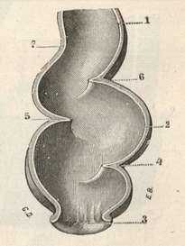
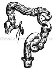
It seems a misnomer, as the rectum of the human species is actually "S" shaped, as seen in the accompanying image. The reason for this discrepancy is that the rectum was named by Galen of Pergamon (129AD - 200 AD) who himself studied this structure in animals such as sheep and goats. In these animals the rectum is indeed straight, and since contradicting Galen was not acceptable (see Michael Servetus), the name has survived until this day. Even Andreas Vesalius has in his 1953 "Fabrica" a depiction of a straight rectum in the human! Click on second image to see a larger depiction of Vesalius' idea of the rectum. Although Vesalius stated that he wanted to show human anatomy as it is, and not as Galen said it should be, here is a demonstration that in 1543 he was still a lukewarm Galenist.
There is an area between the sigmoid and the rectum called the sigmoidorectal junction, although most anatomists call it (wrongly) the rectosigmoid junction (RSJ). This is an anatomically diffuse area with no clear anatomical transition between the sigmoid and the rectum or the RSJ from the rectum.
As the proximal end of the "S" shaped rectum is not clearly discernible from the sigmoidorectal region, there is no clear agreement on the length of the rectum. Authors state that it measures approximately six to seven inches in length (15 - 17 cm), while others measure it as between 8-10 inches. The rectum ends distally at the junction of the rectum with the pelvic diaphragm. It is at this point that the anal canal begins.
The rectum is characterized by three transverse rectal folds, one on the right side, and two on the left side. These folds are know as the "rectal valves" or the "valves of Houston". The middle rectal fold is known to European anatomists as the "valve of Kohlrausch" Their function in maintaining fecal material in place as well as their function in defecation is still under study. The rectal valves also have a high level of anatomical variation and may not be present at all.
Images:
1. "Tratado de Anatomia Humana" Testut et Latarjet 8 Ed. 1931 Salvat Editores, Spain
2. "De Humani Corporis Fabrica, Libri Septem" A. Vesalius 1543 Brussels
Recommended reading: "Transverse Folds of Rectum: Anatomic Study and Clinical Implications" Shafik, A, et al. Clin Anat 14: 196-203 (2001).


