![]()
Medical Terminology Daily (MTD) is a blog sponsored by Clinical Anatomy Associates, Inc. as a service to the medical community. We post anatomical, medical or surgical terms, their meaning and usage, as well as biographical notes on anatomists, surgeons, and researchers through the ages. Be warned that some of the images used depict human anatomical specimens.
You are welcome to submit questions and suggestions using our "Contact Us" form. The information on this blog follows the terms on our "Privacy and Security Statement" and cannot be construed as medical guidance or instructions for treatment.
We have 531 guests and no members online
")
Marcia Crocker Noyes
(1869 – 1946)
Further to my comment on old books and research that started with an interesting bookplate (Ex-Libris). I continued my research and found that the person in charge of the Osler library bookplate was a fascinating individual that today maybe a ghost in the MedChi library and building in Baltimore... This is certainly an article that can be called "A Moment in History"
Marcia Crocker Noyes was the librarian at The Maryland State Medical Society from 1896 to 1946 and was a founding member of the Medical Library Association.[1][2][3]
Sir William Osler, MD. a famous Johns Hopkins surgeon was a noted bibliophile and had a large personal collection of books on various topics. When he became the President of MedChi in 1896, he was dismayed at the condition of the library and knew that with the right person and some stewardship, it could become a significant collection. Sir William asked his friend, Dr. Bernard Steiner, a physician and President of the Enoch Pratt Free Library in Baltimore for suggestions of a librarian, and Dr. Steiner recommended Marcia Crocker Noyes. A native of New York, and a graduate of Hunter College, Marcia had moved to Baltimore for a lengthy visit with her sister, and took a “temporary” position at the Pratt Library, which turned into three years. Although she had no medical experience or background, she was enthusiastic, and most importantly, she was willing to move into the apartment provided for the librarian, who needed to be available 24 hours a day.
The image in this article is Ms. Noyes on her first year on the job. Marcia developed a book classification system for medical books, based on the Index Medicus, and called it the Classification for Medical Literature. The system uses the alphabet with capital letters for the major divisions of medicine and lower-case ones for the sub-sections. The system was used for many years, but it's now dated and the Faculty's original shelving scheme was never changed. The card catalogs still reflect her classification and many of the cards are written in Marcia's back-slanting handwriting.
Marcia knew enough to ask the Faculty's members about medical questions, terminology and literature. She gradually won over the predominantly male membership and they became her greatest allies; Sir William at the start, and then for nearly 40 years, Dr. John Ruhräh, a wealthy pediatrician with no immediate family of his own. She made a point of attending almost every Faculty function, and in 1904, under guidelines from the American Medical Association, Marcia was made the Faculty Secretary. For much of her first 10 years, she was the Faculty's only full-time employee, only being assisted by Mr. Caution, the Faculty's janitor. Later in life Marcia would say that she hired him because of his name!
Within ten years, the library had outgrown its space, and plans, spearheaded by Marcia and Sir William before his move to Oxford, were made to build a headquarters building, mainly to house the library's growing collection of medical books and journals.
Marcia was instrumental in the design and building of the new headquarters. She travelled to Philadelphia, New York and Boston to look at their medical society buildings, and eventually, the Philadelphia architectural firm, Ellicott & Emmart was selected to design and build the new Faculty building. Every detail of the building held her imprimatur, from the graceful staircase, to the light-filled reading room, and all of the myriad details of the millwork, marble tesserae, and most of all, the four-story cast iron stacks. She was on-site, climbing up unfinished staircases, checking out the progress of the building, which was built in less than one year at a cost of $90,000.
Among the features of the new building was a fourth-floor apartment for her. She referred to it as the "first penthouse in Baltimore" and it had a garden and rooftop terrace. The library collection eventually grew to more than 65,000 volumes from medical and specialty societies around the world. Journals were traded back and forth, and physicians eagerly anticipated the arrival of each new issue. At the same time, Marcia was involved in the Medical Library Association as one of eight founding members. The MLA promotes medical libraries and the exchange of information. One of the earliest mandates of the MLA was the Exchange, a distribution and trade service for those who had duplicates or little-used books in their collections. Initially, the Exchange was run out of the Philadelphia medical society, but in 1900 it was moved to Baltimore and Marcia oversaw it. Several hundred periodicals and journals were received and sent each month, a huge amount of work for a tiny staff. In 1904, the Faculty had run out of room to manage the Exchange, so it was moved to the Medical Society of the Kings County (Brooklyn). But without Marcia's excellent administrative skills, it floundered and in 1908, the MLA asked Marcia to take charge once again.
In 1909, when the new Faculty building opened, there was enough room to run the Exchange and with the help of MLA Treasurer, noted bibliophile and close friend, Dr. John Ruhräh, it once again became successful. Additionally, Marcia and Dr. Ruhräh combined forces to revive the MLA's bulletin, which had all but ceased publication in 1908, taking the Exchange with it. This duo maintained editorial control from 1911 until 1926. In 1934, around the time of Dr. Ruhräh's death, Marcia became the first “unmedicated” professional to head the MLA. During her tenure, the MLA incorporated, the first seal was adopted, and the annual meeting was held in Baltimore. Marcia wanted to write the history of the MLA once she retired from full-time work at the Faculty, but her health was beginning to fail. She had back problems and had suffered a serious burn on her shoulder as a young woman, possibly from her time running a summer camp, Camp Seyon, for young ladies in the Adirondack Mountains. In 1946, a celebration was planned to honor Marcia's 50 years at the Faculty. But she was adamant that the physicians wait until November, the actual date of her 50 years. However, they knew she was gravely ill, and might not make it until then, so a huge party was held in April. More than 250 physicians attended the celebration, but the ones she was closest to in the early years, were long gone. She was presented with a suitcase, a sum of money to use for travelling, and her favorite painting of Dr. John Philip Smith, a founder of the Medical College in Winchester, Virginia. It was painted by Edward Caledon Smith, a Virginia painter who had been a student of the painter Thomas Sully.[4] She adored this painting and vowed, jokingly, to take it with her wherever she went.
The painting was not to stay with her for very long, for she died in November 1946, and left it to the Faculty in her will. Her funeral was held in the Faculty's Osler Hall, named for her dear friend. More than 60 physicians served as her pallbearers, and she was buried at Baltimore's Green Mount Cemetery. In 1948, the MLA decided to establish an award in the name of Marcia Crocker Noyes. It was for outstanding achievement in medical library field and was to be awarded every two years, or when a truly worthy candidate was submitted. In 2014, the Faculty began giving a bouquet of flowers to the winner of the award in Marcia's name, and in honor of her work. Much evidence exists for this tradition, as we know that the physicians, especially Drs. Osler and Ruhräh, frequently gave her bouquets of flowers. Marcia also cultivated flower gardens at the Faculty and decorated the rooms with her work.
Today, the MedChi building is open for tours and if the rumors are to be believed Ms. Marcia Crocker Noyes is still at work in her beloved library as the "resident ghost" [1][5]

NOTE: This article has been modified from the original Wikipedia article on Marcia Crocker Noyes. The article itself is well-written with interesting images of the subject. I would encourage you to visit it. The second insert is from book 00736 in my personal library and shows in pencil, the incredibly small handwriting of Marsha C. Noyes.
Sources:
1. "Marcia, Marcia, Marcia" MedChi Archives blog.
2. "Marcia C. Noyes, Medical Librarian" (PDF). Bulletin of the Medical Library Association. 35 (1): 108–109. 1947. PMC 194645
3. Smith, Bernie Todd (1974). "Marcia Crocker Noyes, Medical Librarian: The Shaping of a Career" (PDF). Bulletin of the Medical Library Association. 62 (3): 314–324. PMC 198800Freely accessible. PMID 4619344.
4. Edward Caledon BRUCE (1825-1901)"
5. Behind the scenes tour MedChiBuilding
"Clinical Anatomy Associates, Inc., and the contributors of "Medical Terminology Daily" wish to thank all individuals who donate their bodies and tissues for the advancement of education and research”.
Click here for more information


- Details
This article is part of the series "A Moment in History" where we honor those who have contributed to the growth of medical knowledge in the areas of anatomy, medicine, surgery, and medical research.

Dr. Willem Einthoven
Dr. Willem Einthoven (1860 - 1927). Einthoven was Dutch, born on 1860 in the city of Semarang in the island of Java. His father was a physician working for the Dutch military. He started his medical studies at the University of Utrecht, Holland. Having developed an interest in ophthalmology and physiology, he developed his medicine doctorate thesis on stereoscopic color vision.
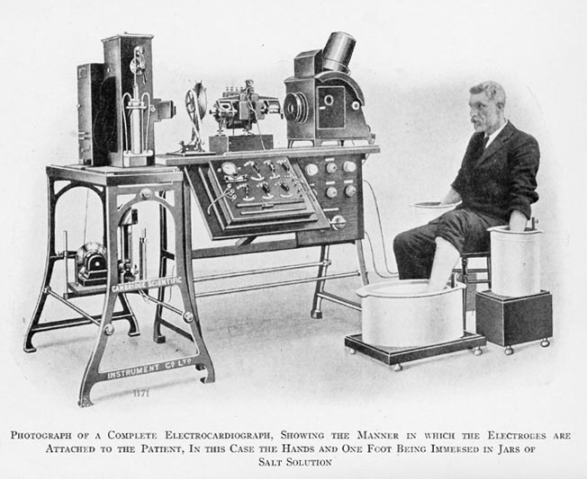
In 1885 Einthoven became a Professor of Physiology at the University of Leiden. Having seen a demonstration of Augustus Waller’s “electrogram” (a device that recorded minute deviations on a mercury column when electrically stimulated) in 1887, he improved it by creating the “string galvanometer”. In 1901 Einthoven published his first recordings of what he called “elektrokardiogramm” (EKG).
The initial device was bulky, heavy, and required the patient to sit with both arms and the left leg in separate buckets of salt water, but it did record the electrical activity of the heart (Click here for an image of one of the first electrocardiographs). Eventually the device was commercialized and history was made. It was Einthoven who used the letter P,Q,R,S, and T in electrocardiography.
In 1924, Willem Einthoven was awarded the Nobel Prize in Physiology.
Sources:
1. "Willem Einthoven (1860-1927): father of electrocardiography". Merritt, C. Tan. SY. Singapore Med J 53:(1) 17
2. "Willem Einthoven (1860-1927)" Davies, M; Hollman, A. Heart. 1997 October; 78(4): 324
3. "Willem Einthoven: The development of the human electrocardiogram" Cajavilcaa, C.,Varonb, J.Resuscitation 76:3 2008; 325–328
Original image courtesy of "Images from the History of Medicine" at www.nih.gov.
- Details

Cerebelum
The word [vermis] is Latin and means "worm".
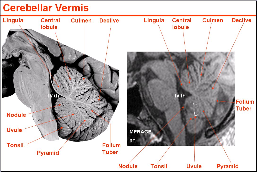
The vermis is the name given by Galen of Pergamon (129AD - 200AD) to the median lobe of the cerebellum, since when seen from the superior aspect, this cerebellar lobe looks like a multisegmented worm. See accompanying image, or click for a larger depiction.
When seen in a median section, the cerebellar vermis looks like a multilobulated leaf with the fourth ventricle of the brain at its base. It is composed of several smaller lobules: Lingula, central, culmen, clivus, tuber vermis, pyramid, uvula, and nodular lobes.
Median section image link courtesy of UCLA Radiology
Sources:
1. "The Origin of Medical Terms" Skinner, HA 1970 Hafner Publishing Co.
2. "Medical Meanings - A Glossary of Word Origins" Haubrich, WD. ACP Philadelphia
3 "Tratado de Anatomia Humana" Testut et Latarjet 8 Ed. 1931 Salvat Editores, Spain
4. "Anatomy of the Human Body" Henry Gray 1918. Philadelphia: Lea & Febiger
Image modified by CAA, Inc. Original image courtesy of bartleby.com
- Details
"Nothing in the human body is colored, or labeled"
")
Click for a larger image
"The Chirurgeon must knowe the Anatomie". Thus states Thomas Vicary (1460 -1561) on the knowledge of Anatomy. He continues: "...for all authors write against those surgeons who work in a man's body not knowing the Anatomie"1. There is no doubt that knowledge must include the awareness of the possibility of anatomical variations. Some anatomical variations, like the "Corona Mortis" can be critical, and in some surgical cases, be the cause for exsanguination!
It is interesting that several medical schools are reducing the total number of hours working on, or moving away from cadaver disection in first year medical school and using computer simulations instead. No computer simulation will give the medical student the detail, variations, and feel of the tissues as actual hands-on experience. I am sure no one wants a surgeon whose first view of the internal aspect of a human body is a living patient...on the surgical table.
It is a fact that "Nothing in the human body is really colored... or labeled" or as someone else said "nothing looks exactly like the anatomy book", unless it is photography, and then each photo is taken after hours of laboring to "Netterize" the organ or area that one is trying to detail. Nothing gives the future professional the exact idea of what to expect in the future patient than the hours and hours of laborious work in the anatomy laboratory.
The same is true with anatomical variations, one "standard" digital cadaver,even with built-in anatomical variations does not give the student the sense of awe and discovery when an anatomical variation is found, interpreted, and analyzed with a group of peers, contributing to the learning process and the formation of future health care professionals.When questioning what is normal or abnormal, Dr. Elizabeth Murray says it most elegantly: "The cadaver is always right"
The image depicts a case of a coronary artery arising from the pulmonary trunk!
Sources:
1. "The Chirurgeon must knowe the Anatomie" R. Shane Tubbs Clin Anat 26:417 (2013)
2. "Two cases of an abnormal coronary artery of the heart arising from the pulmonary artery"Brooks, H; J. Anat. Physiol. 20:26-29, 1886 (anatomyatlases.org)
THIS ARTICLE IS THE THIRD IN A SERIES. TO READ THE FIRST ARTICLE CLICK HERE
- Details
"No anatomical structure has the moral obligation to be where they are supposed to be"
")
Click for a larger image
Not only may an anatomical structure be absent, such as in the case of renal aplasia or agenesis, or in the case of a non-existent circumflex coronary artery, but sometimes extra structures can be found. Such is the case where a kidney can present two or even three ureters, all functional. Double inferior vena cavae, cervical ribs, lumbar ribs, the list goes on and on!
Muscles can be added to this list, again, with absence of a muscle, or with new and completely unexpected attachments. An example of this is the presence of a continuation of the rectus abdominis muscle into the chest region, a variation called a sternalis muscle.
The accompanying image shows the sternalis muscle in one of the "muscle plates" of De Humani Corporis Fabrica Libri Septem, published in 1543 by Andreas Vesalius. This image was criticized by showing a muscle that does not exist, although Vesalius clearly stated in the text of his book that this was an anatomical variation that he had seen.
For many decades surgeons had to operate and "see what they could find". There were the days of the exploratory laparotomy. After the discovery of the application of X-rays by Wilhem Konrad Roentgen (1845 - 1923) and the incredible advances in imaging techniques including CT-scan, MRI, PET, etc, the surgeon is now not usually surprised by anatomical variations.
There are areas in the body that have an high rate of anatomical variation, such as the hepatobiliary region, which includes the "Triangle of Calot". In this area, the standard anatomy is found only in 64% of the cases! In the rest, expect the unexpected. Lahey (1948) states "...the fact that cholecystectomy is a dangerous operation. It is dangerous unless one realizes.... that anomalous anatomy is very common". Today the dangers are less, because of better visualization and technology, but anatomical variations are still there.
Another area where anatomical variations are extremely important is the heart's coronary circulation. Anatomical variations can cause different cardiac dominance. Normal anatomy states that there are two coronary arteries, yet, up to five separate coronary arteries arising directly from the ascending aorta have been described! There is one variation where the left coronary arises from the right coronary artery, effectively having only one artery arise from the aorta and being in charge of all the arterial supply to the heart. What happens if this single artery stenoses? Bear in mind that this is not an "anomalous" vessel, it is just an anatomical variation.
Sources:
1. Lahey DH, discussing the paper "Partial Hepatectomy with Intrahepatic Cholangiojejunostomy" by Wilson H, and Gillespie CE, Ann Surg. 1949 June; 129(6): 756–765
2. "Renal aplasia is the predominant cause of congenital solitary kidneys" Hiraoka, M et al Kidney Int. 2002 May;61(5):1840-4.
This article is the second in a series of three; Click here for the first article.
TO CONTINUE READING: CLICK HERE
- Details
"The only constant in anatomy is variation"

Click for a larger image
This dictum is incredibly powerful and true. Even the so-called "anatomical constants" are subject to it.
One common misconception is that "we are all the same". This could not be further from the truth. Every body is different from every else's body. Anatomical variations range from the minimal to the incredible. One of the most interesting anatomical variations is the one called "situs inversus". In this case the individual is a mirror image of a human. The apex of the heart points to the right side of the body; the duodenum circles to the right, the liver "hangs" from the left side of the respiratory diaphragm, etc. This particular anatomical variation presents in different degrees and can sometimes coexist with some cardiovascular congenital abnormalities.
Of course there are minor anatomical variations that have no effect on daily life at all and are only discovered by accident, or upon autopsy or dissection. One of the most complete resources on this topic is the Illustrated Encyclopedia of Human Anatomic Variations. An excerpt from this site states: "It is clear that textbook writers and teachers over the centuries, even until today, fail to understand or to transmit to their students the crucial concept that anatomical and physiological diversity and variation is a canon of living organisms. This failure leads to the belief that textbooks are conveying immutable facts with only few anomalous exceptions".
Shown here is an extremely rare case of a third kidney. Dixon (1911) describes in his research paper that as of that date, only 10 cases were known, of these only eight were recorded, with 87% of them found on the left side of the body. Click on the image for a larger depiction.
Source and primary image: "Supernumerary kidney: The occurrence of three kidneys in an adult male subject" Dixon, A.F. J. Anat. Physiol. 45:117-121, 1911.
THIS POST IS CONTINUED, CLICK HERE
- Details
The prefix [para-] has a Greek origin and means "beside" or "alongside". Today we add the meaning of "parallel to".
We see the daily application of this prefix in words such as [paramedic], [parajournalism], [paralogism], and [paranormal]. Medical applications of the term include:
- parasternal: alongside the sternum, such as the internal thoracic vessels
- paramedian: alongside the median plane
- parasagittal: parallel to a sagittal plane (synonym with paramedian)
- paraumbilical: alongside the umbilicus, such as paraumbilical visceral extrusion in a gastroschisis
- parathyroid glands: glands that are found besides the thyroid gland, etc.