![]()
Medical Terminology Daily (MTD) is a blog sponsored by Clinical Anatomy Associates, Inc. as a service to the medical community. We post anatomical, medical or surgical terms, their meaning and usage, as well as biographical notes on anatomists, surgeons, and researchers through the ages. Be warned that some of the images used depict human anatomical specimens.
You are welcome to submit questions and suggestions using our "Contact Us" form. The information on this blog follows the terms on our "Privacy and Security Statement" and cannot be construed as medical guidance or instructions for treatment.
We have 164 guests and no members online
")
Marcia Crocker Noyes
(1869 – 1946)
Further to my comment on old books and research that started with an interesting bookplate (Ex-Libris). I continued my research and found that the person in charge of the Osler library bookplate was a fascinating individual that today maybe a ghost in the MedChi library and building in Baltimore... This is certainly an article that can be called "A Moment in History"
Marcia Crocker Noyes was the librarian at The Maryland State Medical Society from 1896 to 1946 and was a founding member of the Medical Library Association.[1][2][3]
Sir William Osler, MD. a famous Johns Hopkins surgeon was a noted bibliophile and had a large personal collection of books on various topics. When he became the President of MedChi in 1896, he was dismayed at the condition of the library and knew that with the right person and some stewardship, it could become a significant collection. Sir William asked his friend, Dr. Bernard Steiner, a physician and President of the Enoch Pratt Free Library in Baltimore for suggestions of a librarian, and Dr. Steiner recommended Marcia Crocker Noyes. A native of New York, and a graduate of Hunter College, Marcia had moved to Baltimore for a lengthy visit with her sister, and took a “temporary” position at the Pratt Library, which turned into three years. Although she had no medical experience or background, she was enthusiastic, and most importantly, she was willing to move into the apartment provided for the librarian, who needed to be available 24 hours a day.
The image in this article is Ms. Noyes on her first year on the job. Marcia developed a book classification system for medical books, based on the Index Medicus, and called it the Classification for Medical Literature. The system uses the alphabet with capital letters for the major divisions of medicine and lower-case ones for the sub-sections. The system was used for many years, but it's now dated and the Faculty's original shelving scheme was never changed. The card catalogs still reflect her classification and many of the cards are written in Marcia's back-slanting handwriting.
Marcia knew enough to ask the Faculty's members about medical questions, terminology and literature. She gradually won over the predominantly male membership and they became her greatest allies; Sir William at the start, and then for nearly 40 years, Dr. John Ruhräh, a wealthy pediatrician with no immediate family of his own. She made a point of attending almost every Faculty function, and in 1904, under guidelines from the American Medical Association, Marcia was made the Faculty Secretary. For much of her first 10 years, she was the Faculty's only full-time employee, only being assisted by Mr. Caution, the Faculty's janitor. Later in life Marcia would say that she hired him because of his name!
Within ten years, the library had outgrown its space, and plans, spearheaded by Marcia and Sir William before his move to Oxford, were made to build a headquarters building, mainly to house the library's growing collection of medical books and journals.
Marcia was instrumental in the design and building of the new headquarters. She travelled to Philadelphia, New York and Boston to look at their medical society buildings, and eventually, the Philadelphia architectural firm, Ellicott & Emmart was selected to design and build the new Faculty building. Every detail of the building held her imprimatur, from the graceful staircase, to the light-filled reading room, and all of the myriad details of the millwork, marble tesserae, and most of all, the four-story cast iron stacks. She was on-site, climbing up unfinished staircases, checking out the progress of the building, which was built in less than one year at a cost of $90,000.
Among the features of the new building was a fourth-floor apartment for her. She referred to it as the "first penthouse in Baltimore" and it had a garden and rooftop terrace. The library collection eventually grew to more than 65,000 volumes from medical and specialty societies around the world. Journals were traded back and forth, and physicians eagerly anticipated the arrival of each new issue. At the same time, Marcia was involved in the Medical Library Association as one of eight founding members. The MLA promotes medical libraries and the exchange of information. One of the earliest mandates of the MLA was the Exchange, a distribution and trade service for those who had duplicates or little-used books in their collections. Initially, the Exchange was run out of the Philadelphia medical society, but in 1900 it was moved to Baltimore and Marcia oversaw it. Several hundred periodicals and journals were received and sent each month, a huge amount of work for a tiny staff. In 1904, the Faculty had run out of room to manage the Exchange, so it was moved to the Medical Society of the Kings County (Brooklyn). But without Marcia's excellent administrative skills, it floundered and in 1908, the MLA asked Marcia to take charge once again.
In 1909, when the new Faculty building opened, there was enough room to run the Exchange and with the help of MLA Treasurer, noted bibliophile and close friend, Dr. John Ruhräh, it once again became successful. Additionally, Marcia and Dr. Ruhräh combined forces to revive the MLA's bulletin, which had all but ceased publication in 1908, taking the Exchange with it. This duo maintained editorial control from 1911 until 1926. In 1934, around the time of Dr. Ruhräh's death, Marcia became the first “unmedicated” professional to head the MLA. During her tenure, the MLA incorporated, the first seal was adopted, and the annual meeting was held in Baltimore. Marcia wanted to write the history of the MLA once she retired from full-time work at the Faculty, but her health was beginning to fail. She had back problems and had suffered a serious burn on her shoulder as a young woman, possibly from her time running a summer camp, Camp Seyon, for young ladies in the Adirondack Mountains. In 1946, a celebration was planned to honor Marcia's 50 years at the Faculty. But she was adamant that the physicians wait until November, the actual date of her 50 years. However, they knew she was gravely ill, and might not make it until then, so a huge party was held in April. More than 250 physicians attended the celebration, but the ones she was closest to in the early years, were long gone. She was presented with a suitcase, a sum of money to use for travelling, and her favorite painting of Dr. John Philip Smith, a founder of the Medical College in Winchester, Virginia. It was painted by Edward Caledon Smith, a Virginia painter who had been a student of the painter Thomas Sully.[4] She adored this painting and vowed, jokingly, to take it with her wherever she went.
The painting was not to stay with her for very long, for she died in November 1946, and left it to the Faculty in her will. Her funeral was held in the Faculty's Osler Hall, named for her dear friend. More than 60 physicians served as her pallbearers, and she was buried at Baltimore's Green Mount Cemetery. In 1948, the MLA decided to establish an award in the name of Marcia Crocker Noyes. It was for outstanding achievement in medical library field and was to be awarded every two years, or when a truly worthy candidate was submitted. In 2014, the Faculty began giving a bouquet of flowers to the winner of the award in Marcia's name, and in honor of her work. Much evidence exists for this tradition, as we know that the physicians, especially Drs. Osler and Ruhräh, frequently gave her bouquets of flowers. Marcia also cultivated flower gardens at the Faculty and decorated the rooms with her work.
Today, the MedChi building is open for tours and if the rumors are to be believed Ms. Marcia Crocker Noyes is still at work in her beloved library as the "resident ghost" [1][5]

NOTE: This article has been modified from the original Wikipedia article on Marcia Crocker Noyes. The article itself is well-written with interesting images of the subject. I would encourage you to visit it. The second insert is from book 00736 in my personal library and shows in pencil, the incredibly small handwriting of Marsha C. Noyes.
Sources:
1. "Marcia, Marcia, Marcia" MedChi Archives blog.
2. "Marcia C. Noyes, Medical Librarian" (PDF). Bulletin of the Medical Library Association. 35 (1): 108–109. 1947. PMC 194645
3. Smith, Bernie Todd (1974). "Marcia Crocker Noyes, Medical Librarian: The Shaping of a Career" (PDF). Bulletin of the Medical Library Association. 62 (3): 314–324. PMC 198800Freely accessible. PMID 4619344.
4. Edward Caledon BRUCE (1825-1901)"
5. Behind the scenes tour MedChiBuilding
"Clinical Anatomy Associates, Inc., and the contributors of "Medical Terminology Daily" wish to thank all individuals who donate their bodies and tissues for the advancement of education and research”.
Click here for more information


- Details
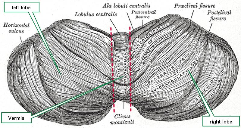
Cerebelum
The word [cerebellum] is Latin and means "little brain". The cerebellum is one of the three main gross components of the brain (encephalon), the other two being the cerebral hemispheres and the brain stem.
The cerebellum is characterized by a tightly folded external cortex where the gyri are long and parallel to each other and the sulci are not very deep. Upon gross examination, the cerebellum presents with two lateral lobes (left and right) and a median lobe known as the vermis. Other authors divide the cerebellum into an anterior and posterior lobe separated by a primary fissure or sulcus, also known as the preclival sulcus.
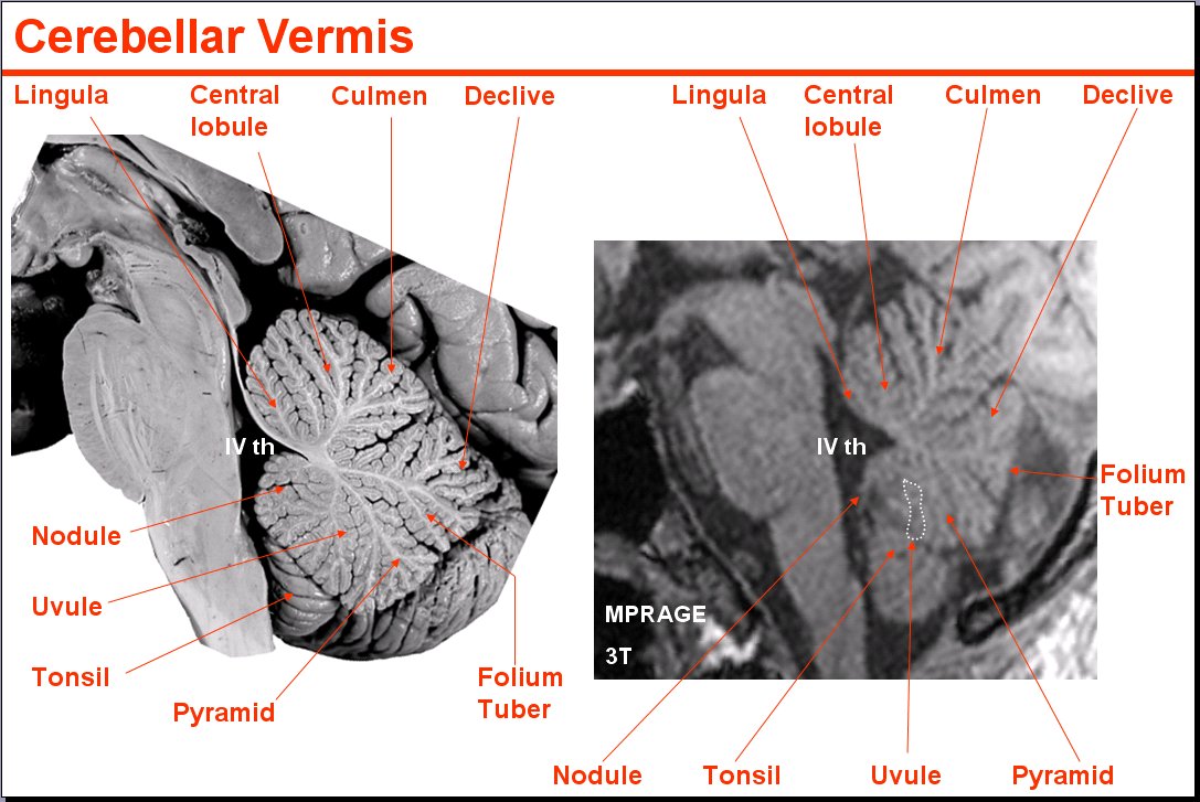
Cerebellar vermis
The cerebellum is located posterior to the brain stem and posteroinferior to the cerebral hemispheres. It is separated from the occipital lobes of the brain by an extension of dura mater called the tentorium cerebelli. Because of its location, the cerebellum serves as a roof for the 4th ventricle, a component of the ventricular system of the brain. Click here to see a median section, of the cerebellum where you can observe its location and relation to the brain stem, 4th ventricle, and occipital lobe.
The cerebellum is part of the motor control of the brain and is involved in motor coordination, precision, balance and accurate motor timing. Cerebellar dysfunction does not cause paralysis, but produces fine motor control disorders.
Median section image link courtesy of UCLA Radiology.
Sources:
1 "Tratado de Anatomia Humana" Testut et Latarjet 8 Ed. 1931 Salvat Editores, Spain
2. "Anatomy of the Human Body" Henry Gray 1918. Philadelphia: Lea & Febiger
Image modified by CAA, Inc. Original image courtesy of bartleby.com
- Details
This article is part of the series "A Moment in History" where we honor those who have contributed to the growth of medical knowledge in the areas of anatomy, medicine, surgery, and medical research.

Abraham Vater
Abraham Vater (1684 - 1751) German anatomist and physician, Abraham Vater was the son of a distinguished physician and was born on the city of Wittenberg. He obtained a Doctorate in Philosophy (PhD) in 1706 and his medical degree in 1710 at the University of Leipzig. He became a professor “extraordinarius” of Anatomy and Botany in 1719, continuing his career in anatomy until he obtained the highest professorial degree at the University.
After the death of his father in 1733 Abraham Vater was appointed to the chair of Anatomy. In 1720 he published the discovery of a “biliary diverticulum” , the hepatopancreatic ampulla, known today as the “Ampulla of Vater” or duodenal papilla. His original article was entitled “Dissertatio anatomica qua novum bilis diverticulum circa orificium ductus choledochi”. He also described a large subcutaneous sensory nerve terminal known as the “Vater–Pacinian corpuscle”.
Known as an anatomist, Vater also wrote on surgery, gynecology, pharmacology, pathology, chemistry, and botany, making him a complete scientist. In a twist of fate, Vater died in 1751 after being afflicted with jaundice, probably a consequence of biliary blockage of his eponymic ampulla.
Sources:
1. "Abraham Vater (1684-1751)" Brit Med J; 2,(4) (1951), 1214
2. "Abraham Vater of the Ampulla (Papilla) of Vater" Lerch, MM; Domschke, W. Gastroenterology (2000) 118: (2) 379
- Details

Click for a larger image
The word [epicardium] is composed by the prefix [epi-], meaning "outer" or "above"; the root term [-card-], meaning "heart"; and the suffix [-ium], meaning "layer" or "membrane". Thus, the word means "outer layer of the heart".
The epicardium is part of a larger structure called the pericardium, in fact, since the pericardium is in contact with a viscus (the heart), it can also be called "visceral pericardium". It must be noted that these two terms are synonyms: "epicardium" and "visceral pericardium".
The epicardium is a type of serosa composed of an outer layer formed by mesothelial cells and an inner layer formed by loose connective tissue containing varying amounts of fat. This inner layer known as the "subepicardial layer" (a misnomer) also contains elastic fibers.
The main arterial and venous components of the coronary circulation are found in the subepicardial layer. When performing a Coronary Artery Bypass Graft (CABG), a surgeon has to open the outer layer of the epicardium, and enter the subepicardial layer to perform the graft anastomosis.
As a serosa, the epicardium is involved in the production and absorption of a clear fluid, the "pericardial fluid". This fluid acts as a lubricant for the pericardium, allowing for the effortless movement of the heart within the pericardial sac.
- Details
The suffix [-iasis] has an original Greek root (such as in [iatros] meaning "healer" or "hospital"). It later became a Latin root meaning "condition, pathology, or disease".
This suffix can be found in many medical terms such as:
- Choledocolithiasis - a condition of stones in the bile duct.
- Elephantiasis - A condiction where a body part grows unusually large, usually lower extremities and/ or scrotum
- Phthiriasis- A condition or infestarion with public or crab lice
- Helminthiasis- A bodily infestation with a type of parasitic worms called helminths (tapeworms)
- Cystolithiasis - Bladder stones
- Details

Anterior view of the sphenoid bone
Both these root terms have their origin from the Greek [πτέρυγα] (ptéryga) and mean "wing".
In human anatomy the most common use of this root term is in the word [pterygoid]. Since the suffix [-oid] means "similar to", the word pterygoid means "similar to a wing", or "wing-like".
On the inferior aspect of the sphenoid bone (os sphenoidale) there are two very thin bat-wing-like bony appendages that are called the lateral and medial pterygoid plates. The medial pterygoid plate has a hook-like bony appendage called the hamulus (Latin: little hook).
Related to the pterygoid plates are the lateral and medial pterygoid muscles, both these muscles aid inthe process of mastication.
The root term [pter-] can be found in words such as [pterodactyl] meaning "winged finger", it refers to a phrehistoric winged animal; [pteranodon], and [pterosaurus].
Sources:
1 "Tratado de Anatomia Humana" Testut et Latarjet 8 Ed. 1931 Salvat Editores, Spain
2. "Anatomy of the Human Body" Henry Gray 1918. Philadelphia: Lea & Febiger
Image in the public domain modified by CAA, Inc. Original image courtesy of bartleby.com
Note: Google Translate includes the symbol (?). Clicking on it will allow you to hear the pronunciation of the word.
- Details
This article is part of the series "A Moment in History" where we honor those who have contributed to the growth of medical knowledge in the areas of anatomy, medicine, surgery, and medical research.

Wilhelm His Jr.
Wilhelm His Jr. (1863-1934). Also known as Wilhelm His the Younger, was born in Switzerland in the city of Basel. His father was a well-known and famous anatomist by the same name who worked at the University of Basel. Wilhelm His Jr. studied in several universities, including Leipzig, Strasbourg, Bern, and Geneva. He received his medical degree from the University of Leipzig in 1889.
Although not an anatomist, his 1893 single and most brilliant contribution to the eventual understanding of the conduction system of the heart was the discovery and description of a muscular bundle that connected the atrial septum with the ventricular septum. Until that moment it was known that no muscles crossed from the atria to the ventricles, leading anatomists and physiologists to wonder how does the heart contract.
In his article His states "after long search I have succeeded in finding a muscle bundle which unites the auricular and ventricular septal walls, and which, up to now, has escaped observation because of incomplete exposure, for it is visible in its entire extent only when the septa are cut exactly in their longitudinal direction".
What is interesting is that His not only found the bundle that today honors his name, but he included in that description the atrioventricular node, the actual structure that crosses the atrioventricular connective tissue barrier known as the "skeleton of the heart". The AV node was later to be clearly identified and researched by Sunao Tawara (1873 - 1952).
Dr. His continued his research on gout and joint diseases. He became a German citizen and joined the German army as a consulting physician. He retired in 1932 and died in 1934.
Although Wilhelm His Jr. did not discover or described the esophagogastric angle, in 1906 JD Cunningham started calling this angle the "Angle of His" in honor of Wilhelm His Jr. This eponym has stayed with us until today.
Sources:
1. "Wilhelm His Jr." JAMA. 1964;187(6):453-454
2. "His, Jr., W.: The Activity of the Embryonic Human Heart and Its Significance for the Understanding of the Heart Movement in the Adult" Arb Med Klin Leipzig, pp 14-49, 1893; Bast, TH,Gardner, WD trans: J Hist Med 4:289-318, 1949
3. "Wilhelm His, Jr. and the Bundle of His" Bast, TH, Gardner, WD J Hist Med All Sci; 1949; 4, (2) 170 -187
4. Firkin BG (1996) Dictionary of medical eponyms.London: Parthenon Publishing Group, 181
Original image in the public domain, courtesy of the National Library of Medicine.