Medical Terminology Daily (MTD) is a blog sponsored by Clinical Anatomy Associates, Inc. as a service to the medical community. We post anatomical, medical or surgical terms, their meaning and usage, as well as biographical notes on anatomists, surgeons, and researchers through the ages. Be warned that some of the images used depict human anatomical specimens.
You are welcome to submit questions and suggestions using our "Contact Us" form. The information on this blog follows the terms on our "Privacy and Security Statement" and cannot be construed as medical guidance or instructions for treatment.
We have 547 guests online

Jean George Bachmann
(1877 – 1959)
French physician–physiologist whose experimental work in the early twentieth century provided the first clear functional description of a preferential interatrial conduction pathway. This structure, eponymically named “Bachmann’s bundle”, plays a central role in normal atrial activation and in the pathophysiology of interatrial block and atrial arrhythmias.
As a young man, Bachmann served as a merchant sailor, crossing the Atlantic multiple times. He emigrated to the United States in 1902 and earned his medical degree at the top of his class from Jefferson Medical College in Philadelphia in 1907. He stayed at this Medical College as a demonstrator and physiologist. In 1910, he joined Emory University in Atlanta. Between 1917 -1918 he served as a medical officer in the US Army. He retired from Emory in 1947 and continued his private medical practice until his death in 1959.
On the personal side, Bachmann was a man of many talents: a polyglot, he was fluent in German, French, Spanish and English. He was a chef in his own right and occasionally worked as a chef in international hotels. In fact, he paid his tuition at Jefferson Medical College, working both as a chef and as a language tutor.
The intrinsic cardiac conduction system was a major focus of cardiovascular research in the late nineteenth and early twentieth centuries. The atrioventricular (AV) node was discovered and described by Sunao Tawara and Karl Albert Aschoff in 1906, and the sinoatrial node by Arthur Keith and Martin Flack in 1907.
While the connections that distribute the electrical impulse from the AV node to the ventricles were known through the works of Wilhelm His Jr, in 1893 and Jan Evangelista Purkinje in 1839, the mechanism by which electrical impulses spread between the atria remained uncertain.
In 1916 Bachmann published a paper titled “The Inter-Auricular Time Interval” in the American Journal of Physiology. Bachmann measured activation times between the right and left atria and demonstrated that interruption of a distinct anterior interatrial muscular band resulted in delayed left atrial activation. He concluded that this band constituted the principal route for rapid interatrial conduction.
Subsequent anatomical and electrophysiological studies confirmed the importance of the structure described by Bachmann, which came to bear his name. Bachmann’s bundle is now recognized as a key determinant of atrial activation patterns, and its dysfunction is associated with interatrial block, atrial fibrillation, and abnormal P-wave morphology. His work remains foundational in both basic cardiac anatomy and clinical electrophysiology.
Sources and references
1. Bachmann G. “The inter-auricular time interval”. Am J Physiol. 1916;41:309–320.
2. Hurst JW. “Profiles in Cardiology: Jean George Bachmann (1877–1959)”. Clin Cardiol. 1987;10:185–187.
3. Lemery R, Guiraudon G, Veinot JP. “Anatomic description of Bachmann’s bundle and its relation to the atrial septum”. Am J Cardiol. 2003;91:148–152.
4. "Remembering the canonical discoverers of the core components of the mammalian cardiac conduction system: Keith and Flack, Aschoff and Tawara, His, and Purkinje" Icilio Cavero and Henry Holzgrefe Advances in Physiology Education 2022 46:4, 549-579.
5. Knol WG, de Vos CB, Crijns HJGM, et al. “The Bachmann bundle and interatrial conduction” Heart Rhythm. 2019;16:127–133.
6. “Iatrogenic biatrial flutter. The role of the Bachmann’s bundle” Constán E.; García F., Linde, A.. Complejo Hospitalario de Jaén, Jaén. Spain
7. Keith A, Flack M. The form and nature of the muscular connections between the primary divisions of the vertebrate heart. J Anat Physiol 41: 172–189, 1907.
"Clinical Anatomy Associates, Inc., and the contributors of "Medical Terminology Daily" wish to thank all individuals who donate their bodies and tissues for the advancement of education and research”.
Click here for more information
- Details
This article is part of the series "A Moment in History" where we honor those who have contributed to the growth of medical knowledge in the areas of anatomy, medicine, surgery, and medical research.

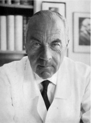
Dr. Rudolph Nissen
Dr. Rudolf Nissen (1896 - 1981). Dr Nissen’s life is extraordinary. Born in the city of Neisse, Germany in 1896, he was the son of a local surgeon. He studied medicine in the Universities of Munich, Marburg, and Breslau. He was the pupil of the famous pathologist Albert Aschoff (discoverer of the heart’s AV node, along with Sunao Tawara).
Nissen became a professor of surgery in Berlin, and in 1933 moved to Turkey where he was placed in charge of the Department of Surgery of the University of Istanbul. In 1939 he moved to the US, first to the Massachusetts General Hospital and later to the Jewish Hospital in Brooklyn, New York. After becoming a US citizen, he moved again in 1952 to Basel, Switzerland as Chief of the Department of Surgery, where he retired in 1967. He died in 1981.
His contributions to surgery are innumerable. He wrote over 30 books and 450 journal articles. Known for the development in 1956 of what is today known as the “Nissen fundoplication” for esophageal hiatus hernia surgery, Nissen also worked with his assistant, Dr. Mario Rossetti to develop the “floppy Nissen fundoplication”, also known as the “Nissen-Rossetti procedure”. This would be enough to honor this man, still, he (with Sauerbruch) performed the first lung lobectomy and the first pneumonectomy (called then a total pneumonectomy). In 1949 he performed the first esophagectomy with a gastroesophagostomy for lower esophageal cancer.
His personal life is even more interesting. Drafted at 20, he fought in WWI and was wounded several times. In 1933, under the Nazi regime, he was ordered to fire all the Jewish-German assistants under his care. Being Jewish himself, he was told that he would keep his job, Nissen could not take this. He resigned his position and moved out of Germany.
Another little known fact is that he operated on Albert Einstein in 1948. He operated on Einstein because of intestinal cysts. Having found a developing abdominal aortic aneurysm, he reinforced it with cellophane, undoubtedly giving his patient a few extra years to live. Einstein died in 1955.
As a personal side note, our good friend Dr. Aaron Ruhalter scrubbed in with Dr. Nissen while serving as a surgical resident at the Brooklyn Jewish Hospital!
Sources:
1. “Rudolf Nissen: The man behind the fundoplication” Schein et al. Surgery 1999;125:347-53
2. “Rudolf Nissen (1896–1981)-Perspective” Liebermann-Meffert, D. J Gastrointest Surg (2010) 14 (Suppl 1):S58–S61
3. “The Life of Rudolf Nissen: Advancing Surgery Through Science and Principle” Fults, DW; Taussky, P. World J Surg (2011) 35:1402–1408
4. “Total Pneumonectomy” Nissen, R. Ann Thorac Surg 1980; 29:390-394
5. “Historical Development of Pulmonary Surgery” Nissen, R. Am J Surg 80: Jan 1955 9- 15
Image in the public domain, courtesy of the Universitat Basel
- Details
The suffix [-oma] means "tumor", "mass", or "growth". It should be noted that the word [tumor] is originally Latin, and means "swelling" or "bulging". Sometimes the plural form [-omata] can be used.
It is a general misconception that the suffix [-oma] or the term [tumor] are synonymous with "cancer". This is not so. [Tumor] only means a mass and the type of mass, benign or malignant is not implied in the term. A cancerous mass will be denoted by the addition of the root term [-carcin-] meaning "cancer" therefore the combined root and suffix will be [-carcinoma]. There are other root-suffix combinations that also mean "cancerous".
The suffix [-oma] can be found in many medical words, such as:
- Hematoma: from the Greek root [-hem-] meaning "blood". A mass of blood
- Myoma: from the root [-my-] meaning "muscle". A mass, growth, or tumor of muscle
- Fibroma: from the root [-fibr-] meaning "fiber". A mass of fibers
- Fibromyoma: the combining form of "muscle" is [myo-], therefore "a mass of fibers and muscle" (Pl. fibromyomata)
- Adenoma: from the Greek [aden-] meaning "gland". A mass that has a glandular look to it or a mass in a gland
Sources:
1. "The Language of Medicine" John H. Dirckx Pub: Harper & Row 1976
2. "Medical Meanings" Haubrich, William S. Am Coll Phys Philadelphia 1997
3. "The origin of Medical Terms" Skinner, AH, 1970
- Details
This article is part of the series "A Moment in History" where we honor those who have contributed to the growth of medical knowledge in the areas of anatomy, medicine, surgery, and medical research.

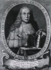
Giovanni Batista Morgagni
Giovanni Battista Morgagni (1682 - 1771). Italian anatomist, physician, and pathologist, Morgagni was born in the city of Forli. He started his medical studies at the University of Bologna, graduating in 1701 with a degree in Medicine and Philosophy. In 1712 he became a professor of anatomy at the University of Padua, Italy, 175 years after Andreas Vesalius. Morgagni was offered and accepted the Chair of Anatomy in 1715 at the University of Padua. Although Morgagni held a position at the anatomy department of the University of Padua, his name is associated mostly with his pathological studies.
Morgagni was interested in the works of Theophile Boneti (1620 - 1689), who started analyzing the correlation between post-mortem anatomical findings and diseases. He tried to establish a relation between the disease and the cause of death. In 1761 Morgagni published his most influential work "De Sedibus et Causis Morburum Per Anatomen Indagatis" (On the Sites and Causes of Diseases, Investigated by Dissection). His work was essential for pathological anatomy to be recognized as a science in itself.
Morgagni was elected to become a member of several Academies of Science and Surgery: The Royal Society of London, The Academy of Science in Paris, The Berlin Academy of Science, and the Imperial Academy of Saint Petersburg in Russia. He is remembered today by several eponyms in anatomy and pathology:
- Morgagni's caruncle or lobe, referring to the middle lobe of the prostate
- Morgagni's columns: the anal (or anorectal) columns
- Morgagni's concha, referring to the superior nasal concha
- Morgagni's foramina: two hiatuses in the respiratory diaphragm allowing for passage of the superior epigastric vessels
- Morgagni's hernia: an hiatal hernia through Morgagni's foramen, in the respiratory diaphragm
- Morgagni's ventricle: an internal pouch or dilation between the true and false vocal cords in the larynx
- Morgagni's nodules: the nodules at the point of coaptation of the leaflets (cusps) of the pulmonary valve. Erroneously called the "nodules of Arantius", which are only found in the aortic valve
Sources:
1. "A Note From History: The First Printed Case Reports of Cancer" Hadju, S.I. Cancer 2010;116:2493–8
2. "Giovanni Battista Morgagni" Klotz, O. Can Med Assoc J 1932 27:3 298-303
3. "Morgagni (1682 -1771)" JAMA 1964 187:12 948-950
Original image in the public domain, courtesy of National Institutes of Health.
- Details

Hover for "Lumen"
The term [lumen] is Latin and means "light". It is an international measure of light intensity. The plural form is either [lumens] or the Latin version [lumina].
The question is how did this term end up meaning the "internal opening or cavity of a hollow organ"? Dr. J. Dirckx (1976) explains that the term [lumen] in gastrointestinal anatomy was not used until the late 19th century, but it was initially used by microscopists looking at cross sections of small vessels. Since the empty interior of the vessel was represented on the histological slide as a clear, lighted region, they started calling that the "light" of the vessel, or [lumen].
The root term for [lumen] is [-lumin-] and can be found in over 30 words in English and related to light, such as luminosity, luminaria, luminescent, luminous, illumination, illuminator, intraluminal, transluminal, endoluminal, etc.
The correct adjective form for [lumen] is [luminal], as stated by Haubrich (1997). The point is that the use of the term "lumenal" as an adjective form for lumen is not correct. This term can be seen wrongly used in many textbooks today. The proper form for the NOTES acronym is "Natural Orifice Transluminal Endoscopic Surgery"
If you hover over the image you will "illuminate" this article!
- Details

Original image courtesy of Wikipedia.org
An esophageal hiatus hernia (also known as a hiatal hernia) eventually may require surgery. In this case, the objective is three-fold: To bring the abdominal viscera to its proper intraabdominal position (reduction) , to create a pseudovalve to prevent gastroesophageal reflux, and to prevent a recurrence of the herniation.
There have been different procedures developed to this effect. One of the most popular has been the Nissen fundoplication either trough the open surgery approach or by way of a minimally invasive laparoscopic procedure.
This procedure was pioneered by Dr. Rudolf Nissen (1896 - 1981) in 1955. After reducing the hiatal hernia and repairing the dilated esophageal hiatus, the surgeon creates a gastric wrap around the abdominal esophagus by bringing the fundus of the stomach through a retroesophageal passage, and suturing the fundus to the stomach. (see image). One of the concerns of the procedure is the ligation and transection of the short gastric vessels that pass within the gastrosplenic ligament to allow greater mobility of the gastric fundus and prevent potential avulsion of the short gastric vessels.
Since the introduction of this open procedure in 1955 there have been several variations, such as the "Nissen-Rosetti" procedure, a "loose" fundoplication; the "Toupet" procedure, an "incomplete" fundic wrap, and others, including laparoscopic procedures.
The advent of NOTES (Natural Orifice Transluminal Endoscopic Surgery) has brought a new procedure: Transoral Incisionless fundoplication (TIF), where a pseudovalve is created using an endoscope inserted into the esophagus and stomach through the oral cavity without abdominal incisions or trocar ports. For more information on this procedure, click here. Clicking on the inferior image will start a six-minute video of the TIF procedure and the EsophX device.
- Details
The term [hiatus] derives from the Latin word [hiare], meaning to "gape" or to "yawn". In human anatomy this term is used to mean an "opening" or a "defect". It must be pointed out that in anatomy (and surgery) the term "defect" does not necessarily mean "defective". In most cases a "defect" is a normal opening in a structure, such as the esophageal hiatus. The plural form is either [hiatus] or [hiatuses].
There are many hiatuses in the human body, such as:
• Hiatus semilunaris: a crescent-shape opening in the lateral aspect of the nasal wall
• Esophageal hiatus: an opening in the muscular posterior aspect of the respiratory diaphragm, bound by two muscular crura
• Aortic hiatus: an opening in the posterior aspect of the respiratory diaphragm, bound by two tendinous crura
• Hiatus Fallopii: the entrance to the facial canal, an opening in the temporal bone allowing for passage of the facial nerve (CN V). Named after Gabrielle Fallopius

