![]()
Medical Terminology Daily (MTD) is a blog sponsored by Clinical Anatomy Associates, Inc. as a service to the medical community. We post anatomical, medical or surgical terms, their meaning and usage, as well as biographical notes on anatomists, surgeons, and researchers through the ages. Be warned that some of the images used depict human anatomical specimens.
You are welcome to submit questions and suggestions using our "Contact Us" form. The information on this blog follows the terms on our "Privacy and Security Statement" and cannot be construed as medical guidance or instructions for treatment.
We have 531 guests and no members online
")
Marcia Crocker Noyes
(1869 – 1946)
Further to my comment on old books and research that started with an interesting bookplate (Ex-Libris). I continued my research and found that the person in charge of the Osler library bookplate was a fascinating individual that today maybe a ghost in the MedChi library and building in Baltimore... This is certainly an article that can be called "A Moment in History"
Marcia Crocker Noyes was the librarian at The Maryland State Medical Society from 1896 to 1946 and was a founding member of the Medical Library Association.[1][2][3]
Sir William Osler, MD. a famous Johns Hopkins surgeon was a noted bibliophile and had a large personal collection of books on various topics. When he became the President of MedChi in 1896, he was dismayed at the condition of the library and knew that with the right person and some stewardship, it could become a significant collection. Sir William asked his friend, Dr. Bernard Steiner, a physician and President of the Enoch Pratt Free Library in Baltimore for suggestions of a librarian, and Dr. Steiner recommended Marcia Crocker Noyes. A native of New York, and a graduate of Hunter College, Marcia had moved to Baltimore for a lengthy visit with her sister, and took a “temporary” position at the Pratt Library, which turned into three years. Although she had no medical experience or background, she was enthusiastic, and most importantly, she was willing to move into the apartment provided for the librarian, who needed to be available 24 hours a day.
The image in this article is Ms. Noyes on her first year on the job. Marcia developed a book classification system for medical books, based on the Index Medicus, and called it the Classification for Medical Literature. The system uses the alphabet with capital letters for the major divisions of medicine and lower-case ones for the sub-sections. The system was used for many years, but it's now dated and the Faculty's original shelving scheme was never changed. The card catalogs still reflect her classification and many of the cards are written in Marcia's back-slanting handwriting.
Marcia knew enough to ask the Faculty's members about medical questions, terminology and literature. She gradually won over the predominantly male membership and they became her greatest allies; Sir William at the start, and then for nearly 40 years, Dr. John Ruhräh, a wealthy pediatrician with no immediate family of his own. She made a point of attending almost every Faculty function, and in 1904, under guidelines from the American Medical Association, Marcia was made the Faculty Secretary. For much of her first 10 years, she was the Faculty's only full-time employee, only being assisted by Mr. Caution, the Faculty's janitor. Later in life Marcia would say that she hired him because of his name!
Within ten years, the library had outgrown its space, and plans, spearheaded by Marcia and Sir William before his move to Oxford, were made to build a headquarters building, mainly to house the library's growing collection of medical books and journals.
Marcia was instrumental in the design and building of the new headquarters. She travelled to Philadelphia, New York and Boston to look at their medical society buildings, and eventually, the Philadelphia architectural firm, Ellicott & Emmart was selected to design and build the new Faculty building. Every detail of the building held her imprimatur, from the graceful staircase, to the light-filled reading room, and all of the myriad details of the millwork, marble tesserae, and most of all, the four-story cast iron stacks. She was on-site, climbing up unfinished staircases, checking out the progress of the building, which was built in less than one year at a cost of $90,000.
Among the features of the new building was a fourth-floor apartment for her. She referred to it as the "first penthouse in Baltimore" and it had a garden and rooftop terrace. The library collection eventually grew to more than 65,000 volumes from medical and specialty societies around the world. Journals were traded back and forth, and physicians eagerly anticipated the arrival of each new issue. At the same time, Marcia was involved in the Medical Library Association as one of eight founding members. The MLA promotes medical libraries and the exchange of information. One of the earliest mandates of the MLA was the Exchange, a distribution and trade service for those who had duplicates or little-used books in their collections. Initially, the Exchange was run out of the Philadelphia medical society, but in 1900 it was moved to Baltimore and Marcia oversaw it. Several hundred periodicals and journals were received and sent each month, a huge amount of work for a tiny staff. In 1904, the Faculty had run out of room to manage the Exchange, so it was moved to the Medical Society of the Kings County (Brooklyn). But without Marcia's excellent administrative skills, it floundered and in 1908, the MLA asked Marcia to take charge once again.
In 1909, when the new Faculty building opened, there was enough room to run the Exchange and with the help of MLA Treasurer, noted bibliophile and close friend, Dr. John Ruhräh, it once again became successful. Additionally, Marcia and Dr. Ruhräh combined forces to revive the MLA's bulletin, which had all but ceased publication in 1908, taking the Exchange with it. This duo maintained editorial control from 1911 until 1926. In 1934, around the time of Dr. Ruhräh's death, Marcia became the first “unmedicated” professional to head the MLA. During her tenure, the MLA incorporated, the first seal was adopted, and the annual meeting was held in Baltimore. Marcia wanted to write the history of the MLA once she retired from full-time work at the Faculty, but her health was beginning to fail. She had back problems and had suffered a serious burn on her shoulder as a young woman, possibly from her time running a summer camp, Camp Seyon, for young ladies in the Adirondack Mountains. In 1946, a celebration was planned to honor Marcia's 50 years at the Faculty. But she was adamant that the physicians wait until November, the actual date of her 50 years. However, they knew she was gravely ill, and might not make it until then, so a huge party was held in April. More than 250 physicians attended the celebration, but the ones she was closest to in the early years, were long gone. She was presented with a suitcase, a sum of money to use for travelling, and her favorite painting of Dr. John Philip Smith, a founder of the Medical College in Winchester, Virginia. It was painted by Edward Caledon Smith, a Virginia painter who had been a student of the painter Thomas Sully.[4] She adored this painting and vowed, jokingly, to take it with her wherever she went.
The painting was not to stay with her for very long, for she died in November 1946, and left it to the Faculty in her will. Her funeral was held in the Faculty's Osler Hall, named for her dear friend. More than 60 physicians served as her pallbearers, and she was buried at Baltimore's Green Mount Cemetery. In 1948, the MLA decided to establish an award in the name of Marcia Crocker Noyes. It was for outstanding achievement in medical library field and was to be awarded every two years, or when a truly worthy candidate was submitted. In 2014, the Faculty began giving a bouquet of flowers to the winner of the award in Marcia's name, and in honor of her work. Much evidence exists for this tradition, as we know that the physicians, especially Drs. Osler and Ruhräh, frequently gave her bouquets of flowers. Marcia also cultivated flower gardens at the Faculty and decorated the rooms with her work.
Today, the MedChi building is open for tours and if the rumors are to be believed Ms. Marcia Crocker Noyes is still at work in her beloved library as the "resident ghost" [1][5]

NOTE: This article has been modified from the original Wikipedia article on Marcia Crocker Noyes. The article itself is well-written with interesting images of the subject. I would encourage you to visit it. The second insert is from book 00736 in my personal library and shows in pencil, the incredibly small handwriting of Marsha C. Noyes.
Sources:
1. "Marcia, Marcia, Marcia" MedChi Archives blog.
2. "Marcia C. Noyes, Medical Librarian" (PDF). Bulletin of the Medical Library Association. 35 (1): 108–109. 1947. PMC 194645
3. Smith, Bernie Todd (1974). "Marcia Crocker Noyes, Medical Librarian: The Shaping of a Career" (PDF). Bulletin of the Medical Library Association. 62 (3): 314–324. PMC 198800Freely accessible. PMID 4619344.
4. Edward Caledon BRUCE (1825-1901)"
5. Behind the scenes tour MedChiBuilding
"Clinical Anatomy Associates, Inc., and the contributors of "Medical Terminology Daily" wish to thank all individuals who donate their bodies and tissues for the advancement of education and research”.
Click here for more information


- Details
This article is part of the series "A Moment in History" where we honor those who have contributed to the growth of medical knowledge in the areas of anatomy, medicine, surgery, and medical research.

Dr. Otto C. Brantigan
Otto C. Brantigan, MD. (1904-1981) An American surgeon and anatomist, Otto Charles Brantigan was born in Chattanooga, TN in 1904. Having dropped out of high school to help his family and working as a first class machinist, he decided to continue with graduate school. He studied at the Northwestern University in Chicago, where he graduated from the Medical School in 1933. In 1948 he became Chief of Surgery, and eventually became Professor of Surgery, Professor of Thoracic Surgery, and Professor of Anatomy at the Maryland School of Medicine. He retired in 1976 having earned many accolades for his profuse surgical work and publications.
As a surgeon of the times, Dr. Brantigan had a wide area of interest. His over 110 publications and surgical work range from thoracoscopy to vascular, plastic, cardiac, and orthopedic surgery. He is most remembered for the pioneer work he did on chronic obstructive pulmonary disease (COPD), emphysema and lung volume reduction surgery (LVRS), which he presented in 1958. The procedure had (at the time) a very high mortality rate (16 -20%) and Brantigan's work was not readily accepted.
It was not until J. Cooper and his team, revisited the operation proposed by Brantigan that the operation was accepted, now with new surgical stapling and staple line buttressing technology. Dr. Brantigan's name was recognized as a pioneer in lung emphysema surgery, unfortunately 14 years after his death. In 1994 his son, Dr Charles O. Brantigan delivered a beautiful biography of Dr. Otto Brantigan in the same meeting where Cooper presented his results with LVRS.
Personal note: I am proud to own one of the copies of Dr. O.C. Brantigan;s "Clinical Anatomy", a book that I use quite frequently. It is listed in my library catalog. Dr. Miranda.
Sources:
1. "Biography of Otto C Brantigan" C.O. Brantigan 1994 Meeting of the American Association for Thoracic Surgery
2. "LVRS in chronic obstructive pulmonary disease" Davies, L; Calverley, P. Thorax 1996;51(Suppl 2):S29-S34
3. ""Bilateral pneumectomy (volume reduction) for chronic obstructive pulmonary disease" Cooper, J.,The Journal of Thoracic and Cardiovascular Surgery Volume 109, Number 1:106-119
4. "The Surgical Approach to Pulmonary Emphysema" Brantigan, OC; Kress, MB; Mueller, EA. Chest. 1961; 39(5):485-499
5. "History of Emphysema Surgery" Naef, AP. Ann Thorac Surg 1997;64:1506-1508
Original image courtesy of National Institutes of Health.Biography of Dr. Otto Brantigan courtesy of Dr. Charles O. Brantigan.
- Details

Click for a larger image
The rectum is the most distal segment of the large intestine, along with the anal canal.
The word [rectum] arises from the Latin [rectus] and means "straight", such as its use in the name "rectus abdominis" for the "straight muscle of the abdomen".
It seems a misnomer, as the rectum of the human species is actually "S" shaped, as seen in the accompanying image. The reason for this discrepancy is that the rectum was named by Galen of Pergamon (129AD - 200 AD) who himself studied this structure in animals such as sheep and goats. In these animals the rectum is indeed straight, and since contradicting Galen was not acceptable (see Michael Servetus), the name has survived until this day. Even Andreas Vesalius has in his 1953 "Fabrica" a depiction of a straight rectum in the human! Click on second image to see a larger depiction of Vesalius' idea of the rectum. Although Vesalius stated that he wanted to show human anatomy as it is, and not as Galen said it should be, here is a demonstration that in 1543 he was still a lukewarm Galenist.
There is an area between the sigmoid and the rectum called the sigmoidorectal junction, although most anatomists call it (wrongly) the rectosigmoid junction (RSJ). This is an anatomically diffuse area with no clear anatomical transition between the sigmoid and the rectum or the RSJ from the rectum.
")
Click for a larger image
As the proximal end of the "S" shaped rectum is not clearly discernible from the sigmoidorectal region, there is no clear agreement on the length of the rectum. Authors state that it measures approximately six to seven inches in length (15 - 17 cm), while others measure it as between 8-10 inches. The rectum ends distally at the junction of the rectum with the pelvic diaphragm. It is at this point that the anal canal begins.
The rectum is characterized by three transverse rectal folds, one on the right side, and two on the left side. These folds are know as the "rectal valves" or the "valves of Houston". The middle rectal fold is known to European anatomists as the "valve of Kohlrausch" Their function in maintaining fecal material in place as well as their function in defecation is still under study. The rectal valves also have a high level of anatomical variation and may not be present at all.
Images:
1. "Tratado de Anatomia Humana" Testut et Latarjet 8 Ed. 1931 Salvat Editores, Spain
2. "De Humani Corporis Fabrica, Libri Septem" A. Vesalius 1543 Brussels
Recommended reading: "Transverse Folds of Rectum: Anatomic Study and Clinical Implications" Shafik, A, et al. Clin Anat 14: 196-203 (2001).
- Details

Click for a larger image
The term "collateral circulation" is generally used to denote a situation where small blood channels dilate and provide blood supply when a pathology creates a stricture and diminishes blood flow (ischemia).
Although the above is correct, the term is also applicable to a normal, non-pathological situation most common in the human body. Please refer to the accompanying image for the following explanation. If needed, click on the image for a larger depiction. In the image, the arrows represent direction of flow.
Most organs or organ segments receive blood supply from more than one source of blood supply. In some cases, like the stomach, there are up to four arteries that provide blood supply to the organ: the right and left gastric arteries, and the right and left gastroepiploic arteries.
In other cases, like the small intestine shown in the image, blood arrives to the organ arising from several arteries (A, B, and C) that themselves arise from a parent structure. Because of hydrodynamics, the vascular territories of each artery (represented by dashed lines) tend not to overlap. If for any reason there is stenosis or blockage in any of these arteries (A,B, or C) blood will flow immediately through an alternate route and the organ will not suffer ischemia or necrosis.
This is extremely important, as these collateral channels maintain blood supply to areas that may be affected by bending, such as the elbow and knee, which have a rich collateral network. Most of the organs in the body, with some exceptions (brain, heart), have collateral circulation.
Collateral circulation is extremely important for surgery, as surgeons can safely remove parts of organs without affecting the blood supply to the organ. This is also true for all gastrointestinal anastomoses.
Image property of: CAA, Inc. Artist: Dr. Miranda
- Details
Click for a larger image
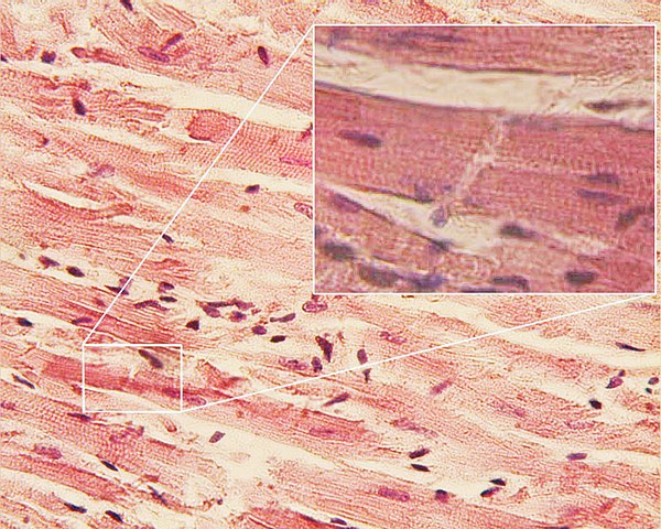
Histology is the scientific branch that studies tissues.
The root term [-hist-] is used to mean "tissues", but how the term came to be used is somewhat convoluted. It arises from the Greek [histos], which indicates the mast of a ship, it then was used to denote a Greek weaver's loom central mast (where the fabric is woven horizontally), and then it was used to indicate that which was woven [histios], the fabric, or the "tissues". The suffix [-ology] also has Greek origin from [logos] meaning a "book", a "treatise" or "to study".
The concept of the body being formed by different tissues was pioneered by Marie-Francois Xavier Bichat (1771-1802) who called them "membranes" Bichat is considered to be the "father of Histology". The image shows a histological slide of cardiac muscle. Click on the image for a larger depiction.
Original image by S. Girod and A. Becker, courtesy of Wikipedia.
- Details

Click for a larger image
The root term [-spondyl-] arises from the Greek [spondylos] meaning "vertebra", and suffix [-osis] means "condition", but with the connotation of "many". The word [spondylosis] means " condition of many vertebrae". This does not add much to the use of this word as an indicator of a pathology, but it does indicate that there is excess bone in a vertebral pathology.
Spondylosis is an osteoarthritic degeneration of the vertebrae and the spine characterized by abnormal bony growths on the vertebrae that can impinge on nerves and other structures causing pain and mobility problems. The definition of spondylosis also includes degenerative changes in the intervertebral discs.
The abnormal growth of portions of the vertebral body, usually forms "bone spurs", also referred to as "spondylophytes". The accompanying image shows a lumbar vertebra with spondylophytes.
Image property of: CAA, Inc. Photographer: David M. Klein
- Details
This article is part of the series "A Moment in History" where we honor those who have contributed to the growth of medical knowledge in the areas of anatomy, medicine, surgery, and medical research.

Dr. Rudolph Nissen
Dr. Rudolf Nissen (1896 - 1981). Dr Nissen’s life is extraordinary. Born in the city of Neisse, Germany in 1896, he was the son of a local surgeon. He studied medicine in the Universities of Munich, Marburg, and Breslau. He was the pupil of the Karl Albert Ludwig Aschoff (1866 - 1942) a German physician and cardiovascular researcher. Another of Aschoff's pupil, Dr. Sunao Tawara (1873 - 1952), discovered the atrioventricular node.
Nissen became a professor of surgery in Berlin, and in 1933 moved to Turkey where he was placed in charge of the Department of Surgery of the University of Istanbul. In 1939 he moved to the US, first to the Massachusetts General Hospital and later to the Jewish Hospital in Brooklyn, New York. After becoming a US citizen, he moved again in 1952 to Basel, Switzerland as Chief of the Department of Surgery, where he retired in 1967. He died in 1981.
His contributions to surgery are innumerable. He wrote over 30 books and 450 journal articles. Known for the development in 1956 of what is today known as the “Nissen fundoplication” for esophageal hiatus hernia surgery, Nissen also worked with his assistant, Dr. Mario Rossetti to develop the “floppy Nissen fundoplication”, also known as the “Nissen-Rossetti procedure”. This would be enough to honor this man, still, he (with Sauerbruch) performed the first lung lobectomy and the first pneumonectomy (called then a total pneumonectomy). In 1949 he performed the first esophagectomy with a gastroesophagostomy for lower esophageal cancer.
His personal life is even more interesting. Drafted at 20, he fought in WWI and was wounded several times. In 1933, under the Nazi regime, he was ordered to fire all the Jewish-German assistants under his care. Being Jewish himself, he was told that he would keep his job, Nissen could not take this. He resigned his position and moved out of Germany.
Another little known fact is that he operated on Albert Einstein in 1948. He operated on Einstein because of intestinal cysts. Having found a developing abdominal aortic aneurysm, he reinforced it with cellophane, undoubtedly giving his patient a few extra years to live. Einstein died in 1955.
As a personal side note, our good friend Dr. Aaron Ruhalter scrubbed in with Dr. Nissen while serving as a surgical resident at the Brooklyn Jewish Hospital!
Sources:
1. “Rudolf Nissen: The man behind the fundoplication” Schein et al. Surgery 1999;125:347-53
2. “Rudolf Nissen (1896–1981)-Perspective” Liebermann-Meffert, D. J Gastrointest Surg (2010) 14 (Suppl 1):S58–S61
3. “The Life of Rudolf Nissen: Advancing Surgery Through Science and Principle” Fults, DW; Taussky, P. World J Surg (2011) 35:1402–1408
4. “Total Pneumonectomy” Nissen, R. Ann Thorac Surg 1980; 29:390-394
5. “Historical Development of Pulmonary Surgery” Nissen, R. Am J Surg 80: Jan 1955 9- 15
Image in the public domain, courtesy of the Universitat Basel