Medical Terminology Daily (MTD) is a blog sponsored by Clinical Anatomy Associates, Inc. as a service to the medical community. We post anatomical, medical or surgical terms, their meaning and usage, as well as biographical notes on anatomists, surgeons, and researchers through the ages. Be warned that some of the images used depict human anatomical specimens.
You are welcome to submit questions and suggestions using our "Contact Us" form. The information on this blog follows the terms on our "Privacy and Security Statement" and cannot be construed as medical guidance or instructions for treatment.
We have 521 guests online

Jean George Bachmann
(1877 – 1959)
French physician–physiologist whose experimental work in the early twentieth century provided the first clear functional description of a preferential interatrial conduction pathway. This structure, eponymically named “Bachmann’s bundle”, plays a central role in normal atrial activation and in the pathophysiology of interatrial block and atrial arrhythmias.
As a young man, Bachmann served as a merchant sailor, crossing the Atlantic multiple times. He emigrated to the United States in 1902 and earned his medical degree at the top of his class from Jefferson Medical College in Philadelphia in 1907. He stayed at this Medical College as a demonstrator and physiologist. In 1910, he joined Emory University in Atlanta. Between 1917 -1918 he served as a medical officer in the US Army. He retired from Emory in 1947 and continued his private medical practice until his death in 1959.
On the personal side, Bachmann was a man of many talents: a polyglot, he was fluent in German, French, Spanish and English. He was a chef in his own right and occasionally worked as a chef in international hotels. In fact, he paid his tuition at Jefferson Medical College, working both as a chef and as a language tutor.
The intrinsic cardiac conduction system was a major focus of cardiovascular research in the late nineteenth and early twentieth centuries. The atrioventricular (AV) node was discovered and described by Sunao Tawara and Karl Albert Aschoff in 1906, and the sinoatrial node by Arthur Keith and Martin Flack in 1907.
While the connections that distribute the electrical impulse from the AV node to the ventricles were known through the works of Wilhelm His Jr, in 1893 and Jan Evangelista Purkinje in 1839, the mechanism by which electrical impulses spread between the atria remained uncertain.
In 1916 Bachmann published a paper titled “The Inter-Auricular Time Interval” in the American Journal of Physiology. Bachmann measured activation times between the right and left atria and demonstrated that interruption of a distinct anterior interatrial muscular band resulted in delayed left atrial activation. He concluded that this band constituted the principal route for rapid interatrial conduction.
Subsequent anatomical and electrophysiological studies confirmed the importance of the structure described by Bachmann, which came to bear his name. Bachmann’s bundle is now recognized as a key determinant of atrial activation patterns, and its dysfunction is associated with interatrial block, atrial fibrillation, and abnormal P-wave morphology. His work remains foundational in both basic cardiac anatomy and clinical electrophysiology.
Sources and references
1. Bachmann G. “The inter-auricular time interval”. Am J Physiol. 1916;41:309–320.
2. Hurst JW. “Profiles in Cardiology: Jean George Bachmann (1877–1959)”. Clin Cardiol. 1987;10:185–187.
3. Lemery R, Guiraudon G, Veinot JP. “Anatomic description of Bachmann’s bundle and its relation to the atrial septum”. Am J Cardiol. 2003;91:148–152.
4. "Remembering the canonical discoverers of the core components of the mammalian cardiac conduction system: Keith and Flack, Aschoff and Tawara, His, and Purkinje" Icilio Cavero and Henry Holzgrefe Advances in Physiology Education 2022 46:4, 549-579.
5. Knol WG, de Vos CB, Crijns HJGM, et al. “The Bachmann bundle and interatrial conduction” Heart Rhythm. 2019;16:127–133.
6. “Iatrogenic biatrial flutter. The role of the Bachmann’s bundle” Constán E.; García F., Linde, A.. Complejo Hospitalario de Jaén, Jaén. Spain
7. Keith A, Flack M. The form and nature of the muscular connections between the primary divisions of the vertebrate heart. J Anat Physiol 41: 172–189, 1907.
"Clinical Anatomy Associates, Inc., and the contributors of "Medical Terminology Daily" wish to thank all individuals who donate their bodies and tissues for the advancement of education and research”.
Click here for more information
- Details
- Written by: Prof. C. Uribe
|
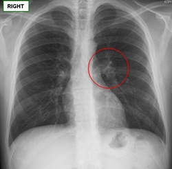
Anatomy:[Hilum] is a generic term which names area of an organ where all the structures that enter or leave the organ are found. In the lung it is depression in the mediastinal surface of the lung through which the vessels and bronchi enter and leave the lungs. This set of structures corresponds to the “root of the lung” or “pulmonary pedicle”. The term has a singular form, [hilum]; and a plural form, [hila]; the adjectival form is [hilar]. Radiographs and CAT Scan: The hilum appears as a composite opacity at the root of each lung produced by bronchi, arteries, veins, lymph nodes, nerves ,and other tissues. Sources: |
 AP thoracic x-ray image showing the pulmonary hilum |
| Back to MTD Main Page | Subscribe to MTD |
- Details
We would like to welcome Professor Cristián Uribe as a contributor to Medical Terminology Daily.
Prof. Uribe is a Physical Therapist, has a Masters in Human Anatomy and is a Professor in the Human Anatomy Department of the Medical School at the University Finis Terrae in Santiago, Chile.
Prof. Uribe is also the Director of the Postgraduate Course on “Anatomical Bases of Normal Imaging”, as well as the Executive Secretary of the Postgraduate Office at the same University.
He published the book “Eponyms in Anatomical Nomenclature” (2011, Ed. U. Finis Terrae). For his LinkedIn page click here.
- Details
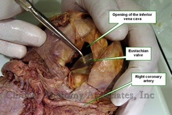
The [valve of the inferior vena cava] is probably better known by its eponym, the [Eustachian valve]. This is an incomplete valve found at the most distal end of the inferior vena cava, at the point where it opens into the right atrium of the heart. The valve is muscular at its base and composed mostly by membranous endocardium, and is normally the only valve present in the inferior vena cava.
It appears as a membranous semilunar fold extending posteriorly from the limbus fossa ovalis, anterior to the inferior caval orifice where it disappears. Its free border is usually membranous, concave, and directed anterosuperiorly. Its base is a transverse muscular ridge continuous with the anterior margin of the caval orifice.
It has no function in the adult, but in the fetus it helps to divert the flow of blood coming from the inferior vena cava towards the foramen ovale [fossa ovalis], as part of fetal circulation.
It has great anatomical variation in size, shape and thickness. On its medial aspect it joins with the Thebesian valve at the orifice of the coronary sinus.
First described by Bartolomeo Eustachius (c1500 - 1574) in 1653, the existence of the valve of the inferior vena cava was disputed by many until Winslow in 1717 confirmed its presence and suggested its function in fetal circulation.
Sources:
1. "The Valve of the Inferior Vena Cava" Hickie, JB Br Heart J. 1956 Jul; 18(3): 320–326
2. "Bartolommeo Eustachio; a great medical genius whose masterpiece remained hidden for 150 years" Wells, WA Arch Otolaring (1925) 48: 58
- Details
The medical word [dysphagia] arises from the suffix [dys-] which means "abnormal", the root term [phag-] which arises from the Greek [φάγος] (phagos), meaning "to swallow", a "glutton" or "eating much", and the suffix [-ia] meaning "condition". The word dysphagia then means "a condition of abnormal swallowing".
- Details
This article is part of the series "A Moment in History" where we honor those who have contributed to the growth of medical knowledge in the areas of anatomy, medicine, surgery, and medical research.

Bartolomeo Eustachius
Bartolomeo Eustachius (c1500 - 1574) Italian physician and anatomist, also known as Bartholomew Eustachius, Esutachi, and e was born in the small town of San Severino Marche, in the province of Macerata, central Italy. His father was a respected physician, and may have attended as a student the University of Sapienza, where he later taught as a professor of practical medicine. Eustachius’ life is mostly unknown and some of his works remained hidden for over 150 years. His birth date is only an estimate and data varies from c1500 AD to c1510 AD.
It is known that Eustachius lived and worked in Rome from 1549 to 1574. He was both an anatomist and a physician to the Vatican. As an anatomist, Eustachius is credited for having been the first to prepare anatomical images for printing using copper plates.
As a hospital physician, Eustachius was adamant on the need of autopsies on patients who died at the hospital, crediting him with being one of the first pathological anatomists.
Eustachius’ is credited with several anatomical discoveries, which he published in short monographs, on topics like the kidneys, the suprarenal glands (which he was the first to describe), the movements of the head, the azygos vein, etc. One of these was entirely dedicated to dentistry, “Libellus de Dentibus”, which have led to some to call Eustachius “The Father of Dental Anatomy”.
Eustachius dedicated himself to prepare a number of anatomical copper plates, apparently getting ready to publish a book to rival Vesalius’ “Fabrica”. It is known that Eustachius disliked Vesalius because of Vesalius’ contempt for the teachings of Galen. Eustachius died before completing his work and the copper plates were forgotten for over a century. After being rediscovered, these plates were published with commentaries by anatomists, until a final publication in Latin by Bernard Siegfried Albinus (1697 – 1770) entitled “Explicatio Tabularum Anatomicarum Bartholomaei Eustachii Anatomici Summi” (An explanation of the Anatomical Picture of Bartholomew Eustachius, Supreme Anatomist”.
Besides the suprarenal gland, Eustachius is credited for having discovered the stapes, the tensor tympani muscle, the valve of the inferior vena cava, the cervical sympathetic chain and the thoracic duct.
Interesting note: Eustachius’ assistant Petrus Matthaeus Pinus, who helped develop the copper plates, voiced his master’s dislike of Vesalius in a poem that was published by Albinus over a hundred years after the death of all of them, it is also an epitaph for a great anatomist:
“Just as the Master from Pergamon (Galen)
Teaching his method of healing, once refuted
The false writings of the ignorant Thessalus,
So also my BARTOLO (Eustachius)
Teaching his method of denoting every detail, position,
Shape, structure, order, number and condition
Has repulsed the shameless arrogance and claims
Of the impudent Vesalius
To him (Eustachius) all future generations
Adhere In reverential admiration
The age to come will envy us on account of you Father,
And historians will wish they had lived sooner
They will extol our era, through you fortunate,
Lucky beyond measure"
Sources:
1. "The root of dental anatomy: a case for naming Eustachius the "Father of Dental Anatomy"" Bennett, G (2009) J Hist Dent (1089-6287), 57 (2) 85 -88
2. “The papal anatomist: Eustachius in renaissance Rome” Simpson, D, ANZ J Surg (2011) 81: (12)905 -910
3. “Bartholomeo Eustachio – The Third Man: Eustachius Published by Albinus” Fahrer, M. (2003) ANS J Surg; 73: 523- 528
4. "Bartolommeo Eustachio; a great medical genius whose masterpiece remained hidden for 150 years" Wells, WA Arch Otolaring (1925) 48: 58
Original image courtesy of: Wikimedia Commons.
- Details
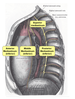
UPDATED: The mediastinum is the median region of the thorax, usually described as the "space"1 between the lungs. This region is divided into a superior and inferior mediastinum by a plane that pases through the sternal angle or Angle of Louis.
The inferior mediastinum is itself divided into three separate regions by the pericardial sac. The region anterior to the pericardial sac is the "anterior mediastinum", the region posterior to the pericardial sac is the "posterior mediastinum", and the region containing and including the pericardial sac is the "middle mediastinum". Thus described the mediastinum comprises four regions as follows:
• Superior mediastinum: It contains the aortic arch, the brachiocephalic trunk, the thoracic segments of the left common carotid and the left subclavian arteries, the brachiocephalic veins, a portion of the superior vena cava, the vagus nerve, phrenic nerve, and left recurrent laryngeal nerve, trachea, esophagus, thoracic duct and the remains of the thymus gland
• Anterior mediastinum: A narrow space, more developed on the left side, anterior to the pericardial sac and contains some lymph nodes and connective tissue
• Middle mediastinum: The largest mediastinal region, it contains the pericardial sac, the heart, the bifurcation of the trachea, the inferior vena cava, and the cardiac end of the great vessels.
• Posterior mediastinum: Contains the descending aorta, the azygos and hemiazygos veins, the esophagus, and thoracic duct.
Personally, I do not use the description of the mediastinum as a "the space beween the lungs", as it conjures the image of literal "open spaces" around or between the organs. The fact is that the mediastinum is tightly packed with no spaces between the organs. This is why I prefer the definition of the mediastinum as an "area" or "region" between the lungs. Dr. Miranda.


