Medical Terminology Daily (MTD) is a blog sponsored by Clinical Anatomy Associates, Inc. as a service to the medical community. We post anatomical, medical or surgical terms, their meaning and usage, as well as biographical notes on anatomists, surgeons, and researchers through the ages. Be warned that some of the images used depict human anatomical specimens.
You are welcome to submit questions and suggestions using our "Contact Us" form. The information on this blog follows the terms on our "Privacy and Security Statement" and cannot be construed as medical guidance or instructions for treatment.
We have 900 guests online

Jean George Bachmann
(1877 – 1959)
French physician–physiologist whose experimental work in the early twentieth century provided the first clear functional description of a preferential interatrial conduction pathway. This structure, eponymically named “Bachmann’s bundle”, plays a central role in normal atrial activation and in the pathophysiology of interatrial block and atrial arrhythmias.
As a young man, Bachmann served as a merchant sailor, crossing the Atlantic multiple times. He emigrated to the United States in 1902 and earned his medical degree at the top of his class from Jefferson Medical College in Philadelphia in 1907. He stayed at this Medical College as a demonstrator and physiologist. In 1910, he joined Emory University in Atlanta. Between 1917 -1918 he served as a medical officer in the US Army. He retired from Emory in 1947 and continued his private medical practice until his death in 1959.
On the personal side, Bachmann was a man of many talents: a polyglot, he was fluent in German, French, Spanish and English. He was a chef in his own right and occasionally worked as a chef in international hotels. In fact, he paid his tuition at Jefferson Medical College, working both as a chef and as a language tutor.
The intrinsic cardiac conduction system was a major focus of cardiovascular research in the late nineteenth and early twentieth centuries. The atrioventricular (AV) node was discovered and described by Sunao Tawara and Karl Albert Aschoff in 1906, and the sinoatrial node by Arthur Keith and Martin Flack in 1907.
While the connections that distribute the electrical impulse from the AV node to the ventricles were known through the works of Wilhelm His Jr, in 1893 and Jan Evangelista Purkinje in 1839, the mechanism by which electrical impulses spread between the atria remained uncertain.
In 1916 Bachmann published a paper titled “The Inter-Auricular Time Interval” in the American Journal of Physiology. Bachmann measured activation times between the right and left atria and demonstrated that interruption of a distinct anterior interatrial muscular band resulted in delayed left atrial activation. He concluded that this band constituted the principal route for rapid interatrial conduction.
Subsequent anatomical and electrophysiological studies confirmed the importance of the structure described by Bachmann, which came to bear his name. Bachmann’s bundle is now recognized as a key determinant of atrial activation patterns, and its dysfunction is associated with interatrial block, atrial fibrillation, and abnormal P-wave morphology. His work remains foundational in both basic cardiac anatomy and clinical electrophysiology.
Sources and references
1. Bachmann G. “The inter-auricular time interval”. Am J Physiol. 1916;41:309–320.
2. Hurst JW. “Profiles in Cardiology: Jean George Bachmann (1877–1959)”. Clin Cardiol. 1987;10:185–187.
3. Lemery R, Guiraudon G, Veinot JP. “Anatomic description of Bachmann’s bundle and its relation to the atrial septum”. Am J Cardiol. 2003;91:148–152.
4. "Remembering the canonical discoverers of the core components of the mammalian cardiac conduction system: Keith and Flack, Aschoff and Tawara, His, and Purkinje" Icilio Cavero and Henry Holzgrefe Advances in Physiology Education 2022 46:4, 549-579.
5. Knol WG, de Vos CB, Crijns HJGM, et al. “The Bachmann bundle and interatrial conduction” Heart Rhythm. 2019;16:127–133.
6. “Iatrogenic biatrial flutter. The role of the Bachmann’s bundle” Constán E.; García F., Linde, A.. Complejo Hospitalario de Jaén, Jaén. Spain
7. Keith A, Flack M. The form and nature of the muscular connections between the primary divisions of the vertebrate heart. J Anat Physiol 41: 172–189, 1907.
"Clinical Anatomy Associates, Inc., and the contributors of "Medical Terminology Daily" wish to thank all individuals who donate their bodies and tissues for the advancement of education and research”.
Click here for more information
- Details
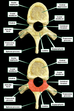
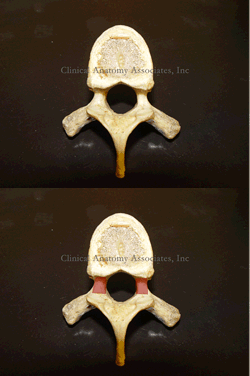
The [vertebral arch] is one of the components of a typical vertebra. It is a bony arch found posterior to the vertebral body, and it is composed by the pedicles, the vertebral laminae, and the root or base of the articular processes or zygapophyses.
The presence of the vertebral arch defines the vertebral foramen, a space that contains the spinal cord with its meninges, spinal arteries and venous plexuses, and epidural fat. The sum of all the vertebral foramina creates the vertebral canal.
Image property of: CAA.Inc. Photographer: David M. Klein
- Details
The root term [-narc-] is a derived from the Greek [ναρκος] (narkos ), meaning “torpor” or “lethargic”. Initially it was used to denote numbness in an extremity, but eventually evolved to its modern meaning. It is used in medical word such as:
- Narcotic: A group of pharmaceuticals that numbs pain
- Narcosis: The suffix [-osis] means “condition”. A condition of generalized numbness or lethargicness
- Narcolepsy: The suffix [-lepsy]refers to a sudden onset or a seizure. Sudden numbness or lethargicness
Note: The links to Google Translate include an icon that will allow you to hear the pronunciation of the word.
- Details

Click on the image for a larger depiction
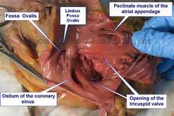
The coronary sinus is a venous structure that receives blood from the coronary circulation and returns it to the right atrium of the heart. It is found in the atrioventricular sulcus and receives all the veins of the heart (small, middle, great and oblique cardiac veins, and others), with the exception of some anterior cardiac veins that may empty directly into the right atrium. There are small venous valves at the point where most of these veins enter the coronary sinus.
Unlike most veins, the coronary sinus in the human has an evident layer of smooth muscle that may become the source of ectopic foci of atrial depolarization, causing atrial fibrillation.
The opening of the coronary sinus into the right atrium is called the "ostium of the coronary sinus".
There is discussion as to where the coronary sinus begins, as there is sometimes a gradual dilation of the great cardiac vein and a clear-cut boundary cannot be seen. Ludinghausen (1992) proposed to use as a boundary the point where the oblique vein of the left atrium (vein of Marshall) enters the coronary sinus. A this point there is a small valve called the "valve of Vieussens".
The ostium of the coronary sinus, where it empties into the right atrium, is characterized by the presence of a semilunar fold or band of tissue called the "valve of Thebesius". This valve may be absent (as in the image), it may be small, large, trabeculated, or cribriform. The presence of a large or cribriform valve of Thebesius may encumber the attempt at retrograde cardioplegia.
The image shows an human heart dissection, where the right atrium has been opened and the following structures exposed and labeled: Fossa ovalis, ostium of the coronary sinus, pectinate muscle of the atrial appendage, and the opening of the tricuspid valve.
Sources:
1. "Tratado de Anatomia Humana" Testut et Latarjet 8 Ed. 1931 Salvat Editores, Spain
2. "Gray's Anatomy" 38th British Ed. Churchill Livingstone 1995
3. "Myocardial coverage of the coronary sinus and related veins" Ludinghausen M, Ohmachi N, Boot C. (1992) Clin Anat 5:1-15
- Details
[UPDATED] The word [pedicle] is a derivative from the Latin [pediculus] meaning “a small foot”, a “stem”, or a “stalk”. The Latin term [pediculus] is itself a derivative of [pes/pedis] meaning “foot”.
[Pedicle] is also used to denote structures that lie at the root of “foot” of an organ, as in the “renal pedicle” (an older anatomical term) or in the pedicle of a sessile tumor. It is also used in surgery, to denote the vascular pedicle or “stalk” of a free tissue graft.
Since a pedicle is also the “foot” of an arch, the term has also been used to denote the base of the vertebral arch. Thus explained, each vertebra has bilateral bony bridges between the vertebral body anteriorly and the laminae posteriorly. These are the vertebral pedicles, which form the lateral walls of the vertebral canal.
The vertebral pedicles have different characteristics (width, length, angulation) depending on their vertebral level. This is important for spine surgery where pedicle screws are used:
• Lumbar vertebra: has a thicker, wider pedicle that tends to angulate posterolaterally
• Thoracic vertebra: has a thinner pedicle that looks almost anteroposteriorly
• Cervical vertebra: the pedicle is very small and thin, angles quite laterally, and forms the medial border of the transverse foramina.
The accompanying image is an inferior view of a thoracic vertebra showing the location of the vertebral pedicles. Click on the image for a larger version.
Additional information: “Vertebral pedicle anatomy in relation to pedicle screw fixation: a cadaver study” Chaynes et al. Surg Rad Anat (2001) 23:2, 85-90
Image property of: CAA.Inc.. Photographer: D.M. Klein.
- Details
The root terms [-stom-] and [-stoma-] both arise from the Greek word [στόμα] (st?ma) meaning “mouth” or “opening”. You can find them in medical terms such as:
- Stomatitis: The suffix [-itis] means inflammation. An inflammation of the mouth
- Stomatognathic: The root term [-gnath] means "jaw". Pertaining to the mouth and jaw
- Ileostomy: Creation of a permanent opening in the ileum for drainage purposes
- Anastomosis: Creation of a common opening between two hollow organs
The word [stoma] is also used as a stand-alone term with the same meaning, as in the creation of a stoma for surgical drainage.
Note: The links to Google Translate include an icon that will allow you to hear the pronunciation of the word.
- Details
|
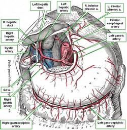
The [cystic artery] (FCAT: arteria cistica) is the artery that provides arterial blood supply to the gallbladder. It is found in the [triangle of Calot], also known as the “cystohepatic triangle” is a triangular region found within the lesser omentum connecting the duodenum, stomach, and liver. It is an area bound superiorly by the inferior surface of the liver, laterally by the cystic duct and the medial border of the gallbladder, and medially by the common hepatic duct. It is usually a branch of the right hepatic artery, which is itself a branch of the proper hepatic artery. After its origin from the right hepatic artery the cystic artery directs towards the neck of the gallbladder where it divides into anterior and posterior branches which then penetrate the gallbladder. These anterior and posterior branches are names "left and right" by Testut and Latarjet (1931) or "right and left" by Morris (1942). The cystic artery, as most of the components of the region of the hepatobiliary tree, has well documented anatomical variations. A detailed explanation of these variations can be found here at the Illustrated Encyclopedia of Human Anatomic Variation, curated by Dr. Ronald Bergman. |
 Hepatobiliary tree and arteries to the stomach. R=right, L= left, a.=artery, Gd a.= gastroduodenal artery |
| Sources: 1. "Tratado de Anatomia Humana" Testut et Latarjet 8 Ed. 1931 Salvat Editores, Spain 2. "Gray's Anatomy" 38th British Ed. Churchill Livingstone 1995 3. "Terminologia Anatomica: International Anatomical Terminology (FCAT)" Thieme, 1998 4. "Morris' Human Anatomy" Pearce, J. (1942) Blakiston Co. Philadlephia USA Image modified from the original by Dr. Henry Vandyke Carter. Public Domain. |
|
| MTD Main Page | Subscribe to MTD |


