![]()
Medical Terminology Daily (MTD) is a blog sponsored by Clinical Anatomy Associates, Inc. as a service to the medical community. We post anatomical, medical or surgical terms, their meaning and usage, as well as biographical notes on anatomists, surgeons, and researchers through the ages. Be warned that some of the images used depict human anatomical specimens.
You are welcome to submit questions and suggestions using our "Contact Us" form. The information on this blog follows the terms on our "Privacy and Security Statement" and cannot be construed as medical guidance or instructions for treatment.
We have 329 guests and no members online
")
Marcia Crocker Noyes
(1869 – 1946)
Further to my comment on old books and research that started with an interesting bookplate (Ex-Libris). I continued my research and found that the person in charge of the Osler library bookplate was a fascinating individual that today maybe a ghost in the MedChi library and building in Baltimore... This is certainly an article that can be called "A Moment in History"
Marcia Crocker Noyes was the librarian at The Maryland State Medical Society from 1896 to 1946 and was a founding member of the Medical Library Association.[1][2][3]
Sir William Osler, MD. a famous Johns Hopkins surgeon was a noted bibliophile and had a large personal collection of books on various topics. When he became the President of MedChi in 1896, he was dismayed at the condition of the library and knew that with the right person and some stewardship, it could become a significant collection. Sir William asked his friend, Dr. Bernard Steiner, a physician and President of the Enoch Pratt Free Library in Baltimore for suggestions of a librarian, and Dr. Steiner recommended Marcia Crocker Noyes. A native of New York, and a graduate of Hunter College, Marcia had moved to Baltimore for a lengthy visit with her sister, and took a “temporary” position at the Pratt Library, which turned into three years. Although she had no medical experience or background, she was enthusiastic, and most importantly, she was willing to move into the apartment provided for the librarian, who needed to be available 24 hours a day.
The image in this article is Ms. Noyes on her first year on the job. Marcia developed a book classification system for medical books, based on the Index Medicus, and called it the Classification for Medical Literature. The system uses the alphabet with capital letters for the major divisions of medicine and lower-case ones for the sub-sections. The system was used for many years, but it's now dated and the Faculty's original shelving scheme was never changed. The card catalogs still reflect her classification and many of the cards are written in Marcia's back-slanting handwriting.
Marcia knew enough to ask the Faculty's members about medical questions, terminology and literature. She gradually won over the predominantly male membership and they became her greatest allies; Sir William at the start, and then for nearly 40 years, Dr. John Ruhräh, a wealthy pediatrician with no immediate family of his own. She made a point of attending almost every Faculty function, and in 1904, under guidelines from the American Medical Association, Marcia was made the Faculty Secretary. For much of her first 10 years, she was the Faculty's only full-time employee, only being assisted by Mr. Caution, the Faculty's janitor. Later in life Marcia would say that she hired him because of his name!
Within ten years, the library had outgrown its space, and plans, spearheaded by Marcia and Sir William before his move to Oxford, were made to build a headquarters building, mainly to house the library's growing collection of medical books and journals.
Marcia was instrumental in the design and building of the new headquarters. She travelled to Philadelphia, New York and Boston to look at their medical society buildings, and eventually, the Philadelphia architectural firm, Ellicott & Emmart was selected to design and build the new Faculty building. Every detail of the building held her imprimatur, from the graceful staircase, to the light-filled reading room, and all of the myriad details of the millwork, marble tesserae, and most of all, the four-story cast iron stacks. She was on-site, climbing up unfinished staircases, checking out the progress of the building, which was built in less than one year at a cost of $90,000.
Among the features of the new building was a fourth-floor apartment for her. She referred to it as the "first penthouse in Baltimore" and it had a garden and rooftop terrace. The library collection eventually grew to more than 65,000 volumes from medical and specialty societies around the world. Journals were traded back and forth, and physicians eagerly anticipated the arrival of each new issue. At the same time, Marcia was involved in the Medical Library Association as one of eight founding members. The MLA promotes medical libraries and the exchange of information. One of the earliest mandates of the MLA was the Exchange, a distribution and trade service for those who had duplicates or little-used books in their collections. Initially, the Exchange was run out of the Philadelphia medical society, but in 1900 it was moved to Baltimore and Marcia oversaw it. Several hundred periodicals and journals were received and sent each month, a huge amount of work for a tiny staff. In 1904, the Faculty had run out of room to manage the Exchange, so it was moved to the Medical Society of the Kings County (Brooklyn). But without Marcia's excellent administrative skills, it floundered and in 1908, the MLA asked Marcia to take charge once again.
In 1909, when the new Faculty building opened, there was enough room to run the Exchange and with the help of MLA Treasurer, noted bibliophile and close friend, Dr. John Ruhräh, it once again became successful. Additionally, Marcia and Dr. Ruhräh combined forces to revive the MLA's bulletin, which had all but ceased publication in 1908, taking the Exchange with it. This duo maintained editorial control from 1911 until 1926. In 1934, around the time of Dr. Ruhräh's death, Marcia became the first “unmedicated” professional to head the MLA. During her tenure, the MLA incorporated, the first seal was adopted, and the annual meeting was held in Baltimore. Marcia wanted to write the history of the MLA once she retired from full-time work at the Faculty, but her health was beginning to fail. She had back problems and had suffered a serious burn on her shoulder as a young woman, possibly from her time running a summer camp, Camp Seyon, for young ladies in the Adirondack Mountains. In 1946, a celebration was planned to honor Marcia's 50 years at the Faculty. But she was adamant that the physicians wait until November, the actual date of her 50 years. However, they knew she was gravely ill, and might not make it until then, so a huge party was held in April. More than 250 physicians attended the celebration, but the ones she was closest to in the early years, were long gone. She was presented with a suitcase, a sum of money to use for travelling, and her favorite painting of Dr. John Philip Smith, a founder of the Medical College in Winchester, Virginia. It was painted by Edward Caledon Smith, a Virginia painter who had been a student of the painter Thomas Sully.[4] She adored this painting and vowed, jokingly, to take it with her wherever she went.
The painting was not to stay with her for very long, for she died in November 1946, and left it to the Faculty in her will. Her funeral was held in the Faculty's Osler Hall, named for her dear friend. More than 60 physicians served as her pallbearers, and she was buried at Baltimore's Green Mount Cemetery. In 1948, the MLA decided to establish an award in the name of Marcia Crocker Noyes. It was for outstanding achievement in medical library field and was to be awarded every two years, or when a truly worthy candidate was submitted. In 2014, the Faculty began giving a bouquet of flowers to the winner of the award in Marcia's name, and in honor of her work. Much evidence exists for this tradition, as we know that the physicians, especially Drs. Osler and Ruhräh, frequently gave her bouquets of flowers. Marcia also cultivated flower gardens at the Faculty and decorated the rooms with her work.
Today, the MedChi building is open for tours and if the rumors are to be believed Ms. Marcia Crocker Noyes is still at work in her beloved library as the "resident ghost" [1][5]

NOTE: This article has been modified from the original Wikipedia article on Marcia Crocker Noyes. The article itself is well-written with interesting images of the subject. I would encourage you to visit it. The second insert is from book 00736 in my personal library and shows in pencil, the incredibly small handwriting of Marsha C. Noyes.
Sources:
1. "Marcia, Marcia, Marcia" MedChi Archives blog.
2. "Marcia C. Noyes, Medical Librarian" (PDF). Bulletin of the Medical Library Association. 35 (1): 108–109. 1947. PMC 194645
3. Smith, Bernie Todd (1974). "Marcia Crocker Noyes, Medical Librarian: The Shaping of a Career" (PDF). Bulletin of the Medical Library Association. 62 (3): 314–324. PMC 198800Freely accessible. PMID 4619344.
4. Edward Caledon BRUCE (1825-1901)"
5. Behind the scenes tour MedChiBuilding
"Clinical Anatomy Associates, Inc., and the contributors of "Medical Terminology Daily" wish to thank all individuals who donate their bodies and tissues for the advancement of education and research”.
Click here for more information


- Details
Click on the image for a larger depiction
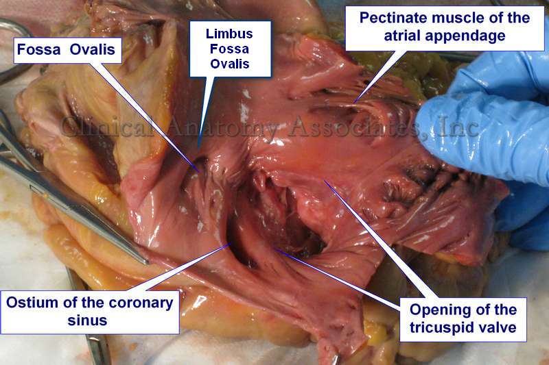
The coronary sinus is a venous structure that receives blood from the coronary circulation and returns it to the right atrium of the heart. It is found in the atrioventricular sulcus and receives all the veins of the heart (small, middle, great and oblique cardiac veins, and others), with the exception of some anterior cardiac veins that may empty directly into the right atrium. There are small venous valves at the point where most of these veins enter the coronary sinus.
Unlike most veins, the coronary sinus in the human has an evident layer of smooth muscle that may become the source of ectopic foci of atrial depolarization, causing atrial fibrillation.
The opening of the coronary sinus into the right atrium is called the "ostium of the coronary sinus".
There is discussion as to where the coronary sinus begins, as there is sometimes a gradual dilation of the great cardiac vein and a clear-cut boundary cannot be seen. Ludinghausen (1992) proposed to use as a boundary the point where the oblique vein of the left atrium (vein of Marshall) enters the coronary sinus. A this point there is a small valve called the "valve of Vieussens".
The ostium of the coronary sinus, where it empties into the right atrium, is characterized by the presence of a semilunar fold or band of tissue called the "valve of Thebesius". This valve may be absent (as in the image), it may be small, large, trabeculated, or cribriform. The presence of a large or cribriform valve of Thebesius may encumber the attempt at retrograde cardioplegia.
The image shows an human heart dissection, where the right atrium has been opened and the following structures exposed and labeled: Fossa ovalis, ostium of the coronary sinus, pectinate muscle of the atrial appendage, and the opening of the tricuspid valve.
Sources:
1. "Tratado de Anatomia Humana" Testut et Latarjet 8 Ed. 1931 Salvat Editores, Spain
2. "Gray's Anatomy" 38th British Ed. Churchill Livingstone 1995
3. "Myocardial coverage of the coronary sinus and related veins" Ludinghausen M, Ohmachi N, Boot C. (1992) Clin Anat 5:1-15
- Details

Click for a larger image
[UPDATED] The word [pedicle] is a derivative from the Latin [pediculus] meaning “a small foot”, a “stem”, or a “stalk”. The Latin term [pediculus] is itself a derivative of [pes/pedis] meaning “foot”.
[Pedicle] is also used to denote structures that lie at the root of “foot” of an organ, as in the “renal pedicle” (an older anatomical term) or in the pedicle of a sessile tumor. It is also used in surgery, to denote the vascular pedicle or “stalk” of a free tissue graft.
Since a pedicle is also the “foot” of an arch, the term has also been used to denote the base of the vertebral arch. Thus explained, each vertebra has bilateral bony bridges between the vertebral body anteriorly and the laminae posteriorly. These are the vertebral pedicles, which form the lateral walls of the vertebral canal.
The vertebral pedicles have different characteristics (width, length, angulation) depending on their vertebral level. This is important for spine surgery where pedicle screws are used:
• Lumbar vertebra: has a thicker, wider pedicle that tends to angulate posterolaterally
• Thoracic vertebra: has a thinner pedicle that looks almost anteroposteriorly
• Cervical vertebra: the pedicle is very small and thin, angles quite laterally, and forms the medial border of the transverse foramina.
The accompanying image is an inferior view of a thoracic vertebra showing the location of the vertebral pedicles. Click on the image for a larger version.
Additional information: “Vertebral pedicle anatomy in relation to pedicle screw fixation: a cadaver study” Chaynes et al. Surg Rad Anat (2001) 23:2, 85-90
Image property of: CAA.Inc.. Photographer: D.M. Klein.
- Details
The root terms [-stom-] and [-stoma-] both arise from the Greek word [στόμα] (st?ma) meaning “mouth” or “opening”. You can find them in medical terms such as:
- Stomatitis: The suffix [-itis] means inflammation. An inflammation of the mouth
- Stomatognathic: The root term [-gnath] means "jaw". Pertaining to the mouth and jaw
- Ileostomy: Creation of a permanent opening in the ileum for drainage purposes
- Anastomosis: Creation of a common opening between two hollow organs
The word [stoma] is also used as a stand-alone term with the same meaning, as in the creation of a stoma for surgical drainage.
Note: The links to Google Translate include an icon that will allow you to hear the pronunciation of the word.
- Details
|
The [cystic artery] (FCAT: arteria cistica) is the artery that provides arterial blood supply to the gallbladder. It is found in the [triangle of Calot], also known as the “cystohepatic triangle” is a triangular region found within the lesser omentum connecting the duodenum, stomach, and liver. It is an area bound superiorly by the inferior surface of the liver, laterally by the cystic duct and the medial border of the gallbladder, and medially by the common hepatic duct. It is usually a branch of the right hepatic artery, which is itself a branch of the proper hepatic artery. After its origin from the right hepatic artery the cystic artery directs towards the neck of the gallbladder where it divides into anterior and posterior branches which then penetrate the gallbladder. These anterior and posterior branches are names "left and right" by Testut and Latarjet (1931) or "right and left" by Morris (1942). The cystic artery, as most of the components of the region of the hepatobiliary tree, has well documented anatomical variations. A detailed explanation of these variations can be found here at the Illustrated Encyclopedia of Human Anatomic Variation, curated by Dr. Ronald Bergman. |
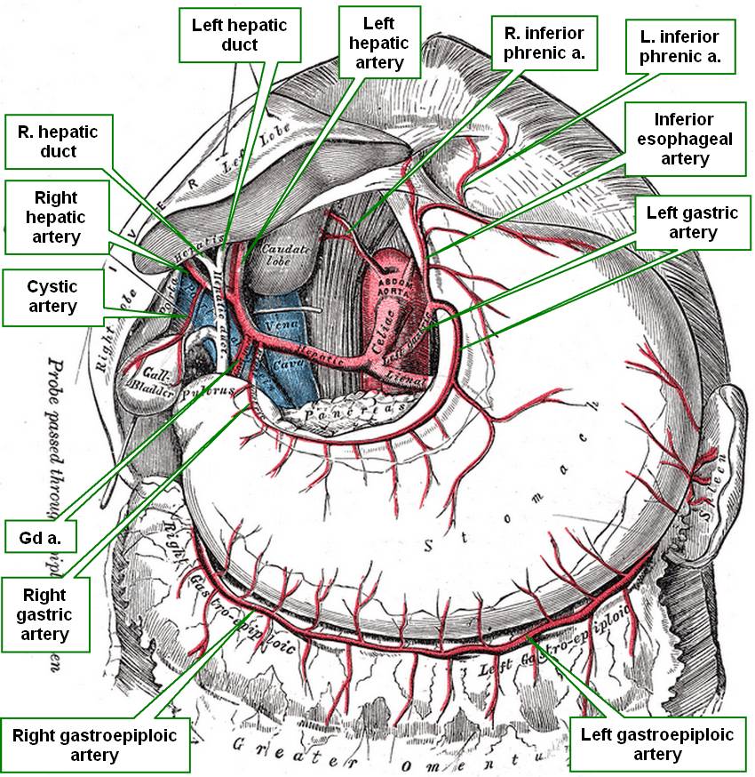 Hepatobiliary tree and arteries to the stomach. R=right, L= left, a.=artery, Gd a.= gastroduodenal artery |
| Sources: 1. "Tratado de Anatomia Humana" Testut et Latarjet 8 Ed. 1931 Salvat Editores, Spain 2. "Gray's Anatomy" 38th British Ed. Churchill Livingstone 1995 3. "Terminologia Anatomica: International Anatomical Terminology (FCAT)" Thieme, 1998 4. "Morris' Human Anatomy" Pearce, J. (1942) Blakiston Co. Philadlephia USA Image modified from the original by Dr. Henry Vandyke Carter. Public Domain. |
|
| MTD Main Page | Subscribe to MTD |
- Details
This article is part of the series "A Moment in History" where we honor those who have contributed to the growth of medical knowledge in the areas of anatomy, medicine, surgery, and medical research.
")
Click for a larger image
Lorem ipsum dolor sit amet, consectetur adipiscing elit, sed do eiusmod tempor incididunt ut labore et dolore magna aliqua.
Henry Vandyke Carter, MD (1831 – 1897). English physician, surgeon, medical artist, and a pioneer in leprosy and mycetoma studies. HV Carter was born in Yorkshire in 1831. He was the son of Henry Barlow Carter, a well-known artist and it is possible that he honed his natural talents with his father. His mother picked his middle name after a famous painter, Anthony Van Dyck. This is probably why his name is sometimes shown as Henry Van Dyke Carter, although the most common presentation of his middle name is Vandyke.
Having problems to finance his medical studies, HV Carter trained as an apothecary and later as an anatomical demonstrator at St. George’s Hospital in London, where he met Henry Gray (1872-1861), who was at the time the anatomical lecturer. Having seen the quality of HV Carter’s drawings, Henry Gray teamed with him to produce one of the most popular and longer-lived anatomy books in history: “Gray’s Anatomy”, which was first published in late 1857. The book itself, about which many papers have been written, was immediately accepted and praised because of the clarity of the text as well as the incredible drawings of Henry Vandyke Carter.
While working on the book’s drawings, HV Carter continued his studies and received his MD in 1856.
In spite of initially being offered a co-authorship of the book, Dr. Carter was relegated to the position of illustrator by Henry Gray and never saw the royalties that the book could have generated for him. For all his work and dedication, Dr. Carter only received a one-time payment of 150 pounds. Dr. Carter never worked again with Gray, who died of smallpox only a few years later.
Frustrated, Dr. Carter took the exams for the India Medical Service. In 1858 he joined as an Assistant Surgeon and later became a professor of anatomy and physiology. Even later he served as a Civil Surgeon. During his tenure with the India Medical Service he attained the ranks of Surgeon, Surgeon-Major, Surgeon-Lieutenant-Colonel, and Brigade-Surgeon.
Dr. Carter dedicated the rest of his life to the study of leprosy, and other ailments typical of India at that time. He held several important offices, including that of Dean of the Medical School of the University of Bombay. In 1890, after his retirement, he was appointed Honorary Physician to the Queen.
Dr. Henry Vandyke Carter died of tuberculosis in 1897.
Personal note: Had history been different, this famous book would have been called “Gray and Carter’s Anatomy” and Dr. Carter never gone to India. His legacy is still seen in the images of the thousands of copies of “Gray’s Anatomy” throughout the world and the many reproductions of his work available on the Internet. We are proud to use some of his images in this blog. The image accompanying this article is a self-portrait of Dr. Carter. Click on the image for a larger depiction. Dr. Miranda
Sources:
1. “Obituary: Henry Vandyke Carter” Br Med J (1897);1:1256-7
2. “The Anatomist: A True Story of ‘Gray’s Anatomy” Hayes W. (2007) USA: Ballantine
3. “A Glimpse of Our Past: Henry Gray’s Anatomy” Pearce, JMS. J Clin Anat (2009) 22:291–295
4. “Henry Gray and Henry Vandyke Carter: Creators of a famous textbook” Roberts S. J Med Biogr (2000) 8:206–212.
5. “Henry Vandyke Carter and his meritorious works in India” Tappa, DM et al. Indian J Dermatol Venereol Leprol (2011) 77:101-3
- Details
This article is part of the series "A Moment in History" where we honor those who have contributed to the growth of medical knowledge in the areas of anatomy, medicine, surgery, and medical research.

Alexander Monro Secundus
Alexander Monro Secundus (1733- 1817). Scottish physician and anatomist, born in Edinburgh. Alexander Monro Secundus (the second), studied anatomy with his father Alexander Monro Primus (the first). He received his doctorate in medicine at 22 years of age. His studies led him to write on the lymphatic system, leading to a public written dispute with William Hunter. In 1753 he demonstrated the communication channels between both lateral and third ventricle of the brain, describing it in a published work in 1797. Since then, these channels have been know as the foramina of Monro. Later analysis of prior publications demonstrate that these foramina were known, although probably not well understood.
The Monro family gave history three anatomists who occupied the chair of Anatomy at the University of Edinburgh for over a century. Alexander Monro Primus (1697 - 1767), Alexander Monro Secundus (1733 - 1817), and Alexander Monro Tertius (1773 - 1859)
Sources:
1. Sharp, J. A. (1961). Alexander Monro secundus and the interventricular foramen. Medical History, 5(1), 83
2. Wu, O. C., Manjila, S., Malakooti, N., & Cohen, A. R. (2012). The remarkable medical lineage of the Monro family: contributions of Alexander primus, secundus, and tertius. Journal of neurosurgery, 116(6), 1337-1346.
3. "The origin of Medical Terms" Skinner, HA; 1970
Original image: Coloured stipple engraving by James Heath (1757–1834), after Henry Raeburn (1756–1823) [Public domain], via Wikimedia Commons