Medical Terminology Daily (MTD) is a blog sponsored by Clinical Anatomy Associates, Inc. as a service to the medical community. We post anatomical, medical or surgical terms, their meaning and usage, as well as biographical notes on anatomists, surgeons, and researchers through the ages. Be warned that some of the images used depict human anatomical specimens.
You are welcome to submit questions and suggestions using our "Contact Us" form. The information on this blog follows the terms on our "Privacy and Security Statement" and cannot be construed as medical guidance or instructions for treatment.
We have 869 guests online

Jean George Bachmann
(1877 – 1959)
French physician–physiologist whose experimental work in the early twentieth century provided the first clear functional description of a preferential interatrial conduction pathway. This structure, eponymically named “Bachmann’s bundle”, plays a central role in normal atrial activation and in the pathophysiology of interatrial block and atrial arrhythmias.
As a young man, Bachmann served as a merchant sailor, crossing the Atlantic multiple times. He emigrated to the United States in 1902 and earned his medical degree at the top of his class from Jefferson Medical College in Philadelphia in 1907. He stayed at this Medical College as a demonstrator and physiologist. In 1910, he joined Emory University in Atlanta. Between 1917 -1918 he served as a medical officer in the US Army. He retired from Emory in 1947 and continued his private medical practice until his death in 1959.
On the personal side, Bachmann was a man of many talents: a polyglot, he was fluent in German, French, Spanish and English. He was a chef in his own right and occasionally worked as a chef in international hotels. In fact, he paid his tuition at Jefferson Medical College, working both as a chef and as a language tutor.
The intrinsic cardiac conduction system was a major focus of cardiovascular research in the late nineteenth and early twentieth centuries. The atrioventricular (AV) node was discovered and described by Sunao Tawara and Karl Albert Aschoff in 1906, and the sinoatrial node by Arthur Keith and Martin Flack in 1907.
While the connections that distribute the electrical impulse from the AV node to the ventricles were known through the works of Wilhelm His Jr, in 1893 and Jan Evangelista Purkinje in 1839, the mechanism by which electrical impulses spread between the atria remained uncertain.
In 1916 Bachmann published a paper titled “The Inter-Auricular Time Interval” in the American Journal of Physiology. Bachmann measured activation times between the right and left atria and demonstrated that interruption of a distinct anterior interatrial muscular band resulted in delayed left atrial activation. He concluded that this band constituted the principal route for rapid interatrial conduction.
Subsequent anatomical and electrophysiological studies confirmed the importance of the structure described by Bachmann, which came to bear his name. Bachmann’s bundle is now recognized as a key determinant of atrial activation patterns, and its dysfunction is associated with interatrial block, atrial fibrillation, and abnormal P-wave morphology. His work remains foundational in both basic cardiac anatomy and clinical electrophysiology.
Sources and references
1. Bachmann G. “The inter-auricular time interval”. Am J Physiol. 1916;41:309–320.
2. Hurst JW. “Profiles in Cardiology: Jean George Bachmann (1877–1959)”. Clin Cardiol. 1987;10:185–187.
3. Lemery R, Guiraudon G, Veinot JP. “Anatomic description of Bachmann’s bundle and its relation to the atrial septum”. Am J Cardiol. 2003;91:148–152.
4. "Remembering the canonical discoverers of the core components of the mammalian cardiac conduction system: Keith and Flack, Aschoff and Tawara, His, and Purkinje" Icilio Cavero and Henry Holzgrefe Advances in Physiology Education 2022 46:4, 549-579.
5. Knol WG, de Vos CB, Crijns HJGM, et al. “The Bachmann bundle and interatrial conduction” Heart Rhythm. 2019;16:127–133.
6. “Iatrogenic biatrial flutter. The role of the Bachmann’s bundle” Constán E.; García F., Linde, A.. Complejo Hospitalario de Jaén, Jaén. Spain
7. Keith A, Flack M. The form and nature of the muscular connections between the primary divisions of the vertebrate heart. J Anat Physiol 41: 172–189, 1907.
"Clinical Anatomy Associates, Inc., and the contributors of "Medical Terminology Daily" wish to thank all individuals who donate their bodies and tissues for the advancement of education and research”.
Click here for more information
- Details
[Dysmorphism] is a medical term formed by the prefix [dys-] meaning "abnormal", the root term [-morph-], arising from the Greek word [μορφος] (morfos), meaning "form" or "shape", and the suffix [-ism], also of Greek origin, in this case meaning "condition". A condition of abnormal shape, or "misshapen".
The term dysmorphism is used to denote an anatomical anomaly, usually superficial, that goes beyond the objective (and sometimes subjective) boundaries of normalcy. An example of these are craniofacial dysmorphisms associated with specific congenital disorders, such as Crouzon syndrome; birth defects such as hemifacial microsomia, or as in the case of the image on this article, Mevalonate Kinase Deficiency, a metabolic disease.
Some craniofacial dysmorphisms can affect articulation and speech, such as cleft palate, cleft lip (harelip), prognathism, and retrognathism.
Dysmorphism is also used in some mental disorders where the patient has an abnormal self-image, seeing his/her body or body parts as abnormal, when they are not. This is known as Body Dysmorphic Disorder (BDD), and can be associated with obsessive-compulsive behavior, anxiety, or depression. In some cases BDD can be associated or found in eating disorders such as Anorexia Nervosa, or bulimia.
The image shows a small child with mevalonic aciduria, a deficiency of mevalonate kinase deficiency. This is a rare genetic autosomal recessive metabolic disorder.
Thanks to Maria E. Gallegos, Chair of the Speech Pathology School, Iberoamerican University, Santiago Chile, for suggesting this article. Dr. Miranda
Image Source: Haas D, Hoffmann GF. Mevalonate kinase deficiencies: from mevalonic aciduria to hyperimmunoglobulinemia D syndrome. Orphanet J Rare Dis. 1, 13. 2006. PMID 16722536. DOI:10.1186/1750-1172-1-13 Image in the public domain
- Details
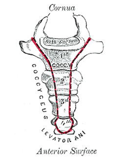
The word [coccyx] arises from the Greek term [κούκος] (pronounced koúkos)and means "cuckoo". It is the name of the lower segment of the spinal column, and was named by Herophilus of Alexandria (325-255BC) because of a resemblance of this structure to the bill of the cuckoo bird. Vesalius also used the same analogy. There is another structure of the body named after the beak of a bird, do you know it? If not, click here.
The coccyx (vernacularly known as "tailbone") is usually represented by four rudimentary vertebrae, although the number varies between 3 to 5 vertebrae. There have been reported cases of "human tails" but these do not have a bony structure and are usually related to congenital abnormalities such as spina bifida.
The coccyx has a well-formed superior component, which usually presents with two cornua (horns) which serve as part of a rudimentary zygapophyseal (facet) joint. The lower coccygeal vertebra is usually a small bony node.
The coccyx has an anterior sacrococcygeal ligament, which is continued with the anoccygeal raphe, a ligamentous structure that serves as a posterior attachment for muscular components of the pelvic diaphragm, and helps anchor the anal canal. The coccygeus muscle, the posterior component of the pelvic diaphragm and part of the sacrospinous ligament also attach to the anterolateral aspect of the coccyx.
Coccygeal pain is referred to as coccydynia.
Sources:
1. "The Origin of Medical Terms" Skinner, HA 1970 Hafner Publishing Co.
2. "Medical Meanings - A Glossary of Word Origins" Haubrich, WD. ACP Philadelphia
3. "Dorlands's Illustrated Medical Dictionary" 26th Ed. W.B. Saunders 1994
5 "Tratado de Anatomia Humana" Testut et Latarjet 8 Ed. 1931 Salvat Editores, Spain
6. "Anatomy of the Human Body" Henry Gray 1918. Philadelphia: Lea & Febiger
Image modified by CAA, Inc. Original image courtesy of bartleby.com
Note: Google Translate includes the symbol (?). Clicking on it will allow you to hear the pronunciation of the word.
- Details
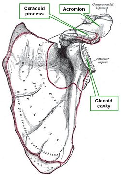
The [coracoid process] is a curved bony projection that attaches to the superior aspect of the neck of the scapula . At its origin it has a broad base continuous with a segment that projects slightly superomedially, it then continues anterolaterally with a narrower distal segment.
The pectoralis minor and coracobrachialis muscles attach to the coracoid process as well as the tendon of the short head of the biceps brachii muscle. The trapezoid and conoid ligaments, which also attach to the clavicle, attach to the coracoid process.
The root term [-corac-] originates from the Greek word [κοράκι] (koríki), meaning “raven” or “crow. The suffix [-oid], also of Greek origin, means “similar to". The word [coracoid] was coined because of the similarity of the coracoid process to a raven’s beak.
Trivia question: What other part of the body is named after a bird’s beak? For the answer, click here. What other parts of the body are named after birds? For the answer, click here.
The image shows the anterior view of the left scapula. Modified from image in Public Domain, by Henry Vandyke Carter - Gray's Anatomy
Source:
“Gray’s Anatomy” Henry Gray, 1918
- Details
The [acromion] is a bony process found at the superolateral end of the spine of the scapula forming the highest point of the shoulder. It is a flattened somewhat triangular bony process that is found superior to the glenoid cavity. The trapezius and deltoid muscles attach to the acromion.
The term [acromion] is formed by two Greek roots; [άκρο] (ákro), meaning “end” or “point”, and [ώμος] (omos), meaning “shoulder”. The term [acromion] is quite descriptive, as it means the “point of the shoulder.
The image shows the anterior view of the left scapula. Modified from image in Public Domain, by Henry Vandyke Carter - Gray's Anatomy
Source:
“Gray’s Anatomy” Henry Gray, 1918
- Details
UPDATED: From the Greek, the prefix [ana-] meaning "trough" or "up", the root term [-tom-], arising from [τομή] (tomi) meaning "to cut", and the suffix [-y], meaning "process". The word [anatomy] means then, "a process of cutting up", a very good description of what anatomists do. What is interesting is that the word anatomy describes an action, and as such can be used as a verb. It is correct to say "to anatomize" when referring to the act of dissecting a body or body part.
The word "anatomy" has the same meaning as "dissection", a word with Latin roots. The prefix [dis-] means "apart", while the root term [-section] means "to cut". [-section] arises from the Latin [sectis] or [secare]. both meaning "to cut".
Today the term [anatomy] is used to describe one of the basic medical sciences; in the Middle Ages the terms to "dissect" or to "anatomize" were interchangeable.
Anatomy is "the study of the human body, its parts and components, and the spatial relationship between this components". For many, anatomy is at the basis of the Science of Surgery
There are many subspecialties in anatomy, including:
• Gross anatomy: That anatomy that can be seen with the naked eye
• Surface anatomy: The correlation between superficial landmarks and internal structures and organs
• Clinical anatomy: The study of anatomy and its relation to physiology, pathology, and surgical treatment
Personal note: The improper pronunciation of the term [dissection] is one of my pet peeves!
- Details
UPDATED: The word [anesthesia] is formed by the prefix [an-] meaning "without" or absence of", and the Greek root term [-esthesia-] meaning "sensation". Skinner (1970) mentions that the suffix [-ia] means "condition". Thus analyzed, the word [anesthesia] means "a condition of absence of sensation". The word was coined and first used by Oliver W. Holmes Sr. (1809 - 1894).
The search for an anesthetic agent that could alleviate pain and help surgery has ancient origins, potions, alcohol, and herbal remedies had been used until the discovery of nitrous oxide (laughing gas) first, and ether second. It was this latter element, pioneered by William T.G. Morton (1819–1868), that started a revolution in surgery.
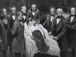
In 1846 Morton working as the anesthetist helped a public demonstration of surgery at the Massachusetts General Hospital. This was the first use of anesthesia in surgery. The event was so important that drawings and paintings were made. See the accompanying image.
After this demonstration, research was continued and the anesthetic properties of many compounds were discovered, leading to modern anesthetics.
The operation took place on October 16, 1846, in the first operating room built at the Massachusetts General Hospital. This room has been preserved and is today known as the "Ether Dome". You can read an article on a visit I made to this historic place. Dr. Miranda
Original image (public domain) courtesy of "Images from the History of Medicine" at www.nih.gov


