Medical Terminology Daily (MTD) is a blog sponsored by Clinical Anatomy Associates, Inc. as a service to the medical community. We post anatomical, medical or surgical terms, their meaning and usage, as well as biographical notes on anatomists, surgeons, and researchers through the ages. Be warned that some of the images used depict human anatomical specimens.
You are welcome to submit questions and suggestions using our "Contact Us" form. The information on this blog follows the terms on our "Privacy and Security Statement" and cannot be construed as medical guidance or instructions for treatment.
We have 1399 guests online

Jean George Bachmann
(1877 – 1959)
French physician–physiologist whose experimental work in the early twentieth century provided the first clear functional description of a preferential interatrial conduction pathway. This structure, eponymically named “Bachmann’s bundle”, plays a central role in normal atrial activation and in the pathophysiology of interatrial block and atrial arrhythmias.
As a young man, Bachmann served as a merchant sailor, crossing the Atlantic multiple times. He emigrated to the United States in 1902 and earned his medical degree at the top of his class from Jefferson Medical College in Philadelphia in 1907. He stayed at this Medical College as a demonstrator and physiologist. In 1910, he joined Emory University in Atlanta. Between 1917 -1918 he served as a medical officer in the US Army. He retired from Emory in 1947 and continued his private medical practice until his death in 1959.
On the personal side, Bachmann was a man of many talents: a polyglot, he was fluent in German, French, Spanish and English. He was a chef in his own right and occasionally worked as a chef in international hotels. In fact, he paid his tuition at Jefferson Medical College, working both as a chef and as a language tutor.
The intrinsic cardiac conduction system was a major focus of cardiovascular research in the late nineteenth and early twentieth centuries. The atrioventricular (AV) node was discovered and described by Sunao Tawara and Karl Albert Aschoff in 1906, and the sinoatrial node by Arthur Keith and Martin Flack in 1907.
While the connections that distribute the electrical impulse from the AV node to the ventricles were known through the works of Wilhelm His Jr, in 1893 and Jan Evangelista Purkinje in 1839, the mechanism by which electrical impulses spread between the atria remained uncertain.
In 1916 Bachmann published a paper titled “The Inter-Auricular Time Interval” in the American Journal of Physiology. Bachmann measured activation times between the right and left atria and demonstrated that interruption of a distinct anterior interatrial muscular band resulted in delayed left atrial activation. He concluded that this band constituted the principal route for rapid interatrial conduction.
Subsequent anatomical and electrophysiological studies confirmed the importance of the structure described by Bachmann, which came to bear his name. Bachmann’s bundle is now recognized as a key determinant of atrial activation patterns, and its dysfunction is associated with interatrial block, atrial fibrillation, and abnormal P-wave morphology. His work remains foundational in both basic cardiac anatomy and clinical electrophysiology.
Sources and references
1. Bachmann G. “The inter-auricular time interval”. Am J Physiol. 1916;41:309–320.
2. Hurst JW. “Profiles in Cardiology: Jean George Bachmann (1877–1959)”. Clin Cardiol. 1987;10:185–187.
3. Lemery R, Guiraudon G, Veinot JP. “Anatomic description of Bachmann’s bundle and its relation to the atrial septum”. Am J Cardiol. 2003;91:148–152.
4. "Remembering the canonical discoverers of the core components of the mammalian cardiac conduction system: Keith and Flack, Aschoff and Tawara, His, and Purkinje" Icilio Cavero and Henry Holzgrefe Advances in Physiology Education 2022 46:4, 549-579.
5. Knol WG, de Vos CB, Crijns HJGM, et al. “The Bachmann bundle and interatrial conduction” Heart Rhythm. 2019;16:127–133.
6. “Iatrogenic biatrial flutter. The role of the Bachmann’s bundle” Constán E.; García F., Linde, A.. Complejo Hospitalario de Jaén, Jaén. Spain
7. Keith A, Flack M. The form and nature of the muscular connections between the primary divisions of the vertebrate heart. J Anat Physiol 41: 172–189, 1907.
"Clinical Anatomy Associates, Inc., and the contributors of "Medical Terminology Daily" wish to thank all individuals who donate their bodies and tissues for the advancement of education and research”.
Click here for more information
- Details
This is a medical root term arising from the Latin word [seco] meaning "to cut". The term [section] is a derivative of the same from the Latin terms [sectio] and [sectionis]. In anatomy and histology the term [section] is used to denote "a slice".
- Section: "A slice"
- Transsection: To "cut across". This is the proper spelling of the word, although 'transection" is also accepted
- Dissection:To "cut apart".
- Resection: To "cut again", used to denote "removal"
- Venesection: To "cut a vein". This term was used in the times of bloodletting
- Details
The term is a mix of two Greex root terms, [Pneum-] meaning "air" and [thorax] for "chest". The term [pneumothorax] means "air in the chest".
A pneumothorax occurs when the parietal pleura that lines the internal aspect of the chest and the visceral pleura that lines a lung are separated by a small amount of air that penetrates the thoracic cavity. This air causes the capillary action of the pleural fluid to fail, allowing for more air to enter the thoracic cavity and the lung to collapse.
Since the pleural cavities are separate, the collapse of one lung does not necessarily failure of the contralateral lung, except in a rare anatomical variation where both pleural cavities are communicated.
A pneumothorax can be spontaneous, with no apparent cause, or caused by trauma that allows the air to enter the thorax. If blood and air enter the thoracic cavity, the condition is then called a "pneumohemothorax".
Copyrighted image property of:CAA.Inc. Artist:D.M. Klein
- Details
This medical term is Greek and is composed of [ίδιος] (idios) meaning "self" or "one's own", something peculiar to a particular individual. This is probably why this root term is used for [idiot] or [idiocy]. The second portion of the word is also Greek [πάθος] (pathos) meaning "suffering", or "disease". The word [idiopathic] literally means "one's own disease".
The term is used today to denote a pathology that has no known cause.
- Details
[UPDATED] The medical term [hemangioma] is formed by two root terms and a suffix. The root term [hem-] arises from the Greek word [αίμα] (a?ma) meaning "blood", the second root term [-angi-] .from the Greek term [αγγείο] (angeio), meaning "vessel” and the suffix [-oma] (ωμα), also Greek, meaning "mass", "growth”, or "tumor". The term [hemangioma] then means a “mass of blood vessels”, a description not far from the truth.
A variation of the term is [hemangiomata], where the suffix [-omata] is plural, indicating multiple hemangiomas.
In general, a hemangioma can be described as a mass of blood vessels that can be convoluted, radiated, or swollen. Usually benign, hemangiomas appear in newborns and tend to evolve, involve, and disappear in the first ten years of life.
Other types of hemangiomas can be found in adults, with a tendency to be more prevalent in females than males. There are three types of hemangiomas described:
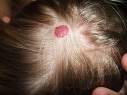
• Capillary hemangioma. Usually form skin patches known as “strawberry hemangiomas”. See image.
• Venous hemangioma. They are characterized by a “knot” or convoluted mass of veins. Since veins are usually more dilated than arteries, they are also known as “cavernous hemangiomas”. They can be found in a subcutaneous location or on internal organs, such as the liver.
• Arterial hemangioma. Formed by arteries, they tend to radiate and form a network of arteries, hence the term “plexiform hemangiomas”
• Vulvar hemangiomata. Multiple hemangiomas in the external female genitalia. For more information, click here
The image shows a small hemangioma in the scalp of a two-year old. Image by Cbheumircanl (Own work) [Public domain], via Wikimedia Commons
- Details
This term describes a structure that is composed of a multinucleated cytoplasm. It is either the result of the fusion of multiple adjoining cells, or the formation (by division) of multiple nuclei in a large cell.
Since cardiac muscle is formed by separate cells that are anatomically, mechanically, chemically, and electrically connected, the cardiac muscle has been called a "functional syncytium". Functionally, cardiac muscle works as a unit.
For those interested in the etymology of the word [syncytium], it arises from the Greek and its components are the prefix [syn-], meaning "together"; the root term [-cyt-], meaning "cell"; and the suffix [-ium], meaning "layer" or "membrane"
- Details
The root terms [-hem-] and [-hemat-] are both derivate from the Greek word [αίμα] (a?ma) meaning "blood". The same word and meaning applies to the suffix [-emia-]. Applications of these root terms include:
- Hematuria: The suffix [-uria] means "related to urine", or "urine". Refers to a condition where there is detectable blood in the urine
- Hematoma: The suffix [-oma] means "mass", "growth: or "tumor". A mass of blood that usually forms after trauma, a welt.
- Hemangioma: The root term [-angi-] means "vessel". The suffix [-oma] means "mass", "growth: or "tumor". A mass or a growth of blood vessels
Note: The links to Google Translate include an icon that will allow you to hear the Greek or Latin pronunciation of the word.


