![]()
Medical Terminology Daily (MTD) is a blog sponsored by Clinical Anatomy Associates, Inc. as a service to the medical community. We post anatomical, medical or surgical terms, their meaning and usage, as well as biographical notes on anatomists, surgeons, and researchers through the ages. Be warned that some of the images used depict human anatomical specimens.
You are welcome to submit questions and suggestions using our "Contact Us" form. The information on this blog follows the terms on our "Privacy and Security Statement" and cannot be construed as medical guidance or instructions for treatment.
We have 320 guests and no members online
")
Marcia Crocker Noyes
(1869 – 1946)
Further to my comment on old books and research that started with an interesting bookplate (Ex-Libris). I continued my research and found that the person in charge of the Osler library bookplate was a fascinating individual that today maybe a ghost in the MedChi library and building in Baltimore... This is certainly an article that can be called "A Moment in History"
Marcia Crocker Noyes was the librarian at The Maryland State Medical Society from 1896 to 1946 and was a founding member of the Medical Library Association.[1][2][3]
Sir William Osler, MD. a famous Johns Hopkins surgeon was a noted bibliophile and had a large personal collection of books on various topics. When he became the President of MedChi in 1896, he was dismayed at the condition of the library and knew that with the right person and some stewardship, it could become a significant collection. Sir William asked his friend, Dr. Bernard Steiner, a physician and President of the Enoch Pratt Free Library in Baltimore for suggestions of a librarian, and Dr. Steiner recommended Marcia Crocker Noyes. A native of New York, and a graduate of Hunter College, Marcia had moved to Baltimore for a lengthy visit with her sister, and took a “temporary” position at the Pratt Library, which turned into three years. Although she had no medical experience or background, she was enthusiastic, and most importantly, she was willing to move into the apartment provided for the librarian, who needed to be available 24 hours a day.
The image in this article is Ms. Noyes on her first year on the job. Marcia developed a book classification system for medical books, based on the Index Medicus, and called it the Classification for Medical Literature. The system uses the alphabet with capital letters for the major divisions of medicine and lower-case ones for the sub-sections. The system was used for many years, but it's now dated and the Faculty's original shelving scheme was never changed. The card catalogs still reflect her classification and many of the cards are written in Marcia's back-slanting handwriting.
Marcia knew enough to ask the Faculty's members about medical questions, terminology and literature. She gradually won over the predominantly male membership and they became her greatest allies; Sir William at the start, and then for nearly 40 years, Dr. John Ruhräh, a wealthy pediatrician with no immediate family of his own. She made a point of attending almost every Faculty function, and in 1904, under guidelines from the American Medical Association, Marcia was made the Faculty Secretary. For much of her first 10 years, she was the Faculty's only full-time employee, only being assisted by Mr. Caution, the Faculty's janitor. Later in life Marcia would say that she hired him because of his name!
Within ten years, the library had outgrown its space, and plans, spearheaded by Marcia and Sir William before his move to Oxford, were made to build a headquarters building, mainly to house the library's growing collection of medical books and journals.
Marcia was instrumental in the design and building of the new headquarters. She travelled to Philadelphia, New York and Boston to look at their medical society buildings, and eventually, the Philadelphia architectural firm, Ellicott & Emmart was selected to design and build the new Faculty building. Every detail of the building held her imprimatur, from the graceful staircase, to the light-filled reading room, and all of the myriad details of the millwork, marble tesserae, and most of all, the four-story cast iron stacks. She was on-site, climbing up unfinished staircases, checking out the progress of the building, which was built in less than one year at a cost of $90,000.
Among the features of the new building was a fourth-floor apartment for her. She referred to it as the "first penthouse in Baltimore" and it had a garden and rooftop terrace. The library collection eventually grew to more than 65,000 volumes from medical and specialty societies around the world. Journals were traded back and forth, and physicians eagerly anticipated the arrival of each new issue. At the same time, Marcia was involved in the Medical Library Association as one of eight founding members. The MLA promotes medical libraries and the exchange of information. One of the earliest mandates of the MLA was the Exchange, a distribution and trade service for those who had duplicates or little-used books in their collections. Initially, the Exchange was run out of the Philadelphia medical society, but in 1900 it was moved to Baltimore and Marcia oversaw it. Several hundred periodicals and journals were received and sent each month, a huge amount of work for a tiny staff. In 1904, the Faculty had run out of room to manage the Exchange, so it was moved to the Medical Society of the Kings County (Brooklyn). But without Marcia's excellent administrative skills, it floundered and in 1908, the MLA asked Marcia to take charge once again.
In 1909, when the new Faculty building opened, there was enough room to run the Exchange and with the help of MLA Treasurer, noted bibliophile and close friend, Dr. John Ruhräh, it once again became successful. Additionally, Marcia and Dr. Ruhräh combined forces to revive the MLA's bulletin, which had all but ceased publication in 1908, taking the Exchange with it. This duo maintained editorial control from 1911 until 1926. In 1934, around the time of Dr. Ruhräh's death, Marcia became the first “unmedicated” professional to head the MLA. During her tenure, the MLA incorporated, the first seal was adopted, and the annual meeting was held in Baltimore. Marcia wanted to write the history of the MLA once she retired from full-time work at the Faculty, but her health was beginning to fail. She had back problems and had suffered a serious burn on her shoulder as a young woman, possibly from her time running a summer camp, Camp Seyon, for young ladies in the Adirondack Mountains. In 1946, a celebration was planned to honor Marcia's 50 years at the Faculty. But she was adamant that the physicians wait until November, the actual date of her 50 years. However, they knew she was gravely ill, and might not make it until then, so a huge party was held in April. More than 250 physicians attended the celebration, but the ones she was closest to in the early years, were long gone. She was presented with a suitcase, a sum of money to use for travelling, and her favorite painting of Dr. John Philip Smith, a founder of the Medical College in Winchester, Virginia. It was painted by Edward Caledon Smith, a Virginia painter who had been a student of the painter Thomas Sully.[4] She adored this painting and vowed, jokingly, to take it with her wherever she went.
The painting was not to stay with her for very long, for she died in November 1946, and left it to the Faculty in her will. Her funeral was held in the Faculty's Osler Hall, named for her dear friend. More than 60 physicians served as her pallbearers, and she was buried at Baltimore's Green Mount Cemetery. In 1948, the MLA decided to establish an award in the name of Marcia Crocker Noyes. It was for outstanding achievement in medical library field and was to be awarded every two years, or when a truly worthy candidate was submitted. In 2014, the Faculty began giving a bouquet of flowers to the winner of the award in Marcia's name, and in honor of her work. Much evidence exists for this tradition, as we know that the physicians, especially Drs. Osler and Ruhräh, frequently gave her bouquets of flowers. Marcia also cultivated flower gardens at the Faculty and decorated the rooms with her work.
Today, the MedChi building is open for tours and if the rumors are to be believed Ms. Marcia Crocker Noyes is still at work in her beloved library as the "resident ghost" [1][5]

NOTE: This article has been modified from the original Wikipedia article on Marcia Crocker Noyes. The article itself is well-written with interesting images of the subject. I would encourage you to visit it. The second insert is from book 00736 in my personal library and shows in pencil, the incredibly small handwriting of Marsha C. Noyes.
Sources:
1. "Marcia, Marcia, Marcia" MedChi Archives blog.
2. "Marcia C. Noyes, Medical Librarian" (PDF). Bulletin of the Medical Library Association. 35 (1): 108–109. 1947. PMC 194645
3. Smith, Bernie Todd (1974). "Marcia Crocker Noyes, Medical Librarian: The Shaping of a Career" (PDF). Bulletin of the Medical Library Association. 62 (3): 314–324. PMC 198800Freely accessible. PMID 4619344.
4. Edward Caledon BRUCE (1825-1901)"
5. Behind the scenes tour MedChiBuilding
"Clinical Anatomy Associates, Inc., and the contributors of "Medical Terminology Daily" wish to thank all individuals who donate their bodies and tissues for the advancement of education and research”.
Click here for more information


- Details
Click for a larger image
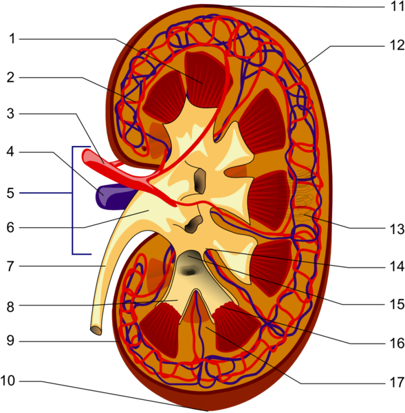
UPDATED: Definition: "A hilum" is the area of an organ where all the structures that enter or leave the organ are found". The term is based on a Latin word meaning something small, or trivial. Also used to describe the small spot on a seed where the seed is attached. The Latin phrase [ne hilum] was used to denote something of no worth or "not at all". In English it would have been similar to "not worth a bean". The plural form for [hilum] is [hila].
In human anatomy the term is used more on the seed attachment meaning. Since a hilum is the area of an organ where all the structures that enter or leave the organ are found, severing the root structures at the level of the hilum detaches the organ from the body. There are several hila in the body:
• Renal hilum: The hilum of the kidney (see item 5 in the accompanying image)
• Lienal hilum: The hilum of the spleen
• Splenic hilum: The hilum of the spleen
• Pulmonary hilum: The hilum of the spleen
• Hepatic hilum: The hilum of the liver. This name is not commonly used and the hepatic hilum is known as the "porta hepatis" meaning the "door to the liver".
There is a wrong version of the term. The word [hilus] is sometimes used and is incorrect. This word was a mistake by the anatomist Bartolomeo Eustachius (c.1520 - 1574) that has continued until today.
Here is the key to the image: Longitudinal section of a kidney. 1-Renal pyramid, 3-Renal artery, 4-Renal vein, 5-Renal hilum, 6-Renal pelvis, 7- Ureter, 8-Minor calyx, 9-Renal capsule, 14-Minor calyx, 15- Major calyx, 16-Renal papilla, 17-Renal column .
Original Image by Piotr Micha? Jaworski; PioM EN DE PL (Own work) [GFDL (http://www.gnu.org/copyleft/fdl.html) or CC-BY-SA-3.0 (http://creativecommons.org/licenses/by-sa/3.0/)], via Wikimedia Commons
- Details
The suffix [-(o)gram] evolved from the Greek word [γράμμα] (gr?mma) , meaning "letter". It was used to denote the written record that was maintained about a patient's ailment. When modern devices that could record an examination, such as an electrocardiograph, the suffix [-ogram] was added to the graphic, so we end up with an electrocardiogram. Today we use it to mean "examination of" with the caveat that some type of written, photographic, digital or drawn record must be left behind. Examples of the use of this suffix are:
- Cholangiogram: Examination of a bile vessel
- Angiogram: Examination of a vessel
- Mammogram: Examination of a breast
- Electrocardiogram: Examination of the electrical activity of the heart. EKG or ECG
- Details
The term [cholecystectomy] is composed by the combined root terms [-chole-] derived from the Greek word [χολή] (cholí) meaning "gall" or "bile, and the root term [-cyst-], also derived from the Greek [κύστη], meaning "bladder". The suffix [-ectomy] results from the combination of two Greek words and means "removal of". For more information of this suffix, click here.
A cholecystectomy is a surgical procedure to remove the gallbladder. It is usually performed because the patient has cholecystolithiasis, a condition where there are calculi (stones) in the gallbladder.
Cholecystectomies were initially made through an "open" procedure, where an incision was done in the abdomen. Today the procedure is performed laparoscopically through small incisions. The first laparoscopic cholecystectomy was performed by Dr. Eric Muhe (1938 - 2005) on September 12, 1985. He was followed by Francois Dubois in 1988, and others. Today with the exception of major emergencies or extremely large gallbladders most cholecystectomies are performed laparoscopically.
The accompanying YouTube video shows a laparoscopic cholecystectomy with a detailed demonstration of the instrumentation used in the procedure, courtesy The Mount Sinai Surgical Film Atlas. Because this video shows a surgical procedure and it is age-restricted. To watch the video you will have to log in to YouTube
Note: The links to Google Translate include an icon that will allow you to hear the Greek or Latin pronunciation of the word.
- Details
This article is part of the series "A Moment in History" where we honor those who have contributed to the growth of medical knowledge in the areas of anatomy, medicine, surgery, and medical research.

Dr. John E. Skandalakis
John E. Skandalakis, MD, PhD, FACS (1920-2009). Born in Greece, Dr. Skandalakis studied Medicine in Athens. During WWII he fought with the Greek Resistance earning a medal from the Greek government. In the USA Dr. Skandalakis obtained an additional degree in Anatomy, becoming one of the few surgeon-anatomists of our age. An extraordinary teacher, Dr. Skandalakis authored over 300 publications, including journal publications and books1. He is well known for his publications on surgical anatomy related to hernia procedures.
I had the pleasure and the opportunity of meeting and speaking with Dr. John Skandalakis a few times. He always impressed me with his dedication to Clinical Anatomy and his passion for the importance of Anatomy in Surgery. During the 1999 meeting of the American Association of Clinical Anatomists.
In 1992, Dr Skandalakis was awarded the title of "An Immortal" by the Academy of Athens, an award reserved for excellent achievement in the Arts, Sciences, and Humanities. This award has its origins from the time of Plato2.
The Journal of Clinical Anatomy published an "In Remembrance" article on Dr. Skandalakis and his life. I strongly recommend it to anyone who would like to know more about the life and works of Dr. Skandalakis. His picture in this page is a link to The Centers for Surgical Anatomy and Technique of the Emory University School of Medicine.
I was saddened by the news of his passing, but he will be remembered by all those he touched in his life: family, patients, students, and peers. I am also sure that his legacy will go on through his writings, and by those who like me, influence and teach others with his knowledge. I am honored to have met him. Dr. Miranda
Original image of Dr. John E. Skandalakis courtesy of the the Centers for Surgical Anatomy and Technique.
Sources:
1. "Obituary: Dr. John Skandalakis, 1920-2009" Jones, G. Hernia (2010) 14: 79-80
2. "In Remembrance: John Elias Skandalakis,MD,PhD,FACS (1920–2009)Loukas,M; Colborn,L; Tubbs: RS Clin Anat 23:332–334 (2010)
- Details

Click for a larger image
In Greek mythology [Atlas] is the son of Iapetus and Clymene, said to bear on his shoulders the weight of the world. Since the depiction of Atlas supporting the world appears in all early cartography books, these books were called atlases. With time, any book with a large number of pictures came to be known as an "atlas".
The [atlas] is a name used for the first cervicalvertebra, since this vertebra bears the weight of the head. The atlas is an atypical vertebra as it does not have a vertebral body and is composed by an anterior and a posterior arch. As a cervical vertebra, the atlas does have two lateral foramina transversaria, for the passage of the vertebral artery. The image depicts a superior view of the atlas and the articular surfaces for the atlantooccipital joint can be seen.
If you hover over the image, a posterior view of the atlas will appear and you will see in the midline the articular surface for the atlantoaxial joint. For a larger version of both images, click on the legends below the image
Images property of: CAA.Inc. Photographer: D.M. Klein
- Details

Click for a larger image
The [gastroduodenal artery] arises from the common hepatic artery, which itself is a branch of the celiac trunk. Immediately after the gastroduodenal artery arises, the common hepatic artery changes its name to proper hepatic artery.
The gastroduodenal artery courses inferiorly, passing posterior to the first portion of the duodenum, and descends dividing into the anterosuperior and posterosuperior pancreaticoduodenal arteries. These branches provide supply to the duonenum and pancreas.
An ulcer of the posterior aspect of the first portion of the duodenum is dangerous, for if undiagnosed and untreated, could perforate into the gastroduodenal artery, which can bleed uncontrolled into the duodenum.
The image is an anteroinferior view of the liver and stomach. The duodenum and stomach are reflected anteriorly. CT= Celiac trunk, CHA= Common hepatic artery, PHA= Proper hepatic artery, GDA= Gastroduodenal artery
Image property of: CAA.Inc. Photographer: David M. Klein