![]()
Medical Terminology Daily (MTD) is a blog sponsored by Clinical Anatomy Associates, Inc. as a service to the medical community. We post anatomical, medical or surgical terms, their meaning and usage, as well as biographical notes on anatomists, surgeons, and researchers through the ages. Be warned that some of the images used depict human anatomical specimens.
You are welcome to submit questions and suggestions using our "Contact Us" form. The information on this blog follows the terms on our "Privacy and Security Statement" and cannot be construed as medical guidance or instructions for treatment.
We have 109 guests and no members online
")
Marcia Crocker Noyes
(1869 – 1946)
Further to my comment on old books and research that started with an interesting bookplate (Ex-Libris). I continued my research and found that the person in charge of the Osler library bookplate was a fascinating individual that today maybe a ghost in the MedChi library and building in Baltimore... This is certainly an article that can be called "A Moment in History"
Marcia Crocker Noyes was the librarian at The Maryland State Medical Society from 1896 to 1946 and was a founding member of the Medical Library Association.[1][2][3]
Sir William Osler, MD. a famous Johns Hopkins surgeon was a noted bibliophile and had a large personal collection of books on various topics. When he became the President of MedChi in 1896, he was dismayed at the condition of the library and knew that with the right person and some stewardship, it could become a significant collection. Sir William asked his friend, Dr. Bernard Steiner, a physician and President of the Enoch Pratt Free Library in Baltimore for suggestions of a librarian, and Dr. Steiner recommended Marcia Crocker Noyes. A native of New York, and a graduate of Hunter College, Marcia had moved to Baltimore for a lengthy visit with her sister, and took a “temporary” position at the Pratt Library, which turned into three years. Although she had no medical experience or background, she was enthusiastic, and most importantly, she was willing to move into the apartment provided for the librarian, who needed to be available 24 hours a day.
The image in this article is Ms. Noyes on her first year on the job. Marcia developed a book classification system for medical books, based on the Index Medicus, and called it the Classification for Medical Literature. The system uses the alphabet with capital letters for the major divisions of medicine and lower-case ones for the sub-sections. The system was used for many years, but it's now dated and the Faculty's original shelving scheme was never changed. The card catalogs still reflect her classification and many of the cards are written in Marcia's back-slanting handwriting.
Marcia knew enough to ask the Faculty's members about medical questions, terminology and literature. She gradually won over the predominantly male membership and they became her greatest allies; Sir William at the start, and then for nearly 40 years, Dr. John Ruhräh, a wealthy pediatrician with no immediate family of his own. She made a point of attending almost every Faculty function, and in 1904, under guidelines from the American Medical Association, Marcia was made the Faculty Secretary. For much of her first 10 years, she was the Faculty's only full-time employee, only being assisted by Mr. Caution, the Faculty's janitor. Later in life Marcia would say that she hired him because of his name!
Within ten years, the library had outgrown its space, and plans, spearheaded by Marcia and Sir William before his move to Oxford, were made to build a headquarters building, mainly to house the library's growing collection of medical books and journals.
Marcia was instrumental in the design and building of the new headquarters. She travelled to Philadelphia, New York and Boston to look at their medical society buildings, and eventually, the Philadelphia architectural firm, Ellicott & Emmart was selected to design and build the new Faculty building. Every detail of the building held her imprimatur, from the graceful staircase, to the light-filled reading room, and all of the myriad details of the millwork, marble tesserae, and most of all, the four-story cast iron stacks. She was on-site, climbing up unfinished staircases, checking out the progress of the building, which was built in less than one year at a cost of $90,000.
Among the features of the new building was a fourth-floor apartment for her. She referred to it as the "first penthouse in Baltimore" and it had a garden and rooftop terrace. The library collection eventually grew to more than 65,000 volumes from medical and specialty societies around the world. Journals were traded back and forth, and physicians eagerly anticipated the arrival of each new issue. At the same time, Marcia was involved in the Medical Library Association as one of eight founding members. The MLA promotes medical libraries and the exchange of information. One of the earliest mandates of the MLA was the Exchange, a distribution and trade service for those who had duplicates or little-used books in their collections. Initially, the Exchange was run out of the Philadelphia medical society, but in 1900 it was moved to Baltimore and Marcia oversaw it. Several hundred periodicals and journals were received and sent each month, a huge amount of work for a tiny staff. In 1904, the Faculty had run out of room to manage the Exchange, so it was moved to the Medical Society of the Kings County (Brooklyn). But without Marcia's excellent administrative skills, it floundered and in 1908, the MLA asked Marcia to take charge once again.
In 1909, when the new Faculty building opened, there was enough room to run the Exchange and with the help of MLA Treasurer, noted bibliophile and close friend, Dr. John Ruhräh, it once again became successful. Additionally, Marcia and Dr. Ruhräh combined forces to revive the MLA's bulletin, which had all but ceased publication in 1908, taking the Exchange with it. This duo maintained editorial control from 1911 until 1926. In 1934, around the time of Dr. Ruhräh's death, Marcia became the first “unmedicated” professional to head the MLA. During her tenure, the MLA incorporated, the first seal was adopted, and the annual meeting was held in Baltimore. Marcia wanted to write the history of the MLA once she retired from full-time work at the Faculty, but her health was beginning to fail. She had back problems and had suffered a serious burn on her shoulder as a young woman, possibly from her time running a summer camp, Camp Seyon, for young ladies in the Adirondack Mountains. In 1946, a celebration was planned to honor Marcia's 50 years at the Faculty. But she was adamant that the physicians wait until November, the actual date of her 50 years. However, they knew she was gravely ill, and might not make it until then, so a huge party was held in April. More than 250 physicians attended the celebration, but the ones she was closest to in the early years, were long gone. She was presented with a suitcase, a sum of money to use for travelling, and her favorite painting of Dr. John Philip Smith, a founder of the Medical College in Winchester, Virginia. It was painted by Edward Caledon Smith, a Virginia painter who had been a student of the painter Thomas Sully.[4] She adored this painting and vowed, jokingly, to take it with her wherever she went.
The painting was not to stay with her for very long, for she died in November 1946, and left it to the Faculty in her will. Her funeral was held in the Faculty's Osler Hall, named for her dear friend. More than 60 physicians served as her pallbearers, and she was buried at Baltimore's Green Mount Cemetery. In 1948, the MLA decided to establish an award in the name of Marcia Crocker Noyes. It was for outstanding achievement in medical library field and was to be awarded every two years, or when a truly worthy candidate was submitted. In 2014, the Faculty began giving a bouquet of flowers to the winner of the award in Marcia's name, and in honor of her work. Much evidence exists for this tradition, as we know that the physicians, especially Drs. Osler and Ruhräh, frequently gave her bouquets of flowers. Marcia also cultivated flower gardens at the Faculty and decorated the rooms with her work.
Today, the MedChi building is open for tours and if the rumors are to be believed Ms. Marcia Crocker Noyes is still at work in her beloved library as the "resident ghost" [1][5]

NOTE: This article has been modified from the original Wikipedia article on Marcia Crocker Noyes. The article itself is well-written with interesting images of the subject. I would encourage you to visit it. The second insert is from book 00736 in my personal library and shows in pencil, the incredibly small handwriting of Marsha C. Noyes.
Sources:
1. "Marcia, Marcia, Marcia" MedChi Archives blog.
2. "Marcia C. Noyes, Medical Librarian" (PDF). Bulletin of the Medical Library Association. 35 (1): 108–109. 1947. PMC 194645
3. Smith, Bernie Todd (1974). "Marcia Crocker Noyes, Medical Librarian: The Shaping of a Career" (PDF). Bulletin of the Medical Library Association. 62 (3): 314–324. PMC 198800Freely accessible. PMID 4619344.
4. Edward Caledon BRUCE (1825-1901)"
5. Behind the scenes tour MedChiBuilding
"Clinical Anatomy Associates, Inc., and the contributors of "Medical Terminology Daily" wish to thank all individuals who donate their bodies and tissues for the advancement of education and research”.
Click here for more information


- Details
Click for a larger image
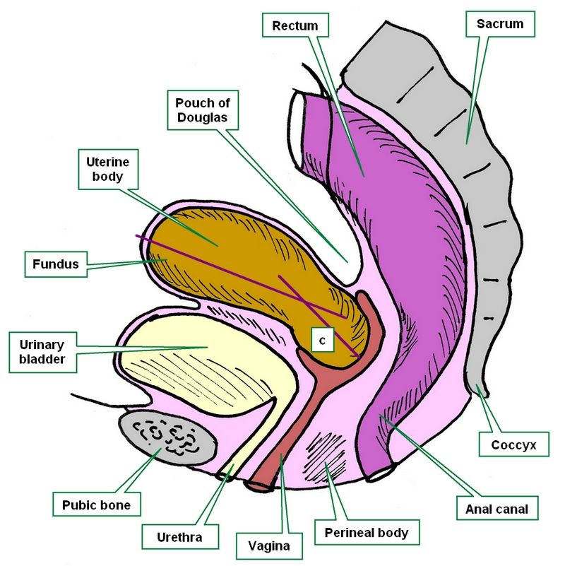
[UPDATED]These three root tems [-hyster-], [-metr-], and [-uter-] refer to the same organ, the [uterus]. There is even a fourth word (not a root term) used to denote the uterus; the matrix. This name originates from the Latin word for "mother".
The word [uterus] and the corresponding root [-uter-] comes from the Latin [uteris] which initially referred to a leather-made water bottle. The use of this term probably originated from the shape of the organ and the fact that during pregnancy it indeed is full of amniotic fluid, called vernacularly "uterine water". The root term [-uter-] can be seen in the words uterine, and uteritis.
The root tem [-metr-] is related to the Greek word [μητέρα] (mit?ra) meaning "mother" and the variation [μήτρα] (mitra) meaning "womb". This root term can be seen in the words endometrium, myometrium, and endometritis.
The root tem [-hyster-] is related to the Greek word [hystera] meaning "womb". This root term can be seen in the words hysterectomy, and hysteria.
Images property of: CAA.Inc. Artist: Dr. E. Miranda
- Details
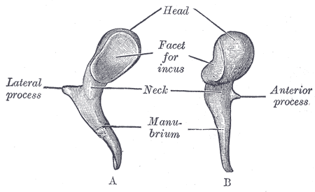
Left malleus. A. Posterior view, B. Medial view
The word [malleus] is Latin word [malleus] meaning "hammer". It is the name of an ossicle in the middle ear.
The manubrium (handle) of the malleus is attached to the tympanic membrane. The head has a ligament, the superior malleolar ligament, which suspends the malleus in place. The head of the malleus has an articular surface for the incus, which itself articulates with the stapes. Malleus, incus, and stapes are the three ossicles found in the middle ear. This chain of bones allows mechanical transmission of the movement of the tympanic membrane through to the inner ear. At the base of the malleolar manubrium there is a site for attachment of the tensor tympani muscle which tenses and relaxes the tympanic membrane.
The Latin word [malleus] is the base for the root term [-mall-], which we can see in the English words [mallet] and even [malleable], which is used in the sense of "something able to be hammered into shape".
Interesting fact: The word [mall] as in "shopping mall" arises from an old Italian game similar to croquet called [pallamaglio]. [palla] means "ball" and [maglio] (from the Latin malleus) means "hammer". The game was quite popular in England where the word evolved from [pallamaglio] into [paille-maille] and then [pall-mall]. Since the game was played in long alleys and streets, some of these began to be referred as [malls], eventually these streets began to be lined with stores, evolving the term into [Shopping Mall].
Sources:
1 "Tratado de Anatomia Humana" Testut et Latarjet 8 Ed. 1931 Salvat Editores, Spain
2. "Anatomy of the Human Body" Henry Gray 1918. Philadelphia: Lea & Febiger
Image modified by CAA, Inc, Original image courtesy of bartleby.com
- Details

Click for a larger image
Today was the closing ceremony of the 2015 Summer Human Gross Anatomy Course and Laboratory for the Doctorate in Physical Therapy of the Mount Saint Joseph University. Gifts were exchanged and the Silver and Gold Scalpel Award were announced for two of the groups. Congratulations!!
The course professor is my good friend Elizabeth Murrray, Ph.D., D-ABFA, a world-renown forensic anthropologist. I have been adjunct faculty for this course for many years and I love to be able to share my knowledge with the students of this course. Unfortunately, this year my commitments did not allow me to be at the lab as much as I would have liked. Sorry.
To the happy faces in this picture my congratulations for having completed the course. Your cadaver was your first patient and I saw many grieve for the person and the pathologies found, and also saw them be thankful for the gift of their bodies for their learning. Congratulations on finishing this demanding course and my best wishes in your professional and personal lives. Dr. Miranda+
Picture by Prof. Doug Miller.
- Details
The word [malleolus] derives from the Latin word [malleus] meaning "hammer". Malleolus is a diminutive form of "malleus" therefore it mean "little hammer". The plural form of [malleolus] is [malleoli].
The term describes two inferior bony prominences found in the lateral and medial aspect of the ankle, the lateral and medial malleoli.
The medial malleolus is a knobby bony prominence of the tibia. It articulates with the talus bone and has a groove, the malleolar sulcus. The lateral malleolus is a bony prominence of the fibula.
- Details

Click for a larger image
The [vertebral arch] is one of the components of a typical vertebra. It is a bony arch found posterior to the vertebral body, and it is composed by the pedicles, the vertebral laminae, and the root or base of the articular processes or zygapophyses.
The presence of the vertebral arch defines the vertebral foramen, a space that contains the spinal cord with its meninges, spinal arteries and venous plexuses, and epidural fat. The sum of all the vertebral foramina creates the vertebral canal.
Image property of: CAA.Inc. Photographer: David M. Klein
- Details
The root term [-narc-] is a derived from the Greek [ναρκος] (narkos ), meaning “torpor” or “lethargic”. Initially it was used to denote numbness in an extremity, but eventually evolved to its modern meaning. It is used in medical word such as:
- Narcotic: A group of pharmaceuticals that numbs pain
- Narcosis: The suffix [-osis] means “condition”. A condition of generalized numbness or lethargicness
- Narcolepsy: The suffix [-lepsy]refers to a sudden onset or a seizure. Sudden numbness or lethargicness
Note: The links to Google Translate include an icon that will allow you to hear the pronunciation of the word.