Medical Terminology Daily (MTD) is a blog sponsored by Clinical Anatomy Associates, Inc. as a service to the medical community. We post anatomical, medical or surgical terms, their meaning and usage, as well as biographical notes on anatomists, surgeons, and researchers through the ages. Be warned that some of the images used depict human anatomical specimens.
You are welcome to submit questions and suggestions using our "Contact Us" form. The information on this blog follows the terms on our "Privacy and Security Statement" and cannot be construed as medical guidance or instructions for treatment.
We have 954 guests online

Jean George Bachmann
(1877 – 1959)
French physician–physiologist whose experimental work in the early twentieth century provided the first clear functional description of a preferential interatrial conduction pathway. This structure, eponymically named “Bachmann’s bundle”, plays a central role in normal atrial activation and in the pathophysiology of interatrial block and atrial arrhythmias.
As a young man, Bachmann served as a merchant sailor, crossing the Atlantic multiple times. He emigrated to the United States in 1902 and earned his medical degree at the top of his class from Jefferson Medical College in Philadelphia in 1907. He stayed at this Medical College as a demonstrator and physiologist. In 1910, he joined Emory University in Atlanta. Between 1917 -1918 he served as a medical officer in the US Army. He retired from Emory in 1947 and continued his private medical practice until his death in 1959.
On the personal side, Bachmann was a man of many talents: a polyglot, he was fluent in German, French, Spanish and English. He was a chef in his own right and occasionally worked as a chef in international hotels. In fact, he paid his tuition at Jefferson Medical College, working both as a chef and as a language tutor.
The intrinsic cardiac conduction system was a major focus of cardiovascular research in the late nineteenth and early twentieth centuries. The atrioventricular (AV) node was discovered and described by Sunao Tawara and Karl Albert Aschoff in 1906, and the sinoatrial node by Arthur Keith and Martin Flack in 1907.
While the connections that distribute the electrical impulse from the AV node to the ventricles were known through the works of Wilhelm His Jr, in 1893 and Jan Evangelista Purkinje in 1839, the mechanism by which electrical impulses spread between the atria remained uncertain.
In 1916 Bachmann published a paper titled “The Inter-Auricular Time Interval” in the American Journal of Physiology. Bachmann measured activation times between the right and left atria and demonstrated that interruption of a distinct anterior interatrial muscular band resulted in delayed left atrial activation. He concluded that this band constituted the principal route for rapid interatrial conduction.
Subsequent anatomical and electrophysiological studies confirmed the importance of the structure described by Bachmann, which came to bear his name. Bachmann’s bundle is now recognized as a key determinant of atrial activation patterns, and its dysfunction is associated with interatrial block, atrial fibrillation, and abnormal P-wave morphology. His work remains foundational in both basic cardiac anatomy and clinical electrophysiology.
Sources and references
1. Bachmann G. “The inter-auricular time interval”. Am J Physiol. 1916;41:309–320.
2. Hurst JW. “Profiles in Cardiology: Jean George Bachmann (1877–1959)”. Clin Cardiol. 1987;10:185–187.
3. Lemery R, Guiraudon G, Veinot JP. “Anatomic description of Bachmann’s bundle and its relation to the atrial septum”. Am J Cardiol. 2003;91:148–152.
4. "Remembering the canonical discoverers of the core components of the mammalian cardiac conduction system: Keith and Flack, Aschoff and Tawara, His, and Purkinje" Icilio Cavero and Henry Holzgrefe Advances in Physiology Education 2022 46:4, 549-579.
5. Knol WG, de Vos CB, Crijns HJGM, et al. “The Bachmann bundle and interatrial conduction” Heart Rhythm. 2019;16:127–133.
6. “Iatrogenic biatrial flutter. The role of the Bachmann’s bundle” Constán E.; García F., Linde, A.. Complejo Hospitalario de Jaén, Jaén. Spain
7. Keith A, Flack M. The form and nature of the muscular connections between the primary divisions of the vertebrate heart. J Anat Physiol 41: 172–189, 1907.
"Clinical Anatomy Associates, Inc., and the contributors of "Medical Terminology Daily" wish to thank all individuals who donate their bodies and tissues for the advancement of education and research”.
Click here for more information
- Details
[UPDATED]The root term [-cephal-] is Greek, arising from the word [κεφάλι] (kef?li) , meaning "head". It was later latinized as [cephalicus]. The applications of this root term are multiple, as follows:
• Cephalic: The suffix [-ic] means "pertaining to", therefore, "pertaining to the head"
• Encephalon: The prefix [en-] means "inside or within", therefore "inside the head". Encephalon is another term for brain
• Anencephalic: The prefix [an-] means "without" Anencephalia is a condition where the brain does not develop.
• Cephalalgia: The suffix [-algia] means "pain". Cephalalgia is the medical term for "headache"
• Cephalad: The suffix [-ad] means "toward".
• Hydrocephaly: Process or condition of "water in the head"
• Dolichocephaly: From the Greek prefix [dolich-] meaning "long". Dolichocephaly is a condition where the head is abnormally large and long
Note: The links to Google Translate include an icon that will allow you to hear the pronunciation of the word.
- Details
[UPDATED] The word is derived from the Greek; the prefix [hydr-] [υδρος]means "water", while the root term [-cephal-] means "head". The term [hydrocephalus] means "water in the head"; of course the "water" is cerebrospinal fluid (CSF) which accumulates excessively in the ventricular system of the brain.
There are several reasons why the amount of CSF within the brain can be excessive, increasing the intracranial pressure: An imbalance between production and absorption of CSF (excessive production or reduced absorption), or a blockage in the ventricular system causing a dilation of the ventricles. The excessive pressure can and will damage the delicate brain tissue. In the case of hydrocephaly (another form of the term) in a newborn the soft cartilage between the cranial bones will distend allowing for the head to dilate and reduce the damage to the brain tissues. A hydrocephalus shunt can allow the patient to reduce the size of the head and eventually lead a normal life.
In 1964 Dr. Salom?n Hakim described, in what was then considered a controversial publication, a condition known as "normal pressure hydrocephalus", usually associated with old-age, Alzheimer's and Parkinson's disease. This condition is now widely accepted as a specific type of hydrocephalus.
The image depicts the ventricular system of the brain in a normal state. If you click on the image, a secondary image of a hydrocephalic baby courtesy of Wikipedia will appear. WARNING! This image is potentially disturbing. For a YouTube video of the insertion of a CSF shunt into the brain, click here.
- Details
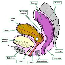
[UPDATED]These three root tems [-hyster-], [-metr-], and [-uter-] refer to the same organ, the [uterus]. There is even a fourth word (not a root term) used to denote the uterus; the matrix. This name originates from the Latin word for "mother".
The word [uterus] and the corresponding root [-uter-] comes from the Latin [uteris] which initially referred to a leather-made water bottle. The use of this term probably originated from the shape of the organ and the fact that during pregnancy it indeed is full of amniotic fluid, called vernacularly "uterine water". The root term [-uter-] can be seen in the words uterine, and uteritis.
The root tem [-metr-] is related to the Greek word [μητέρα] (mit?ra) meaning "mother" and the variation [μήτρα] (mitra) meaning "womb". This root term can be seen in the words endometrium, myometrium, and endometritis.
The root tem [-hyster-] is related to the Greek word [hystera] meaning "womb". This root term can be seen in the words hysterectomy, and hysteria.
Images property of: CAA.Inc. Artist: Dr. E. Miranda
- Details

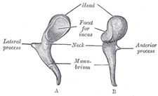
Left malleus. A. Posterior view, B. Medial view
The word [malleus] is Latin word [malleus] meaning "hammer". It is the name of an ossicle in the middle ear.
The manubrium (handle) of the malleus is attached to the tympanic membrane. The head has a ligament, the superior malleolar ligament, which suspends the malleus in place. The head of the malleus has an articular surface for the incus, which itself articulates with the stapes. Malleus, incus, and stapes are the three ossicles found in the middle ear. This chain of bones allows mechanical transmission of the movement of the tympanic membrane through to the inner ear. At the base of the malleolar manubrium there is a site for attachment of the tensor tympani muscle which tenses and relaxes the tympanic membrane.
The Latin word [malleus] is the base for the root term [-mall-], which we can see in the English words [mallet] and even [malleable], which is used in the sense of "something able to be hammered into shape".
Interesting fact: The word [mall] as in "shopping mall" arises from an old Italian game similar to croquet called [pallamaglio]. [palla] means "ball" and [maglio] (from the Latin malleus) means "hammer". The game was quite popular in England where the word evolved from [pallamaglio] into [paille-maille] and then [pall-mall]. Since the game was played in long alleys and streets, some of these began to be referred as [malls], eventually these streets began to be lined with stores, evolving the term into [Shopping Mall].
Sources:
1 "Tratado de Anatomia Humana" Testut et Latarjet 8 Ed. 1931 Salvat Editores, Spain
2. "Anatomy of the Human Body" Henry Gray 1918. Philadelphia: Lea & Febiger
Image modified by CAA, Inc, Original image courtesy of bartleby.com
- Details
Today was the closing ceremony of the 2015 Summer Human Gross Anatomy Course and Laboratory for the Doctorate in Physical Therapy of the Mount Saint Joseph University. Gifts were exchanged and the Silver and Gold Scalpel Award were announced for two of the groups. Congratulations!!
The course professor is my good friend Elizabeth Murrray, Ph.D., D-ABFA, a world-renown forensic anthropologist. I have been adjunct faculty for this course for many years and I love to be able to share my knowledge with the students of this course. Unfortunately, this year my commitments did not allow me to be at the lab as much as I would have liked. Sorry.
To the happy faces in this picture my congratulations for having completed the course. Your cadaver was your first patient and I saw many grieve for the person and the pathologies found, and also saw them be thankful for the gift of their bodies for their learning. Congratulations on finishing this demanding course and my best wishes in your professional and personal lives. Dr. Miranda+
Picture by Prof. Doug Miller.
- Details
The word [malleolus] derives from the Latin word [malleus] meaning "hammer". Malleolus is a diminutive form of "malleus" therefore it mean "little hammer". The plural form of [malleolus] is [malleoli].
The term describes two inferior bony prominences found in the lateral and medial aspect of the ankle, the lateral and medial malleoli.
The medial malleolus is a knobby bony prominence of the tibia. It articulates with the talus bone and has a groove, the malleolar sulcus. The lateral malleolus is a bony prominence of the fibula.


