Medical Terminology Daily (MTD) is a blog sponsored by Clinical Anatomy Associates, Inc. as a service to the medical community. We post anatomical, medical or surgical terms, their meaning and usage, as well as biographical notes on anatomists, surgeons, and researchers through the ages. Be warned that some of the images used depict human anatomical specimens.
You are welcome to submit questions and suggestions using our "Contact Us" form. The information on this blog follows the terms on our "Privacy and Security Statement" and cannot be construed as medical guidance or instructions for treatment.
We have 1295 guests online

Jean George Bachmann
(1877 – 1959)
French physician–physiologist whose experimental work in the early twentieth century provided the first clear functional description of a preferential interatrial conduction pathway. This structure, eponymically named “Bachmann’s bundle”, plays a central role in normal atrial activation and in the pathophysiology of interatrial block and atrial arrhythmias.
As a young man, Bachmann served as a merchant sailor, crossing the Atlantic multiple times. He emigrated to the United States in 1902 and earned his medical degree at the top of his class from Jefferson Medical College in Philadelphia in 1907. He stayed at this Medical College as a demonstrator and physiologist. In 1910, he joined Emory University in Atlanta. Between 1917 -1918 he served as a medical officer in the US Army. He retired from Emory in 1947 and continued his private medical practice until his death in 1959.
On the personal side, Bachmann was a man of many talents: a polyglot, he was fluent in German, French, Spanish and English. He was a chef in his own right and occasionally worked as a chef in international hotels. In fact, he paid his tuition at Jefferson Medical College, working both as a chef and as a language tutor.
The intrinsic cardiac conduction system was a major focus of cardiovascular research in the late nineteenth and early twentieth centuries. The atrioventricular (AV) node was discovered and described by Sunao Tawara and Karl Albert Aschoff in 1906, and the sinoatrial node by Arthur Keith and Martin Flack in 1907.
While the connections that distribute the electrical impulse from the AV node to the ventricles were known through the works of Wilhelm His Jr, in 1893 and Jan Evangelista Purkinje in 1839, the mechanism by which electrical impulses spread between the atria remained uncertain.
In 1916 Bachmann published a paper titled “The Inter-Auricular Time Interval” in the American Journal of Physiology. Bachmann measured activation times between the right and left atria and demonstrated that interruption of a distinct anterior interatrial muscular band resulted in delayed left atrial activation. He concluded that this band constituted the principal route for rapid interatrial conduction.
Subsequent anatomical and electrophysiological studies confirmed the importance of the structure described by Bachmann, which came to bear his name. Bachmann’s bundle is now recognized as a key determinant of atrial activation patterns, and its dysfunction is associated with interatrial block, atrial fibrillation, and abnormal P-wave morphology. His work remains foundational in both basic cardiac anatomy and clinical electrophysiology.
Sources and references
1. Bachmann G. “The inter-auricular time interval”. Am J Physiol. 1916;41:309–320.
2. Hurst JW. “Profiles in Cardiology: Jean George Bachmann (1877–1959)”. Clin Cardiol. 1987;10:185–187.
3. Lemery R, Guiraudon G, Veinot JP. “Anatomic description of Bachmann’s bundle and its relation to the atrial septum”. Am J Cardiol. 2003;91:148–152.
4. "Remembering the canonical discoverers of the core components of the mammalian cardiac conduction system: Keith and Flack, Aschoff and Tawara, His, and Purkinje" Icilio Cavero and Henry Holzgrefe Advances in Physiology Education 2022 46:4, 549-579.
5. Knol WG, de Vos CB, Crijns HJGM, et al. “The Bachmann bundle and interatrial conduction” Heart Rhythm. 2019;16:127–133.
6. “Iatrogenic biatrial flutter. The role of the Bachmann’s bundle” Constán E.; García F., Linde, A.. Complejo Hospitalario de Jaén, Jaén. Spain
7. Keith A, Flack M. The form and nature of the muscular connections between the primary divisions of the vertebrate heart. J Anat Physiol 41: 172–189, 1907.
"Clinical Anatomy Associates, Inc., and the contributors of "Medical Terminology Daily" wish to thank all individuals who donate their bodies and tissues for the advancement of education and research”.
Click here for more information
- Details

Hover for another image
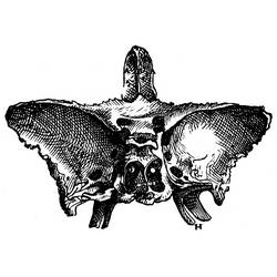
This word has a Greek root [-sphen-] meaning "wedge", and the suffix [-oid] meaning "similar to" or "resembling". [Sphenoid] means "resembling a wedge" or "wedge-like".
The sphenoid bone (os sphenoidale) has been described as butterfly or bat-shaped, and is a complex bone situated as a wedge or keystone in the base of the skull. Hover over the image1 for a view of the location of this bone.
The sphenoid bone has a superior depression called the sella turcica, Latin for "turkish chair" where the hypophysis or pituitary gland is found.
On its inferior aspect the sphenoid bone presents with two thin wing-like plates called the lateral and medial pterygoid plates or lamina (see the area with the letter "H" in the accompanying image)

Image1 modified from "De Humani Corporis Fabrica" 1543 1st Ed.by Andreas Vesalius. Animation via Wikimedia Commons, public domain. Polygon data generated by Database Center for Life Science (DBCLS), CC BY-SA 2.1 JP.
- Details

UPDATED: The term [cholangiogram] is composed by the combined root terms [-chole-] derived from the Greek word [χολή] (cholí) meaning "gall" or "bile, and the root term [-angi-], also derived from the Greek [αγγείο] (angeío), meaning "vase", or "vessel"letter. The suffix [-(o)gram] evolved from the Greek word [γράμμα] (grámma) , meaning "letter", although today we use it to mean "examination of". For more information on this suffix, click here. The term [cholangiogram] therefore means "examination of a bile vessel".
A cholangiogram is the fluoroscopic imaging of a bile duct. To do this a radio-opaque dye is introduced in the bile system and a series of X-ray images are taken of the hepatobiliary tree. Today the examination can be performed intraoperative in conjunction with a cholecystectomy using a C-arm fluoroscope
The accompanying video (without sound) shows an intraoperative normal cholangiogram.
Video courtesy of YouTube, Mr. Andrew Smith and the Yorkshire Gallstone Clinic.
- Details
This article is part of the series "A Moment in History" where we honor those who have contributed to the growth of medical knowledge in the areas of anatomy, medicine, surgery, and medical research.

Raymond de Vieussens
Raymond de Vieussens (c.1635 – 1715). French anatomist and physician. His exact date and place of birth are uncertain, some place him being born in the area of Le Vigan in France and the date for some authors as late as 1641.
What we do know is that he studied at the University of Montpellier where he graduated from his medical studies in 1670. He became a physician at the Hôtel Dieu Saint-Eloi in Montpellier. He later became head physician at the same hospital and apparently maintained this position for the rest of his life. His studies on the anatomy of the heart and lymphatic system were pioneers for the time, as were his studies on the anatomy of the nervous system.
Vieussens was a prolific writer. Among his works in 1706, he published “Nouvelles Découvertes sur le Coeur” (New Discoveries on the Heart) followed by “Traité Nouveau de la Structure et Des causes du Mouvement Naturel du Coeur” (New Treaties on the Structure and Cause of the Natural Movement of the Heart) in 1715. In these books he presented detailed anatomy of the lymphatic system and blood vessels of the heart, as well as his theories on the movement of the heart. In his work, he did the first accurate description of mitral stenosis and aortic disease.
One of his greatest works was “Neurographia Universalis”, published in 1684 in Lyons, France. In this book Vieussens describes the structure of the nervous system with emphasis on the pathways of the white substance, which we know today is formed by bundles of neuronal axons. He accurately described the internal structure of the cerebellum and other structures that today bear his name. Unfortunately Vieussens attempted to describe the physiology of the brain with little factual support, developing wild theories, including the statement that he had found the “fluid of the nerves”.
Some of Vieussens’ work was published posthumously by his family and colleagues. Today, many eponyms remember Vieussens’ name, here are some of them:
- Valve of Vieussens: A valve found at the distal end of the great cardiac vein, where it empties into the coronary sinus
- Ring of Vieussens: Name for and anatomical variation in the heart, an anastomotic communication between two conal arteries, one arising from the right coronary artery, the other arising from the left anterior descending artery (LAD)
- Centrum of Vieussens: A term that describes the mass of white matter at the center of each cerebral hemisphere
- Ring of Vieussens: Eponymic term for the limbus fossa ovalis, a raised muscular ring surrounding the fossa ovalis in the heart
- Valve of Vieussens: A thin veil of tissue between the superior cerebellar peduncles, forming part of the roof of the 4th ventricle. This is known as the superior medullary vellum and may have some sparse cerebellar tissue on it
- The ventricle of Vieussens: The name of a cavity found in the case where the septum pellucidum is double. The septum pellucidum is a membrane that separates the lateral ventricles of the brain in the midline
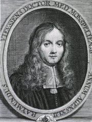
If you hover with your mouse over the image of young Vieussens you will see another image of Vieussens at 65.
Original images courtesy of National Library of Medicine.
- Details
The suffix [-(o)rrhaphy] is derived from the Greek language and means "surgical repair". Initially, the meaning was that of "suturing" but nowadays surgical repair can be accomplished with sutures, staples, tacks, tissue glue, and other medical devices.
Some applications of this suffix are:
- Herniorrhaphy: Surgical repair of a hernia
- Enterorrhaphy: Surgical intestinal repair
- Aneurysmorrhaphy: Surgical repair of an aneurysm
- Gastrorraphy: Surgical repair of the stomach
- Details
Latin term probably derived from [varus] meaning "bent or crooked". The term is used in medicine to denote a single dilation of a vein. The plural form of [varix] is [varices], the adjective [varicose] denotes an area affected by varices.
Peripheral varicose veins are usually caused by failure of the venous valves found in them.
Note that the dilation of a vein is called a varix, while the dilation of an artery is called an aneurysm.
- Details

Anterior view of the stomach
This is a German word composed of [magen] meaning "stomach" and [strasse] meaning "road or street", therefore [magenstrasse] means "stomach road".
Most everybody that studies anatomy or gastrointestinal surgery knows that the stomach has several regions that include the fundus, body (corpus), antrum, pyloric canal , and pylorus. The anatomical term [magenstrasse] refers to a tubular and thicker area of the stomach close to the lesser curvature (see image).
This old anatomical German term has come back en vogue because of bariatric surgery. In a sleeve gastrectomy, the stomach is stapled and separated along the left lateral border of the magenstrasse, leaving only a tubular, less distensible portion of the stomach.
Here is an article from the "Journal of Biomechanics" on the physiological magenstrasse and gastric emptying. For a video of this procedure CLICK HERE
Video available on YouTube, published by Realize TM

