![]()
Medical Terminology Daily (MTD) is a blog sponsored by Clinical Anatomy Associates, Inc. as a service to the medical community. We post anatomical, medical or surgical terms, their meaning and usage, as well as biographical notes on anatomists, surgeons, and researchers through the ages. Be warned that some of the images used depict human anatomical specimens.
You are welcome to submit questions and suggestions using our "Contact Us" form. The information on this blog follows the terms on our "Privacy and Security Statement" and cannot be construed as medical guidance or instructions for treatment.
We have 162 guests and no members online
")
Marcia Crocker Noyes
(1869 – 1946)
Further to my comment on old books and research that started with an interesting bookplate (Ex-Libris). I continued my research and found that the person in charge of the Osler library bookplate was a fascinating individual that today maybe a ghost in the MedChi library and building in Baltimore... This is certainly an article that can be called "A Moment in History"
Marcia Crocker Noyes was the librarian at The Maryland State Medical Society from 1896 to 1946 and was a founding member of the Medical Library Association.[1][2][3]
Sir William Osler, MD. a famous Johns Hopkins surgeon was a noted bibliophile and had a large personal collection of books on various topics. When he became the President of MedChi in 1896, he was dismayed at the condition of the library and knew that with the right person and some stewardship, it could become a significant collection. Sir William asked his friend, Dr. Bernard Steiner, a physician and President of the Enoch Pratt Free Library in Baltimore for suggestions of a librarian, and Dr. Steiner recommended Marcia Crocker Noyes. A native of New York, and a graduate of Hunter College, Marcia had moved to Baltimore for a lengthy visit with her sister, and took a “temporary” position at the Pratt Library, which turned into three years. Although she had no medical experience or background, she was enthusiastic, and most importantly, she was willing to move into the apartment provided for the librarian, who needed to be available 24 hours a day.
The image in this article is Ms. Noyes on her first year on the job. Marcia developed a book classification system for medical books, based on the Index Medicus, and called it the Classification for Medical Literature. The system uses the alphabet with capital letters for the major divisions of medicine and lower-case ones for the sub-sections. The system was used for many years, but it's now dated and the Faculty's original shelving scheme was never changed. The card catalogs still reflect her classification and many of the cards are written in Marcia's back-slanting handwriting.
Marcia knew enough to ask the Faculty's members about medical questions, terminology and literature. She gradually won over the predominantly male membership and they became her greatest allies; Sir William at the start, and then for nearly 40 years, Dr. John Ruhräh, a wealthy pediatrician with no immediate family of his own. She made a point of attending almost every Faculty function, and in 1904, under guidelines from the American Medical Association, Marcia was made the Faculty Secretary. For much of her first 10 years, she was the Faculty's only full-time employee, only being assisted by Mr. Caution, the Faculty's janitor. Later in life Marcia would say that she hired him because of his name!
Within ten years, the library had outgrown its space, and plans, spearheaded by Marcia and Sir William before his move to Oxford, were made to build a headquarters building, mainly to house the library's growing collection of medical books and journals.
Marcia was instrumental in the design and building of the new headquarters. She travelled to Philadelphia, New York and Boston to look at their medical society buildings, and eventually, the Philadelphia architectural firm, Ellicott & Emmart was selected to design and build the new Faculty building. Every detail of the building held her imprimatur, from the graceful staircase, to the light-filled reading room, and all of the myriad details of the millwork, marble tesserae, and most of all, the four-story cast iron stacks. She was on-site, climbing up unfinished staircases, checking out the progress of the building, which was built in less than one year at a cost of $90,000.
Among the features of the new building was a fourth-floor apartment for her. She referred to it as the "first penthouse in Baltimore" and it had a garden and rooftop terrace. The library collection eventually grew to more than 65,000 volumes from medical and specialty societies around the world. Journals were traded back and forth, and physicians eagerly anticipated the arrival of each new issue. At the same time, Marcia was involved in the Medical Library Association as one of eight founding members. The MLA promotes medical libraries and the exchange of information. One of the earliest mandates of the MLA was the Exchange, a distribution and trade service for those who had duplicates or little-used books in their collections. Initially, the Exchange was run out of the Philadelphia medical society, but in 1900 it was moved to Baltimore and Marcia oversaw it. Several hundred periodicals and journals were received and sent each month, a huge amount of work for a tiny staff. In 1904, the Faculty had run out of room to manage the Exchange, so it was moved to the Medical Society of the Kings County (Brooklyn). But without Marcia's excellent administrative skills, it floundered and in 1908, the MLA asked Marcia to take charge once again.
In 1909, when the new Faculty building opened, there was enough room to run the Exchange and with the help of MLA Treasurer, noted bibliophile and close friend, Dr. John Ruhräh, it once again became successful. Additionally, Marcia and Dr. Ruhräh combined forces to revive the MLA's bulletin, which had all but ceased publication in 1908, taking the Exchange with it. This duo maintained editorial control from 1911 until 1926. In 1934, around the time of Dr. Ruhräh's death, Marcia became the first “unmedicated” professional to head the MLA. During her tenure, the MLA incorporated, the first seal was adopted, and the annual meeting was held in Baltimore. Marcia wanted to write the history of the MLA once she retired from full-time work at the Faculty, but her health was beginning to fail. She had back problems and had suffered a serious burn on her shoulder as a young woman, possibly from her time running a summer camp, Camp Seyon, for young ladies in the Adirondack Mountains. In 1946, a celebration was planned to honor Marcia's 50 years at the Faculty. But she was adamant that the physicians wait until November, the actual date of her 50 years. However, they knew she was gravely ill, and might not make it until then, so a huge party was held in April. More than 250 physicians attended the celebration, but the ones she was closest to in the early years, were long gone. She was presented with a suitcase, a sum of money to use for travelling, and her favorite painting of Dr. John Philip Smith, a founder of the Medical College in Winchester, Virginia. It was painted by Edward Caledon Smith, a Virginia painter who had been a student of the painter Thomas Sully.[4] She adored this painting and vowed, jokingly, to take it with her wherever she went.
The painting was not to stay with her for very long, for she died in November 1946, and left it to the Faculty in her will. Her funeral was held in the Faculty's Osler Hall, named for her dear friend. More than 60 physicians served as her pallbearers, and she was buried at Baltimore's Green Mount Cemetery. In 1948, the MLA decided to establish an award in the name of Marcia Crocker Noyes. It was for outstanding achievement in medical library field and was to be awarded every two years, or when a truly worthy candidate was submitted. In 2014, the Faculty began giving a bouquet of flowers to the winner of the award in Marcia's name, and in honor of her work. Much evidence exists for this tradition, as we know that the physicians, especially Drs. Osler and Ruhräh, frequently gave her bouquets of flowers. Marcia also cultivated flower gardens at the Faculty and decorated the rooms with her work.
Today, the MedChi building is open for tours and if the rumors are to be believed Ms. Marcia Crocker Noyes is still at work in her beloved library as the "resident ghost" [1][5]

NOTE: This article has been modified from the original Wikipedia article on Marcia Crocker Noyes. The article itself is well-written with interesting images of the subject. I would encourage you to visit it. The second insert is from book 00736 in my personal library and shows in pencil, the incredibly small handwriting of Marsha C. Noyes.
Sources:
1. "Marcia, Marcia, Marcia" MedChi Archives blog.
2. "Marcia C. Noyes, Medical Librarian" (PDF). Bulletin of the Medical Library Association. 35 (1): 108–109. 1947. PMC 194645
3. Smith, Bernie Todd (1974). "Marcia Crocker Noyes, Medical Librarian: The Shaping of a Career" (PDF). Bulletin of the Medical Library Association. 62 (3): 314–324. PMC 198800Freely accessible. PMID 4619344.
4. Edward Caledon BRUCE (1825-1901)"
5. Behind the scenes tour MedChiBuilding
"Clinical Anatomy Associates, Inc., and the contributors of "Medical Terminology Daily" wish to thank all individuals who donate their bodies and tissues for the advancement of education and research”.
Click here for more information


- Details
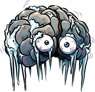
BRRRRRRRR!!
A reminder of one of the joys of summer! The term [sphenopalatine ganglioneuralgia] is a fancy medical term for "brain freeze". which happens when we eat or drink very cold food.
The etymology of the term is complex. [Sphen-] is a term meaning "wedge" and refers to the sphenoid bone. [-palatine-] means "pertaining to the palate" (and to the bones related to the hard palate].
The root term [-gangli-] refer to a ganglion, which is a concentration of neuronal bodies, neurons being the main cells of the nervous system. [-neur-] means "nerve", and the suffix [-algia] means "pain". Simply said, the term [sphenopalatine ganglioneuralgia] means "nerve pain of the sphenopalatine ganglion".
The sphenopalatine ganglion (Meckel's ganglion, nasal ganglion or pterygopalatine ganglion) is a parasympathetic ganglion found in the pterygopalatine fossa. It is largely innervated by the greater petrosal nerve (a branch of the facial nerve); and its neuronal axons innervate the lacrimal glands and nasal mucosa.
Not everybody accepts this theory. Some state that "brain freeze" occurs because of rapid cooling of the blood in the pharynx, causing a drop of temperature of the internal carotid artery, which in turn causes cooling and pain in the meninges related to the base of the cranium.
My thanks to Gina Burg, for bringing this term to my attention. Dr. Miranda
Thanks to Forrest J. Bonjo for the image and additional information. The article was originally stored at pdu.edu, but the server was closed. If you click on the image, this will take you to the article stored at web.archive.org.
- Details
Click for a larger image
There are many in the world that are fascinated by the life and works of Andreas Vesalius (1514 -1564). This has created a market for “Vesaliana”. These are books, art, medals, and works are related to Vesalius. As an example, an original 1543 Fabrica sells today for 400 thousand dollars! Even the “New Fabrica” by Drs. Garrison and Hast has cuadrupled its value in only two years since its publication!
Some of the most coveted items are stamps that celebrate the illustrious anatomist. Probably the most detailed research on the topic was made by Prof. Omer Steeno and Dr. Maurits Biersbrouck, both contributors to this website. Their research is constantly updated and the latest iteration of their work is “Andreas Vesalius in Philately” published in WordPress.com.
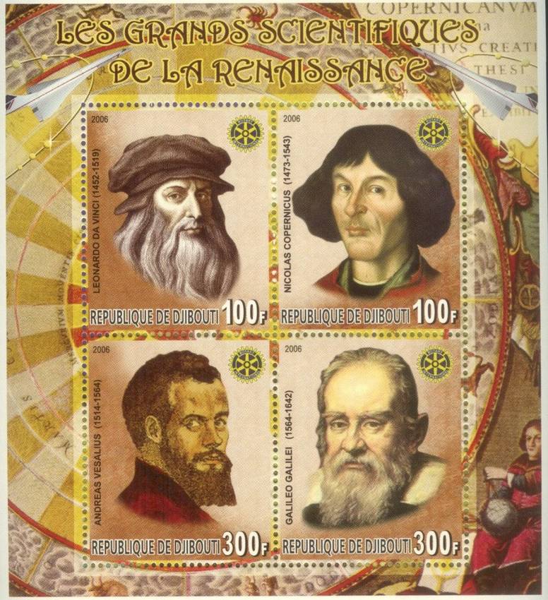
In a recent private communication Prof. Steeno regretted that unscrupulous individuals have taken to forge and falsify stamps. A clear case of this is the stamp collection “Les Grands Scientifiques de la Rennaissance” published in November 23, 2006 by the Republic of Djibouti. The stamps (shown in the accompanying image) depict Leonardo da Vinci, Nicolas Copernicus, Galileo Galilei, and Andreas Vesalius. As a Vesaliana collector, who would not want this set of stamps placing Vesalius in such company?
Djibouti is an African country that gained its independence from France in 2007 and is located in the horn of East Africa and the opening of the Red Sea into the Gulf of Aden.
Drs. Steeno and Beisbrocuk contacted the Djibouti postal service and were able to confirm in February, 2016 that indeed these stamps are false and collectors should be aware.
Sources:
1. “Andreas Veslius in Plhilately” Steeno, O; Biesbrouck, M 2016
2. Private communication. Steeno, O. 2016
3. “On the falsification of a Vesalius Stamp wrongfully ascribed to the postal service of Djibouti” Steeno, O; Biesbrouck, M 2016. EMediTheme 2016 Editor: Menzies, S.
- Details
This is a series of articles on depression and published as a community service. The information in these articles follow our Privacy and Security Guidelines and cannot be construed as medical guidance. For additional information and counseling, consult with your physician or the appropriate health care professional of your choice. You can also find information on Transcranial Magnetic Stimulation (TMS) here. For the initial article on this series click here.
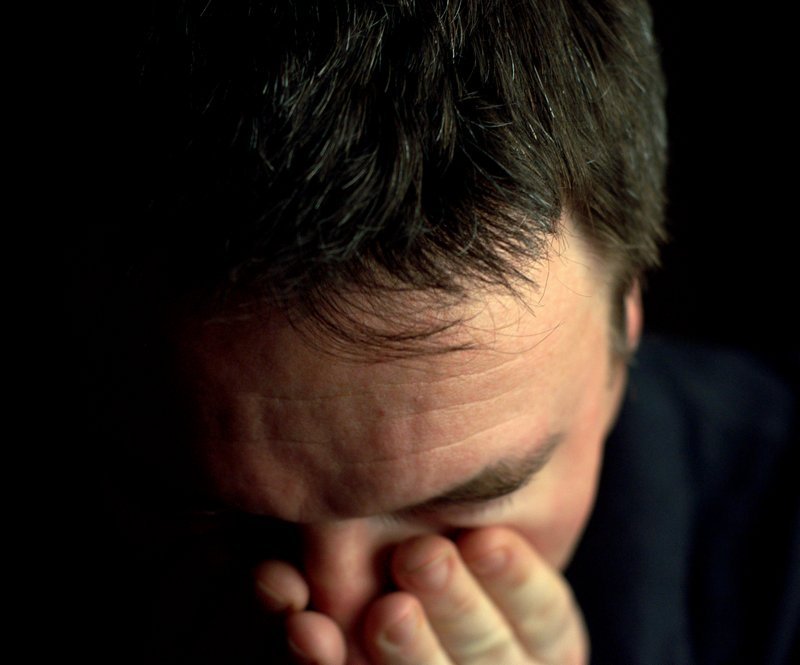
UPDATED: People with depressive pathology do not all experience the same symptoms. The severity, frequency, and duration of symptoms vary depending on the individual and his or her particular illness. Because symptoms are subjective, some patients will not express or hide them, making the diagnosis of depressive disorder difficult.
Following are the description of the symptoms by two different patients:
Patient 1
"It was really hard to get out of bed in the morning. I just wanted to hide under the covers and not talk to anyone. I didn't feel much like eating and I lost a lot of weight. Nothing seemed fun anymore. I was tired all the time, and I wasn't sleeping well at night. But I knew I had to keep going because I've got kids and a job. It just felt so impossible, like nothing was going to change or get better."
Patient 2
"I felt dirty and unwashed. All my surroundings felt dirty and I spent hours cleaning the house with no results. I took long baths and even after them I still felt dirty. My sleep was broken with horrible nightmares with gore and destruction. I felt tired, mostly because I could not sleep. I cried every morning because I felt like a total failure. I felt ugly and no amount of makeup could cover this feeling. I did not want to go out in public at all"
Signs and symptoms of depression may include:
• Persistent sad, anxious, or "empty" feelings
• Feelings of hopelessness or pessimism
• Feelings of guilt, worthlessness, or helplessness
• Irritability, restlessness
• Loss of interest in activities or hobbies once pleasurable, including sex
• Fatigue and decreased energy
• Difficulty concentrating, remembering details, and making decisions
• Insomnia, early-morning wakefulness, or excessive sleeping
• Overeating, or appetite loss
• Thoughts of suicide, suicide attempts
• Aches or pains, headaches, cramps, or digestive problems that do not ease even with treatment
Next article: Causes of Depression
- Details
This is a series of articles on depression published as a community service. The information in these articles follow our Privacy and Security Guidelines and cannot be construed as medical guidance. For additional information and counseling, consult with your physician or the appropriate health care professional of your choice. You can also find information on TMS here.
Click for a larger image
UPDATED: Everyone occasionally feels blue or sad. There are also those dreaded "winter blues". But these feelings are usually short-lived and pass within a couple of days, usually with no problems or persistent symptoms. Some people may even say that they are "depressed". Although this is true, that person is not clinically depressed.
When an individual has clinical depression, there are physical changes that happen within the brain which reflect in attitudes, mood, symptoms, and actions.
Clinical depression is a common but serious mental disorder that affects over 20 million people in the United States, many of which will never seek diagnosis or treatment. Patients present with depressed mood, loss of interest or pleasure, decreased energy, feelings of guilt or low self-worth, abnormal patterns of sleep or appetite, gruesome nightmares, and poor concentration. Moreover, depression may often come with symptoms of anxiety and varying complex presentations of bipolar disorder.
These problems can become chronic or recurrent and lead to substantial impairment in an individual’s ability to take care of his or her everyday responsibilities. At its worst, depression can lead to a patient's attempt on their life. Clinical Depression interferes with daily life and causes pain for both the individual, their families, and loved ones. Patients with depressive disorder often go from one job to another, cannot work, or eventually end in disability, being maintained by their family or loved ones.
Many people afflicted with Major Depressive Disorder (MDD) never seek treatment. This is specially true in males, where the World Health Organization (WHO) estimates that ”fewer than 25% of male sufferers worldwide will seek treatment because of the social stigma associated with mental disorders including depression.”
Properly and timely treated, even those with the most severe depression, can get better. Medications, psychotherapy, and electroconvulsive therapy (ECT) are the most common methods to treat depression. As patients move from one medication to the next level medication as well as augmentation medication, the annual cost for medication can be staggering, as well as the common, insidious, and problematic systemic side effects of both the drug therapy and ECT therapy.
The main objective of all treatments for MDD is to attain remission, but in many cases just reducing the symptoms of MDD and reducing the amount and types of medication used is enough to bring the patient back to a productive life and enhance the relationship with their families and loved ones.
Next article: Symptoms of Depression
- Details
Click for a larger image
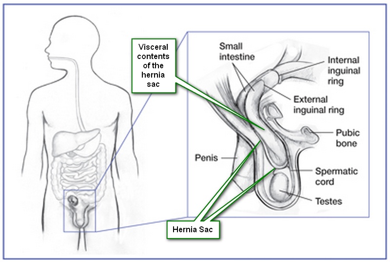
UPDATED: The definition of hernia is "the protrusion of a deep structure through a superficial weakness of defect".
Herniation has many etiologies, but in all cases a weakness of a superficial containing wall (usually layered) or a normal or abnormal opening (defect) must be present. A true hernia usually has a deep sac or hernia sac which contains the herniated viscus or viscera. Repair of a hernia is called a hernioplasty or a herniorrhaphy.
Although with exceptions, a herniation with only weakening of the walls and no hernia sac can be called a "prolapse", the suffix for prolapse (or hernia sometimes) is [-ocele].
• Omphalocele: From the Greek [omphalos] meaning "umbilicus", an omphalocele is a herniation through the umbilicus.
• Cystourethocele: A prolapse of the urinary bladder and urethra with a weakened vaginal wall
There are also "internal' hernias, between bodily compartments. Examples are:
• Esophageal hiatus hernia: Known as a "hiatal hernia", this hernia is a protrusion of a peritoneal sac with abdominal visceral content into the thorax.
• Perineal hernia: The protrusion of abdominopelvic content into the perineal region through a defect in the pelvic diaphragm (levator ani)
A hernia is usually named for the superficial region where it protrudes. An example of this would be a femoral hernia, which starts as an abdominopelvic extrusion, but it ends protruding in the area of the thigh (femoral region). Abdominal or ventral hernias are named according to the abdominal region through which they protrude.
in older times the word "rupture" was used as a synonym for "hernia", as can be seen in a letter written by Dr. Ephraim McDowell in 1829. The image shows an example of an indirect inguinal hernia.
Original Image public domain courtesy of: nih.gov.
- Details

Click for a larger image
UPDATED: The word [pecten] originates from the Latin [pectine] meaning "to comb", the adjective [pectinate] means "resembling a comb". The term denotes structures that have well-formed parallel shapes, such as the pectinate muscle of the heart. The pectinate muscle can be clearly seen in the internal aspect of the atrial appendages. (see image, pointer "B")
The term [pecten] meaning "comb" is an old word used for the superior aspect of the pubic bone (os pubis) where the pectineus muscle attaches. The root term [-pectin-] can be seen then in terms such as the iliopectineal line, and the pectineal ligament, also known as "Cooper's ligament".
The origin of the use of the term [pecten os pubis] to denote the area of attachment of the pectineus muscle to the bony ridge in the superior aspect of the pubic bone is obscure, but the pectineus muscle has well-marked parallel striations resembling a comb.
The image shows a human heart where the right atrium has been opened. The red arrow points to the pectinated muscle characteristic of the right atrial appendage (right auricle). Keep in mind that the distribution and shape of the muscle of the left atrial appendage is completely different.
Note: The links to Google Translate include an icon that will allow you to hear the pronunciation of the word.