![]()
Medical Terminology Daily (MTD) is a blog sponsored by Clinical Anatomy Associates, Inc. as a service to the medical community. We post anatomical, medical or surgical terms, their meaning and usage, as well as biographical notes on anatomists, surgeons, and researchers through the ages. Be warned that some of the images used depict human anatomical specimens.
You are welcome to submit questions and suggestions using our "Contact Us" form. The information on this blog follows the terms on our "Privacy and Security Statement" and cannot be construed as medical guidance or instructions for treatment.
We have 167 guests and no members online
")
Marcia Crocker Noyes
(1869 – 1946)
Further to my comment on old books and research that started with an interesting bookplate (Ex-Libris). I continued my research and found that the person in charge of the Osler library bookplate was a fascinating individual that today maybe a ghost in the MedChi library and building in Baltimore... This is certainly an article that can be called "A Moment in History"
Marcia Crocker Noyes was the librarian at The Maryland State Medical Society from 1896 to 1946 and was a founding member of the Medical Library Association.[1][2][3]
Sir William Osler, MD. a famous Johns Hopkins surgeon was a noted bibliophile and had a large personal collection of books on various topics. When he became the President of MedChi in 1896, he was dismayed at the condition of the library and knew that with the right person and some stewardship, it could become a significant collection. Sir William asked his friend, Dr. Bernard Steiner, a physician and President of the Enoch Pratt Free Library in Baltimore for suggestions of a librarian, and Dr. Steiner recommended Marcia Crocker Noyes. A native of New York, and a graduate of Hunter College, Marcia had moved to Baltimore for a lengthy visit with her sister, and took a “temporary” position at the Pratt Library, which turned into three years. Although she had no medical experience or background, she was enthusiastic, and most importantly, she was willing to move into the apartment provided for the librarian, who needed to be available 24 hours a day.
The image in this article is Ms. Noyes on her first year on the job. Marcia developed a book classification system for medical books, based on the Index Medicus, and called it the Classification for Medical Literature. The system uses the alphabet with capital letters for the major divisions of medicine and lower-case ones for the sub-sections. The system was used for many years, but it's now dated and the Faculty's original shelving scheme was never changed. The card catalogs still reflect her classification and many of the cards are written in Marcia's back-slanting handwriting.
Marcia knew enough to ask the Faculty's members about medical questions, terminology and literature. She gradually won over the predominantly male membership and they became her greatest allies; Sir William at the start, and then for nearly 40 years, Dr. John Ruhräh, a wealthy pediatrician with no immediate family of his own. She made a point of attending almost every Faculty function, and in 1904, under guidelines from the American Medical Association, Marcia was made the Faculty Secretary. For much of her first 10 years, she was the Faculty's only full-time employee, only being assisted by Mr. Caution, the Faculty's janitor. Later in life Marcia would say that she hired him because of his name!
Within ten years, the library had outgrown its space, and plans, spearheaded by Marcia and Sir William before his move to Oxford, were made to build a headquarters building, mainly to house the library's growing collection of medical books and journals.
Marcia was instrumental in the design and building of the new headquarters. She travelled to Philadelphia, New York and Boston to look at their medical society buildings, and eventually, the Philadelphia architectural firm, Ellicott & Emmart was selected to design and build the new Faculty building. Every detail of the building held her imprimatur, from the graceful staircase, to the light-filled reading room, and all of the myriad details of the millwork, marble tesserae, and most of all, the four-story cast iron stacks. She was on-site, climbing up unfinished staircases, checking out the progress of the building, which was built in less than one year at a cost of $90,000.
Among the features of the new building was a fourth-floor apartment for her. She referred to it as the "first penthouse in Baltimore" and it had a garden and rooftop terrace. The library collection eventually grew to more than 65,000 volumes from medical and specialty societies around the world. Journals were traded back and forth, and physicians eagerly anticipated the arrival of each new issue. At the same time, Marcia was involved in the Medical Library Association as one of eight founding members. The MLA promotes medical libraries and the exchange of information. One of the earliest mandates of the MLA was the Exchange, a distribution and trade service for those who had duplicates or little-used books in their collections. Initially, the Exchange was run out of the Philadelphia medical society, but in 1900 it was moved to Baltimore and Marcia oversaw it. Several hundred periodicals and journals were received and sent each month, a huge amount of work for a tiny staff. In 1904, the Faculty had run out of room to manage the Exchange, so it was moved to the Medical Society of the Kings County (Brooklyn). But without Marcia's excellent administrative skills, it floundered and in 1908, the MLA asked Marcia to take charge once again.
In 1909, when the new Faculty building opened, there was enough room to run the Exchange and with the help of MLA Treasurer, noted bibliophile and close friend, Dr. John Ruhräh, it once again became successful. Additionally, Marcia and Dr. Ruhräh combined forces to revive the MLA's bulletin, which had all but ceased publication in 1908, taking the Exchange with it. This duo maintained editorial control from 1911 until 1926. In 1934, around the time of Dr. Ruhräh's death, Marcia became the first “unmedicated” professional to head the MLA. During her tenure, the MLA incorporated, the first seal was adopted, and the annual meeting was held in Baltimore. Marcia wanted to write the history of the MLA once she retired from full-time work at the Faculty, but her health was beginning to fail. She had back problems and had suffered a serious burn on her shoulder as a young woman, possibly from her time running a summer camp, Camp Seyon, for young ladies in the Adirondack Mountains. In 1946, a celebration was planned to honor Marcia's 50 years at the Faculty. But she was adamant that the physicians wait until November, the actual date of her 50 years. However, they knew she was gravely ill, and might not make it until then, so a huge party was held in April. More than 250 physicians attended the celebration, but the ones she was closest to in the early years, were long gone. She was presented with a suitcase, a sum of money to use for travelling, and her favorite painting of Dr. John Philip Smith, a founder of the Medical College in Winchester, Virginia. It was painted by Edward Caledon Smith, a Virginia painter who had been a student of the painter Thomas Sully.[4] She adored this painting and vowed, jokingly, to take it with her wherever she went.
The painting was not to stay with her for very long, for she died in November 1946, and left it to the Faculty in her will. Her funeral was held in the Faculty's Osler Hall, named for her dear friend. More than 60 physicians served as her pallbearers, and she was buried at Baltimore's Green Mount Cemetery. In 1948, the MLA decided to establish an award in the name of Marcia Crocker Noyes. It was for outstanding achievement in medical library field and was to be awarded every two years, or when a truly worthy candidate was submitted. In 2014, the Faculty began giving a bouquet of flowers to the winner of the award in Marcia's name, and in honor of her work. Much evidence exists for this tradition, as we know that the physicians, especially Drs. Osler and Ruhräh, frequently gave her bouquets of flowers. Marcia also cultivated flower gardens at the Faculty and decorated the rooms with her work.
Today, the MedChi building is open for tours and if the rumors are to be believed Ms. Marcia Crocker Noyes is still at work in her beloved library as the "resident ghost" [1][5]

NOTE: This article has been modified from the original Wikipedia article on Marcia Crocker Noyes. The article itself is well-written with interesting images of the subject. I would encourage you to visit it. The second insert is from book 00736 in my personal library and shows in pencil, the incredibly small handwriting of Marsha C. Noyes.
Sources:
1. "Marcia, Marcia, Marcia" MedChi Archives blog.
2. "Marcia C. Noyes, Medical Librarian" (PDF). Bulletin of the Medical Library Association. 35 (1): 108–109. 1947. PMC 194645
3. Smith, Bernie Todd (1974). "Marcia Crocker Noyes, Medical Librarian: The Shaping of a Career" (PDF). Bulletin of the Medical Library Association. 62 (3): 314–324. PMC 198800Freely accessible. PMID 4619344.
4. Edward Caledon BRUCE (1825-1901)"
5. Behind the scenes tour MedChiBuilding
"Clinical Anatomy Associates, Inc., and the contributors of "Medical Terminology Daily" wish to thank all individuals who donate their bodies and tissues for the advancement of education and research”.
Click here for more information


- Details
Click for a larger image
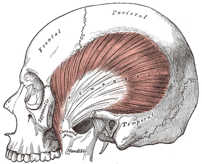
The temporal muscle (Lat:Temporalis) is a bilateral muscle located on the side of the head. It belongs to a subgroup of head muscles called Masticatory Muscles, named after their function elevating the mandible to produce the mandible movements (1,2). Masticatory muscles are four per side: Temporalis, Masseter, Pterygoideus medialis and Pterygoideus lateralis (1,2).
The temporalis muscle is a fan-shaped muscle which occupies the temporal fossa from which its fascicles (fibers) converge to the coronoid process of the mandible. Classic description for this muscle recognizes three main muscular bodies (anterior, midle, and posterior) originated from the temporal fossa up to the lower temporalis line and the temporalis fascia, fascicles which descend through the inner part de the zygomatic arch converging to be inserted on the coronoid process of the mandible, its temporalis crest and anterior margin of the mandibular branch through thick tendons (1,2).
Click for a larger image
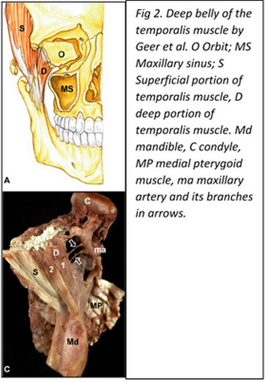
In 1996 Dunn et al. (3) reported the discovery of a so far unknown masticatory muscle called the “sphenomandibularis”, originated from the greater wing of the sphenoid bone medial to the temporalis muscle and descends on an oblique (lateral and slightly posterior) fashion reaching distally the coronoid process of the mandible. This muscular portion has been recognized as the “deep belly of the temporalis muscle” and has been described by several authors since then (4,5,6,8). The importance that has been given to this particular bundle lies on the fact that its medial insertion can reach a close relationship to the foramen rotundum, place of emergency from the cranium of the maxillary nerve, which has been hypothesized, could lead to eventual alteration of this nerve if it got trapped by this part of the muscle (6, 7).
The temporalis muscle receives innervation fundamentally from branches of the mandibular nerve: Deep temporal nerve (N. temporalis profundus)through its anterior middle and posterior branches.
The temporalis muscle is covered by a thick fascia layer: the temporalis fascia.
Article written by: Maria F. Cortés, DDS, MSc.
Images from:
Fig 1. Public domain, by Henry Vandyke Carter, MD - Gray's Anatomy, 1918
Fig 2. Geers C, Nyssen-Behets C, Cosnard G, Lengelé B. The deep belly of the temporalis muscle: an anatomical, histological and MRI study. Surg Radiol Anat. 2005 Aug;27(3):184-91. Epub 2005 Apr 9
Sources:
1. “Anatomía humana” V.2. Latarjet- Ruiz Liard, 4ª ed. 6ª reimp. 2008 Médica Panamericana, Buenos Aires, Argentina.
2. “Anatomía humana: descriptiva, topográfica y funcional. Tomo 1. Cabeza y Cuello, Rouviere H – Delmas A, 11° ed. 2005 MASSON, S.A., Barcelona, Spain.
3. Dunn GF, Hack GD, Robinson WL, Koritzer RT. Anatomical observation of a craniomandibular muscle originating from the skull base: the sphenomandibularis. Cranio. 1996 Apr;14(2):97-103; discussion 104-5.
4. Shimokawa T, Akita K, Soma K, Sato T. Innervation analysis of the small muscle bundles attached to the temporalis: truly new muscles or merely derivatives of the temporalis? Surg Radiol Anat. 1998;20(5):329-34.
5. Akita K, Shimokawa T, Sato T. Aberrant muscle between the temporalis and the lateral pterygoid muscles: M. pterygoideus proprius (Henle). Clin Anat. 2001 Jul;14(4):288-91.
6. Schön Ybarra MA, Bauer B. Medial portion of M. Temporalis and its potential involvement in facial pain. Clin Anat. 2001;14(1):25-30.
7. Fuentes E, Llanos S, Gómez R, Llanos P, Llanos F, Cortés-Sylvester MF, Solaria P, Melian A, Asfura J, Santos M, Zamorano E. Discovery of deep temporalis muscle belly close to maxillary nerve in a patient with trigeminal neuralgia: hypothesis of muscular compression and case report treated by Botox® Onabotulinum toxin tipe-A. Chirurgia 2016 June;29(3):99-102
8. Geers C, Nyssen-Behets C, Cosnard G, Lengelé B. The deep belly of the temporalis muscle: an anatomical, histological and MRI study. Surg Radiol Anat. 2005 Aug;27(3):184-91. Epub 2005 Apr 9.
- Details

Click for a larger image
We would like to welcome María Fernanda Cortés DDS, MSc. as a contributor to Medical Terminology Daily.
Dr. Cortés has a degree in Dentistry and is a Specialist in Dentomaxilofacial Radiology. She also has a Master’s degree in Temporomandibular disorders and orofacial pain.
Currently she is a lecturer of Human Anatomy at the Faculty of Dentistry of the Finis Terrae University. She also collaborates as a Postgraduate teacher for students of the “Anatomical Bases of Normal Imaging” diploma program at the Medical school of the same university.
Dr. Cortés has her own private practice in Santiago, Chile.
Clinical Anatomy Associates, Inc is proud to have Dr. Cortés as a contributor to "Medical Terminology Daily" and as a consultant to our team.
- Details


Click for a larger image
Today I received the bust of Andreas Vesalius which will be displayed in my office in a place of honor.
This bust is a small version of a bronze bust made by the Belgian artist Pascale Pollier, who is a contributor to "Medical Terminology Daily". Copies of this original bronze bust can be found is different libraries and museums around the world. Pascale, along with Theo Dirix, Dr. Sylviane Déderix, and others are on a quest to find the cemetery where Andreas Vesalius was buried in the island of Zakynthos in Greece. Eventually, the final quest is to find the body of this illustrius anatomists.
To fund this private research Pascale and other artists have donated their work to a GoFundMe page whose objective is to raise €9,900, roughly US$10,800. You can reach the GoFundMe page here.
This bust that sits in my office today is the sixth in a run of twelve copies. If you go to the GoFundMe page you can opt to aquire other artistic works or one of the last copies of this bust. To acquire the bust there is a minumum required donation of US$350.
As of this publication, the research is 51% funded. We need your help to achieve our goal!!
- Details
Click for a larger image
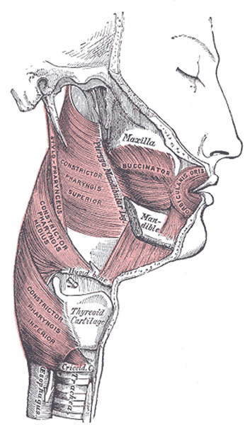
The [buccinator] muscle is a flat, thin quadrilateral muscle, that closes the space between the maxillary bone superiorly and the mandible inferiorly. It forms the side of the face and is the main muscular component of the “cheeks”. Etymologically [buccinator] means "trumpeter".
The superior and inferior boundaries of the muscle are the external surfaces of the alveolar processes of the maxilla and mandible on its posterior region, related to the posterior three molars. The posterior boundary is the anterior border of the pterygomandibular raphe, where posteriorly the middle pharyngeal constrictor also attaches. Anteriorly, its fibers appear to continue with the orbicularis oris muscle, but this is not so, as these two muscles (orbicularis oris and buccinator) are separate.
The fibers of the buccinator muscle are divided into three groups: the horizontal group continues anteriorly horizontally. The superior fibers have an anteroinferior direction and converge toward the angle of the mouth. The inferior fibers have an anterosuperior direction. These fibers appear to be continuous with the orbicularis oris, although they terminate in the mucosa, skin and some intermix with the muscular fibers of the orbicularis oris.
The buccinator muscle is covered by the buccopharyngeal fascia, and is in relation by its superficial surface and posteriorly, with a mass of fat (Bichat’s fat pad or suctorial pad), which separates it from the ramus of the mandible, the masseter, and a small portion of the temporalis muscle.
The parotid duct (Stensen’s duct) pierces the buccinator muscle opposite the second molar tooth of the maxilla.
The buccinator muscle receives innervation from the temporofacial and cervicofacial branches of the facial nerve (7th cranial nerve)
Sources:
1. “Gray’s Anatomy” Henry Gray, 1918
2. "Tratado de Anatomia Humana" Testut et Latarjet 8th Ed. 1931 Salvat Editores, Spain
3. "Gray's Anatomy" 38th British Ed. Churchill Livingstone 1995
Image in Public Domain, by Henry Vandyke Carter, MD - Gray's Anatomy, 1918
- Details
Click for a larger image
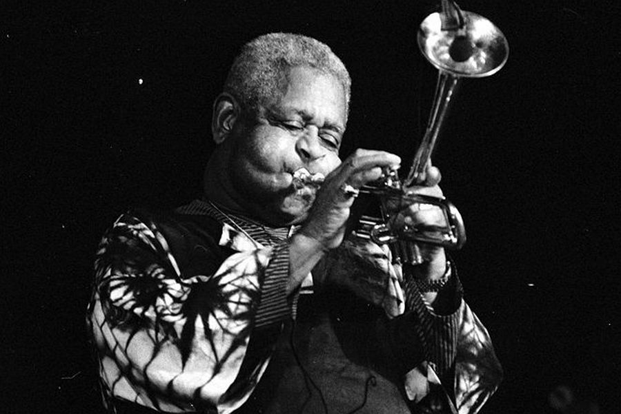
The anatomical, term [buccinator] refers to a quadrilateral muscle that forms the side of the mouth and the face, vernacularly known as “mouth cheeks”.
Its origin is from the Latin word [bucca] meaning mouth (in Spanish the term meaning mouth is "boca"). Further evolution of the word leads to the Latin term [buccina] meaning “trumpet” or “horn”, and [buccinator] which means “trumpeter”
The term takes real meaning when you look at a trumpet player distending the buccinator muscle to obtain longer note, such as the well-known Louis Armstrong or Dizzy Gillespie. (see image).
For the anatomical description of this muscle, click here
Image source: By Roland Godefroy - Own work, CC BY 3.0, https://commons.wikimedia.org/w/index.php?curid=4612877
- Details
Click for a larger image
While working on my library catalog, I made a very nice discovery!
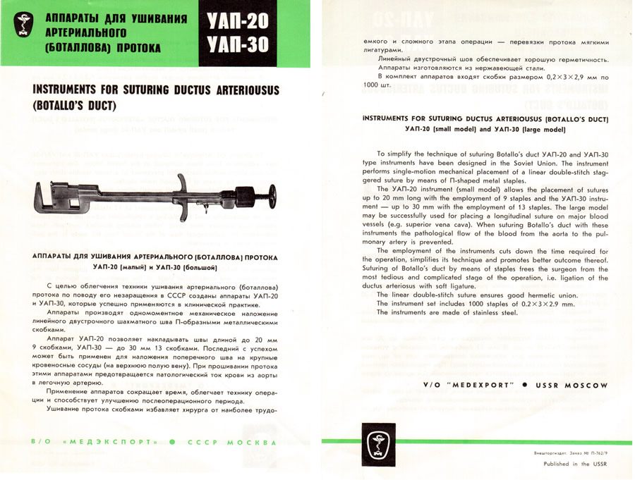
I found a set of 22 medical brochures circa 1950, from MedExport, an USSR government-sponsored company. These brochures depict instruments that are not in use now, or have been modernized.
The brochures were inside one of the books that Dr. Mark Ravitch's family donated to my library.
Some of these are surgical staplers which I have mentioned in my series of articles "History of Surgical Stapling" which includes a video on the topic.
What is interesting is that some of these instruments where discarded and thought of as non-practical, but they are coming back in modern versions such as the coronary artery bypass graft (CABG) vascular devices manufactured by Cardica, a division of Dextera Surgical.
An example of this is shown in the image attached to this article (you can click on the image for a larger view).
This is the YAN-30, used to close the ductus arteriosus, also known as the Duct of Botallus, named after Leonardo Botallus.
This duct has a function in the fetus, and it closed shortly after birth. Patency of the ductus arteriosus causes deoxygenated blood to mix with the oxygenated blood in the thoracic aorta giving rise to a condition known as PDA or Patent Ductus Arteriosus.
Needless to say, I am not selling or trading these priceless collector's items!!