![]()
Medical Terminology Daily (MTD) is a blog sponsored by Clinical Anatomy Associates, Inc. as a service to the medical community. We post anatomical, medical or surgical terms, their meaning and usage, as well as biographical notes on anatomists, surgeons, and researchers through the ages. Be warned that some of the images used depict human anatomical specimens.
You are welcome to submit questions and suggestions using our "Contact Us" form. The information on this blog follows the terms on our "Privacy and Security Statement" and cannot be construed as medical guidance or instructions for treatment.
We have 879 guests and no members online
")
Marcia Crocker Noyes
(1869 – 1946)
Further to my comment on old books and research that started with an interesting bookplate (Ex-Libris). I continued my research and found that the person in charge of the Osler library bookplate was a fascinating individual that today maybe a ghost in the MedChi library and building in Baltimore... This is certainly an article that can be called "A Moment in History"
Marcia Crocker Noyes was the librarian at The Maryland State Medical Society from 1896 to 1946 and was a founding member of the Medical Library Association.[1][2][3]
Sir William Osler, MD. a famous Johns Hopkins surgeon was a noted bibliophile and had a large personal collection of books on various topics. When he became the President of MedChi in 1896, he was dismayed at the condition of the library and knew that with the right person and some stewardship, it could become a significant collection. Sir William asked his friend, Dr. Bernard Steiner, a physician and President of the Enoch Pratt Free Library in Baltimore for suggestions of a librarian, and Dr. Steiner recommended Marcia Crocker Noyes. A native of New York, and a graduate of Hunter College, Marcia had moved to Baltimore for a lengthy visit with her sister, and took a “temporary” position at the Pratt Library, which turned into three years. Although she had no medical experience or background, she was enthusiastic, and most importantly, she was willing to move into the apartment provided for the librarian, who needed to be available 24 hours a day.
The image in this article is Ms. Noyes on her first year on the job. Marcia developed a book classification system for medical books, based on the Index Medicus, and called it the Classification for Medical Literature. The system uses the alphabet with capital letters for the major divisions of medicine and lower-case ones for the sub-sections. The system was used for many years, but it's now dated and the Faculty's original shelving scheme was never changed. The card catalogs still reflect her classification and many of the cards are written in Marcia's back-slanting handwriting.
Marcia knew enough to ask the Faculty's members about medical questions, terminology and literature. She gradually won over the predominantly male membership and they became her greatest allies; Sir William at the start, and then for nearly 40 years, Dr. John Ruhräh, a wealthy pediatrician with no immediate family of his own. She made a point of attending almost every Faculty function, and in 1904, under guidelines from the American Medical Association, Marcia was made the Faculty Secretary. For much of her first 10 years, she was the Faculty's only full-time employee, only being assisted by Mr. Caution, the Faculty's janitor. Later in life Marcia would say that she hired him because of his name!
Within ten years, the library had outgrown its space, and plans, spearheaded by Marcia and Sir William before his move to Oxford, were made to build a headquarters building, mainly to house the library's growing collection of medical books and journals.
Marcia was instrumental in the design and building of the new headquarters. She travelled to Philadelphia, New York and Boston to look at their medical society buildings, and eventually, the Philadelphia architectural firm, Ellicott & Emmart was selected to design and build the new Faculty building. Every detail of the building held her imprimatur, from the graceful staircase, to the light-filled reading room, and all of the myriad details of the millwork, marble tesserae, and most of all, the four-story cast iron stacks. She was on-site, climbing up unfinished staircases, checking out the progress of the building, which was built in less than one year at a cost of $90,000.
Among the features of the new building was a fourth-floor apartment for her. She referred to it as the "first penthouse in Baltimore" and it had a garden and rooftop terrace. The library collection eventually grew to more than 65,000 volumes from medical and specialty societies around the world. Journals were traded back and forth, and physicians eagerly anticipated the arrival of each new issue. At the same time, Marcia was involved in the Medical Library Association as one of eight founding members. The MLA promotes medical libraries and the exchange of information. One of the earliest mandates of the MLA was the Exchange, a distribution and trade service for those who had duplicates or little-used books in their collections. Initially, the Exchange was run out of the Philadelphia medical society, but in 1900 it was moved to Baltimore and Marcia oversaw it. Several hundred periodicals and journals were received and sent each month, a huge amount of work for a tiny staff. In 1904, the Faculty had run out of room to manage the Exchange, so it was moved to the Medical Society of the Kings County (Brooklyn). But without Marcia's excellent administrative skills, it floundered and in 1908, the MLA asked Marcia to take charge once again.
In 1909, when the new Faculty building opened, there was enough room to run the Exchange and with the help of MLA Treasurer, noted bibliophile and close friend, Dr. John Ruhräh, it once again became successful. Additionally, Marcia and Dr. Ruhräh combined forces to revive the MLA's bulletin, which had all but ceased publication in 1908, taking the Exchange with it. This duo maintained editorial control from 1911 until 1926. In 1934, around the time of Dr. Ruhräh's death, Marcia became the first “unmedicated” professional to head the MLA. During her tenure, the MLA incorporated, the first seal was adopted, and the annual meeting was held in Baltimore. Marcia wanted to write the history of the MLA once she retired from full-time work at the Faculty, but her health was beginning to fail. She had back problems and had suffered a serious burn on her shoulder as a young woman, possibly from her time running a summer camp, Camp Seyon, for young ladies in the Adirondack Mountains. In 1946, a celebration was planned to honor Marcia's 50 years at the Faculty. But she was adamant that the physicians wait until November, the actual date of her 50 years. However, they knew she was gravely ill, and might not make it until then, so a huge party was held in April. More than 250 physicians attended the celebration, but the ones she was closest to in the early years, were long gone. She was presented with a suitcase, a sum of money to use for travelling, and her favorite painting of Dr. John Philip Smith, a founder of the Medical College in Winchester, Virginia. It was painted by Edward Caledon Smith, a Virginia painter who had been a student of the painter Thomas Sully.[4] She adored this painting and vowed, jokingly, to take it with her wherever she went.
The painting was not to stay with her for very long, for she died in November 1946, and left it to the Faculty in her will. Her funeral was held in the Faculty's Osler Hall, named for her dear friend. More than 60 physicians served as her pallbearers, and she was buried at Baltimore's Green Mount Cemetery. In 1948, the MLA decided to establish an award in the name of Marcia Crocker Noyes. It was for outstanding achievement in medical library field and was to be awarded every two years, or when a truly worthy candidate was submitted. In 2014, the Faculty began giving a bouquet of flowers to the winner of the award in Marcia's name, and in honor of her work. Much evidence exists for this tradition, as we know that the physicians, especially Drs. Osler and Ruhräh, frequently gave her bouquets of flowers. Marcia also cultivated flower gardens at the Faculty and decorated the rooms with her work.
Today, the MedChi building is open for tours and if the rumors are to be believed Ms. Marcia Crocker Noyes is still at work in her beloved library as the "resident ghost" [1][5]

NOTE: This article has been modified from the original Wikipedia article on Marcia Crocker Noyes. The article itself is well-written with interesting images of the subject. I would encourage you to visit it. The second insert is from book 00736 in my personal library and shows in pencil, the incredibly small handwriting of Marsha C. Noyes.
Sources:
1. "Marcia, Marcia, Marcia" MedChi Archives blog.
2. "Marcia C. Noyes, Medical Librarian" (PDF). Bulletin of the Medical Library Association. 35 (1): 108–109. 1947. PMC 194645
3. Smith, Bernie Todd (1974). "Marcia Crocker Noyes, Medical Librarian: The Shaping of a Career" (PDF). Bulletin of the Medical Library Association. 62 (3): 314–324. PMC 198800Freely accessible. PMID 4619344.
4. Edward Caledon BRUCE (1825-1901)"
5. Behind the scenes tour MedChiBuilding
"Clinical Anatomy Associates, Inc., and the contributors of "Medical Terminology Daily" wish to thank all individuals who donate their bodies and tissues for the advancement of education and research”.
Click here for more information


- Details
Hypophyseal infundibulum
[Infundibulum] is a Latin word and it means "funnel". The plural form is [infundibula]. Variations of the word include [infundibuliform] meaning "with the shape or form of a funnel], and [infundibular] meaning "pertaining to a funnel". This word is widely used in human anatomy and embryology:
- Infundibuliform fascia: Funnel-shaped portion of the transversalis fascia that is directed toward and forming the internal inguinal ring.
- Hypophyseal infundibulum: An inferior extension of the hypothalamus forming a funnel-shaped stalk connected to the hypophysis or pituitary gland. (see image)
- Cystic infundibulum: The funnel-shaped portion of the gallbladder
- Ethmoidal infundibulum: a funnel-shaped extension of the middle meatus of the ethmoid bone, etc.
- Uterine infundibulum: Refers to the funnel-shaped distal opening of the uterine tube
The term infundibulum is also found in heart anatomy. It refers to funnel-shaped extensions of the cardiac chambers. This is well-illustrated by both the cone-like right and left ventricular outflow tracts toward the semilunar valves (aortic and pulmonary). In the case of the atrioventricular valves (tricuspid and mitral) there is also described an infundibular region. In all cases, these funnel-shaped regions allow for smooth, non-turbulent blood flow towards their respective valves.
Word suggested by: J. Estrada. Original image in the public domain, courtesy of bartleby.com
- Details

Click for a larger image
The term [larynx] originates from the Greek [λάρυγξ] meaning "upper windpipe or throat". Known vernacularly as "Adam's apple" or the "voice box" (not proper clinical terms), the larynx is the organ of phonation, and one of the organs found in the cervical visceral compartment. It is found immediately superior to the trachea, and anterior to the pharynx and esophagus.
It is formed by nine cartilages, three of which are median and single (epiglottis, thyroid, and cricoid cartilages), the rest being paired (arytenoid, corniculate, and cuneiform cartilages). In the accompanying image, the thyroid cartilage is depicted in blue, and the cricoid cartilage in green.
Within the larynx is a pair of musculomembranous folds, the vocal cords, which are innervated by the recurrent laryngeal nerves, branches of the Xth cranial nerve, also known as the vagus nerve.
The thyroid gland (in purple) is related to the inferior aspect of the larynx. The gland receives its name from the thyroid cartilage of the larynx, as the Greek term [θυροειδής] (thyreoeidís) means "in the shape of an oblong-shield".
It was Andrea Vesalius who named the cricoid cartilage because of its shape. The Greek term [κρικοειδή] (krikoeidí) refers to a structure "shaped like a ring". The cricoid cartilage is a complete ring, and thus is different from the incomplete or "C" shaped rings of the trachea.
Image property of CAA Inc. Artist: Dr. Miranda
- Details
Click for a larger image
Dr. Maurits Biesbrouck was born in Roeselare (Belgium) on February 15th, 1946, studied medicine at the Catholic University Leuven and became a MD in 1972. He devoted his professional career to clinical pathology (City Hospital Roeselare) and transfusion medicine He became adjunct medical director of the Dienst voor het Bloed (Brussels) and was the president-founder of the Scientific Association Transfusion in Flanders (WVTV).
Having a lifelong interest in Andreas Vesalius he translated the first book of the De Humani Corporis Fabrica Libri Septem into Dutch, compiled an annually updated Vesalius-bibliography and wrote many articles on his life and works, many as a co-author with Omer Steeno (Leuven, Belgium) and Theodoor Goddeeris (Kortrijk, Belgium). For the moment he is working on an overview of the editions of Vesalius’s works. See www.andreasvesalius.be.
Thanks to Dr. Bisbrouck for collaborating with "Medical Terminology Daily" with the article "Andreas Vesalius' Fatal Voyage to Jerusalem", a 2016 updated version of an original presentation at the 2014 Vesalius Continuum Meeting in Zakynthos, Greece.
He was part of the research team that uncovered the "false Vesalius postage stamps".
- Details
By Maurits Biesbrouck, MD. Continued from "Andreas Vesalius’s fatal voyage to Jerusalem (5)".
For the first page of this article, click here.
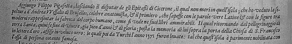
Passage in Abraham Ortelius’s Teatro del Mondo with Pigafetta’s report on Vesalius’s burial place. In Italian
“Leaving aside the discussion on the epitaph of Cicero, who did not die on this island [Zakynthos], I, Filippo Pigafetta, add having seen the grave of Andreas Vesalius from Brussels, famous anatomist and the first one to render in appropriate Latin wordings and accompanied with modern illustrations the fabric of the human body, as can be seen in his marvelous books. While returning from a pilgrimage to the Holy Land he ended his life here after glorious years. An inscription to his memory was placed above the door to the church of Saint Franciscus, in golden letters on black velvet, that was taken away by the Turcs in 1571. Thus this island was ennobled likewise by the bones of such famous persons” (16).
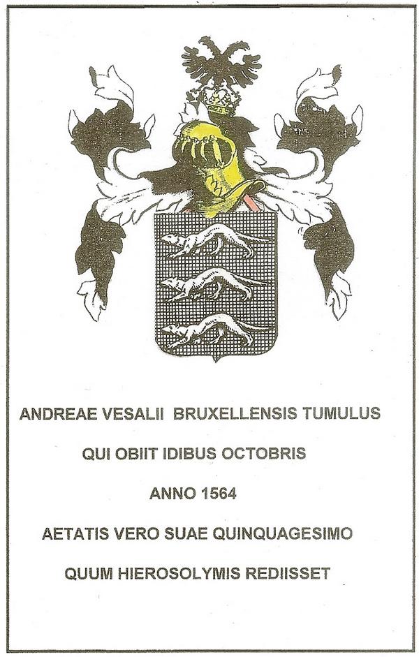
Reconstruction of Vesalius’ epitaph
by Theodoor Goddeeris (2012)
Pigafetta saw the grave in July 1586, almost at the same time as Jean Zuallart, but he may have seen it yet in 1568, on his way to Cyprus, or during his travelling to Egypt and Mount Sion in 1576-1577. When his information dates from 1568, or was based on reliable sources later on, then the Turcs could have taken two inscriptions: his epitaph and a plaque at the entrance of the church (17).
The church S. Maria delle Grazie
As far as is known, Fürer, Zuallart, and Pigafetta were the only three travellers to Jerusalem, to have seen Vesalius’s grave in the S. Maria delle Grazie with their own eyes. Thanks to them we are sure that Vesalius was buried in that church, built in 1488. Regrettably, it was destroyed on October 18th 1840 by a severe earthquake. It was then rebuilt, but moved a few meters inland. In 1893 the church was again destroyed by an earthquake. This time it was not restored, and the rubble was used by the fishermen along the coast to rebuild their houses, as told by Barbiani (18).
Vesalius’s epitaph
So Fürer saw Vesalius’s epitaph before it was stolen, but he gave an incorrect description of Vesalius’ coat of arms on it. Anyway, it follows that Vesalius’ grave was indeed at this church, and that he must have been buried there and not by the side of a road somewhere. An effort was immediately made to give him a worthy burial place. The epitaph with the coat of arms, which was already present so soon after his death, is proof of this. The people of Zakynthos were well aware of who this person was. To correct the errors in the epitaph and in the coat of arms as well, some years ago, Dr. Theodoor Goddeeris, made a graphic reconstruction of both.
Conclusions
I can summarize with four conclusions (19):
1° Andreas Vesalius' trip to Jerusalem had nothing to do with the Inquisition: he went to Jerusalem for religious reasons, and the King took the opportunity to send with him his financial support for the Holy Places, as he used to do.
2° Vesalius did not die in a shipwreck. He died most probably of a combination of exhaustion and illness.
3° He was not buried on some desolate spot, but in the church Santa Maria delle Grazie. His remains are not yet found, however. (see here for additional information on the search for Vesalius' grave).
4° His grave had an epitaph (and we know exactly what it said and looked like).
Personal note: My sincere thanks to Dr. Maurits Biesbrouck for contributing this article to "Medical Terminology Daily". His clear and factual analysis gives us an insight on the last months of Andreas Vesalius' life and the problems that eventually led to his death on Zakynthos, Greece. I am proud to have been one of the many international attendees to the 2014 meeting in the island of Zakynthos where I saw Dr. Biesbrouck deliver the presentation and research on which this article is based. Dr. Miranda.
Sources and author's comments:
16. Teatro del Mondo di Abrahamo Ortelio: da lui poco inanzi la sua morte riveduto, e di tavole nuove, et commenti adorno, e arricchito con la vita dell’Autore. Traslato in Lingua Toscana dal Sigr. Filippo Pigafetta, in Anversa, si vende nella Libraria Plantiniana, 1608 and 1612.
17. Maurits BIESBROUCK, Theodoor GODDEERIS, Omer STEENO. ‘Post Mortem Andreae Vesalii (1514-1564). Deel I: De laatste reis van Andreas Vesalius en de omstandigheden van zijn dood’ [After Vesalius’ Death: The Last Travel of Andreas Vesalius and the Circumstances of his Death] and ‘Deel II: Het graf van Andreas Vesalius op Zakynthos’ [Vesalius’ Grave in Zakynthos] in A. Vesalius, 2015, 27 (no. 3): 154-161 and (no. 4): 193-200, ill.
18. Nicolas Ant. BARBIANI, O André Vésale kai è proodos tès anatomias [Andreas Vesalius and the evolution of anatomy] – L’évolution de l’anatomie et André Vésale, Athens, 1953, 32 pp., ill.; in Greek with a French summary on pp. 7-8.
19. For more details see the papers mentioned in ref. 1.
- Details
By Maurits Biesbrouck, MD. Continued from "Andreas Vesalius’s fatal voyage to Jerusalem (4)".
For the first page of this article, click here.
The story by Solenander
An interesting story, told by Reiner Solenander (1524-1601), deserves our attention here (12). His report is important, but it was hard to find, because it is included in a work of Thomas Theodor Crusius, namely his Vergnügung müssiger Stunden, from 1722. Dr. Theodoor Goddeeris found a copy of this in the Herzog August Bibliothek in Wolfenbüttel (Germany). In it, the story can be found of Vesalius’s end according to Solenander, written in Augsburg, and dated May 1566. This means: one year and seven months after Vesalius’ death. I only translate the most important elements:
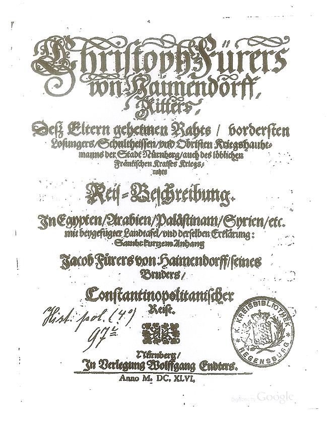
Title page of the Reis-Beschreibung by
Christoph Fürer von Haimendorf (1646)
“…, they returned to the ship. Initially, owing to stormy weather, they were driven off course, and when they had been driven into the open sea, the air became so peaceful, that the ship drifted around for several weeks, in virtually the same place. It was high summer and baking hot. Then most of the passengers fell ill, and many died. When he saw them being thrown into the sea, for several days in succession, Vesalius became dispirited, and began to suffer from sickness himself, but did not say anything about it. …, the provisions began to run out. There was a general shortage and a severe lack of drinking water. A daily ration was given to each, and not a drop more than was deemed necessary. Having ended up in this desperate situation, Vesalius, who was taciturn by nature, melancholy, and not provided for such an eventuality, received no care, as the necessary provisions had by now run out, and he started to become more seriously ill. … After they had been drifting around for a long time, the wind finally began to pick up, and they were able to sail on, with a favourable wind. In the meantime, Vesalius lay sick in the hold…. When land was sighted, everyone became frenzied, but he became even more seriously ill. Only then did the travellers arrive at Zakynthos, they called to him, and when they entered the port and struck sail, Vesalius expired, amid the creaking of the ropes and the noise of camels. But he obtained what he had most desired, namely that he should be carried ashore, and buried on land, at a chapel or shrine, near the port of Zakynthos"(13).
Vesalius’s burial place
This is how Vesalius died most probably: from illness and deprivation. But the question of his eventual burial place also arises. Numerous researchers have tried hard to gain clear information on this subject. The first person to see the grave with his own eyes was Christoph Fürer von Haimendorf (1541-1610), who in his Itinerarium (1621) states that he stopped off on Zakynthos and saw Vesalius’ grave. It is the German version of Fürer’s account of his journey, that contains the most details about Vesalius’ burial place. Not the Latin one, that appeared 25 years earlier (14).
Fürer himself, set out on a journey in July 1565, also from Venice, and headed first for Alexandria in Egypt. They sailed past Corfu, and on the 6th of August they disembarked on Zakynthos, a good seven months after Vesalius' death. He writes: “On this island there is a closter named S. Maria della Gratia, where Vesalius has been buried.” Fürer gives also a description of Vesalius’s epitaph here, because he continues: “In a grave with an epitaph carrying his coat of arms with three whippets on a shield in red, a yellow eagle, with two crowned heads, and the inscription: Tomb of Andreas Vesalius from Brussels, who died in the year 1564 on the 10th of October, on his way back from Jerusalem, at the age of 58...” This should of course be “the 15th of October” instead of the 10th, and “at the age of 50” instead of “58”. The date must be a typographical error, because his Latin text has indeed “the 15th of October”, and his abbreviation “ID” for the ides can easily be misread as a ‘ten’. Also, the description of the coat of arms is incorrect: as everyone knows, there were no whippets on it but weasels.
Solenander gives the epitaph also. According to him it reads: “Tomb of Andreas Vesalius from Brussels, who died October the 15th, 1564, on his way back from Jerusalem, at the age of 58...” Here again, the age is wrong, as Vesalius was born on the 31st of December 1514: thus he was ten weeks short of 50. As both Fürer and Solenander had the wrong age for Vesalius, this points to the fact that the epitaph might have been wrong, and not their reports.
Jean Zuallart, another Jerusalem traveller, writes that Zakynthos is an island which is highly susceptible to earthquakes, and he also mentions the grave in the church of Santa Maria delle Grazie. He saw the tomb in 1586, and tells that Vesalius was buried, on the same spot as Marcus Tullius Cicero, the famous roman writer. According to Zuallart in 1586, that is twenty-two years after Vesalius’ death, he reported that the copper memorial plaque had disappeared, having been stolen by the Turks when they plundered the island in 1571.
Pavlos Plessas was the first one to point, some years ago, to a third eye-witness of Vesalius’s grave (15). He found proof of this in an Italian translation of the worldatlas by Abraham Ortelius (1527-1598) Theatrum Orbis Terrarum. That translation was made by Filippo Pigafetta, member of an italian family of nobility from Vicenza, who added several personal experiences to the text, such as his own visit to Zakynthos. To Ortelius’s text on the Ionian Islands, accompanying Map 217, he adds in the section devoted to Zakynthos: (continued)...
Article continued here: Andreas Vesalius’s fatal voyage to Jerusalem (6).
Sources and author's comments:
12. Maurits BIESBROUCK, Theodoor GODDEERIS, Omer STEENO. ‘Reiner Solenander (1524-1601): an important 16th Century Medical Practioner and his Original Report of Vesalius’ Death in 1564 - Reiner Solenander (1524.-1601.): značajan medicinski praktičar iz 16. stoljeća i njegov izvorni izvještaj o Vezalovoj smrti 1564. godine’ in Acta medico-historica Adriatica, 2015, 13 (no. 2): 265-286, ill.
13. Reiner SOLENANDER. ‘Kurze Nachricht von des Andreae Vesalii Todt und Begräbnisz - Historia de Obitu Andreae Vesalii ex Literis Reineri Solenandri ex Comitiis August. 1566. Mense Majo’ in Thomas Theodor CRUSIUS, Vergnügung müssiger Stunden, oder allerhand nützliche zur heutigen galanten Gelehrsamkeit dienende Anmerckungen, M. Rohrlachs Wittib und Erben, 1722, pp. 483-490. The ‘work’ Vergnügung müssiger Stunden was in fact a journal that was published by Theodor Crusius in Leipzig for 20 years from 1713 to 1732; Solenander’s contribution about Vesalius’ death appeared in volume 18, 1722.
14. Christoph FÜRER VON HAIMENDORF, Reis-Beschreibung in Egypten, Arabien, Palästinam, Syrien, etc.: M. beygef. Landtafel u. ders. Erkl. Sambt Kurtzem Anh. Jacob Fürers von Haimendorff, s. Brüders, Constantinopolitanischer Reise, Nürnberg: Endter, 1646, 384 pp.
15. Pavlos [PLESSAS], ‘The tomb of Vesalius and Filippo Pigafetta’s testimony’ in Pampalaia Zakunthinès, April 3th, 2013, 4 pp., ill.; see <http://pampalaia.blogspot.com>.
- Details
By Maurits Biesbrouck, MD. Continued from "Andreas Vesalius’s fatal voyage to Jerusalem (3)".
For the first page of this article, click here.

Stephanus BONIFACIUS, Liber
de Perenni Cultu Terrae Sanctae
et de Fructuosa eius Peregrinatione,
Venice: Guerra, 1573
Letter 4: from Brother Bonifacio to Philip II
The last letter is from Fra Bonifacio de Ragusa, guardian of the monastery of Mount Sion, and Custodian of the Holy Places, and is dated May 29th, 1564 in Jerusalem. That is two months later and it is likewise addressed to Philip II. As much in this letter is of a religious nature, I only translate what is important to our story: “Holy and Royal Majesty, … , Doctor Vesalius, a devout pilgrim in Jerusalem, and the bearer of this letter, has taken the risk of bringing to us, … , the royal gift of five hundred ducats, intended for the Catholic worshippers in the kingdom of Jerusalem, as a contribution for its shrines, … , etc.”
He ends with:
“… Here I conclude by kissing your royal hands. From … the Holy Mount Sion in Jerusalem, May the 29th, 1564. The pious chaplain, Brother Bonifacio, Guardian of the Holy Land and Apostolic Vicar.”
The letter is, on its address side, sealed with an oval seal, around which run the words (in Latin) “Seal of the guardian of the monastery of the holy Mount Sion”.
Thus, Bonifacio de Ragusa confirms that Vesalius was in Jerusalem on 29 May 1564, and mentions that Vesalius had brought five hundred ducats with him as a contribution for the holy places from the King. Next, Bonifacio states in this letter, that it (the letter) was given to Vesalius to deliver it personally to the King. This is very important! As we shall see, the letter reached its destination although Vesalius himself remained behind on Zakynthos, and died there shortly afterwards, as we all know. Most importantly that letter does not only still exist, but it is preserved in good shape, and there are no signs of damage by seawater (thus debunking the theory that Vesalius shipwrecked and fell into the sea).
As Bonifacio specifies that Vesalius will deliver the letter himself, this shows that Vesalius had not yet made it known on 29 May 1564 that he did not wish to return to Madrid. It may only have been afterwards, that he received the invitation, from the Venetian Senate, to return to Padua, to which various sources refer. Then he might have changed his mind, but we found no authentic documents to support this. At the time Bonifacio de Ragusa wrote this letter, Vesalius clearly was in the favor of the King and intended to return to Spain.
Vesalius’ stay in the Holy Land
Vesalius was in Palestine for at least five months: from the end of March to the end of August 1564. So we must ask what he did there during this months. Well, we don’t know very much about that. The only thing is that Bonifacio writes elsewhere, that he visited the planes in Jericho, together with Vesalius, and that they were interested in the medical virtues of the various herbs and fruits of that region. This point is confirmed by a passage in Bonifacio’s book Liber de Perenni Cultu Terrae Sanctae (11).
Thanks to the four letters, found by José Barón Fernández, the purpose of the journey is now clear: a pilgrimage by Vesalius to the Holy Land with a monetary contribution for the holy places, from the king of Spain. The letters do not give the impression that the primary purpose of the journey was to send Vesalius on ‘a diplomatic mission’. Rather, it seems that the king simply took the opportunity provided by this pilgrimage to send the sum of money with his physician. Such a gift, was not an extraordinary one, as the catholic king used to do this each year.
His return voyage
Now, let us look at his return voyage. The voyage to Jerusalem was usually made, according to Jean Zuallart, in April, May or June, and was accompanied by fine, warm weather, whereas the return trip was made in the autumn, when the weather was usually not so fine and could be windy or even stormy. During the return trip, the traditional stops after Jerusalem were Tripoli, Cyprus, Candia in Crete, Zakynthos and then to Corfu and finally Venice. This represented a route without too many detours. Zuallart also refers to Jaffa and Ramma as stops. According to him, the route took anything from between ten and thirty days, depending on the wind. Interesting perhaps, to illustrate how adventurous and uncertain the trip was, is that Zuallart himself did not reach Zakynthos while returning, and sailed past the island, because of adverse winds.
The causes of Vesalius’s death
We all know that Vesalius died in Zakynthos. The reasons and circumstances of his death remain mysterious however. Most authors refer to a shipwreck due to a storm. Also, the literature consistently refers to Vesalius as the only victim when arriving at Zakynthos. When there is a shipwreck, there are multiple victims. But – as already told - the fact that Vesalius had the letter with him, from Bonifacio for the King, and that this letter still exists, rules out definitely the possibility of a shipwreck. Exhaustion from lack of food or drinking water and from drifting aimlessly over a long period due to adverse winds, as some have suggested, is certainly possible. But Vesalius was a man of means, and if there was a general shortage on board, he would certainly not have been the only one with problems. The most plausible explanation does indeed seem to be illness, possibly exacerbated by general weakness, from whatever cause.
Article continued here: Andreas Vesalius’s fatal voyage to Jerusalem (5).
Sources and author's comments:
11. Stephanus BONIFACIUS, Liber de Perenni Cultu Terrae Sanctae et de Fructuosa eius Peregrinatione, Venetië: Guerra, 1573; see p. 235.