Medical Terminology Daily (MTD) is a blog sponsored by Clinical Anatomy Associates, Inc. as a service to the medical community. We post anatomical, medical or surgical terms, their meaning and usage, as well as biographical notes on anatomists, surgeons, and researchers through the ages. Be warned that some of the images used depict human anatomical specimens.
You are welcome to submit questions and suggestions using our "Contact Us" form. The information on this blog follows the terms on our "Privacy and Security Statement" and cannot be construed as medical guidance or instructions for treatment.
We have 550 guests online

Jean George Bachmann
(1877 – 1959)
French physician–physiologist whose experimental work in the early twentieth century provided the first clear functional description of a preferential interatrial conduction pathway. This structure, eponymically named “Bachmann’s bundle”, plays a central role in normal atrial activation and in the pathophysiology of interatrial block and atrial arrhythmias.
As a young man, Bachmann served as a merchant sailor, crossing the Atlantic multiple times. He emigrated to the United States in 1902 and earned his medical degree at the top of his class from Jefferson Medical College in Philadelphia in 1907. He stayed at this Medical College as a demonstrator and physiologist. In 1910, he joined Emory University in Atlanta. Between 1917 -1918 he served as a medical officer in the US Army. He retired from Emory in 1947 and continued his private medical practice until his death in 1959.
On the personal side, Bachmann was a man of many talents: a polyglot, he was fluent in German, French, Spanish and English. He was a chef in his own right and occasionally worked as a chef in international hotels. In fact, he paid his tuition at Jefferson Medical College, working both as a chef and as a language tutor.
The intrinsic cardiac conduction system was a major focus of cardiovascular research in the late nineteenth and early twentieth centuries. The atrioventricular (AV) node was discovered and described by Sunao Tawara and Karl Albert Aschoff in 1906, and the sinoatrial node by Arthur Keith and Martin Flack in 1907.
While the connections that distribute the electrical impulse from the AV node to the ventricles were known through the works of Wilhelm His Jr, in 1893 and Jan Evangelista Purkinje in 1839, the mechanism by which electrical impulses spread between the atria remained uncertain.
In 1916 Bachmann published a paper titled “The Inter-Auricular Time Interval” in the American Journal of Physiology. Bachmann measured activation times between the right and left atria and demonstrated that interruption of a distinct anterior interatrial muscular band resulted in delayed left atrial activation. He concluded that this band constituted the principal route for rapid interatrial conduction.
Subsequent anatomical and electrophysiological studies confirmed the importance of the structure described by Bachmann, which came to bear his name. Bachmann’s bundle is now recognized as a key determinant of atrial activation patterns, and its dysfunction is associated with interatrial block, atrial fibrillation, and abnormal P-wave morphology. His work remains foundational in both basic cardiac anatomy and clinical electrophysiology.
Sources and references
1. Bachmann G. “The inter-auricular time interval”. Am J Physiol. 1916;41:309–320.
2. Hurst JW. “Profiles in Cardiology: Jean George Bachmann (1877–1959)”. Clin Cardiol. 1987;10:185–187.
3. Lemery R, Guiraudon G, Veinot JP. “Anatomic description of Bachmann’s bundle and its relation to the atrial septum”. Am J Cardiol. 2003;91:148–152.
4. "Remembering the canonical discoverers of the core components of the mammalian cardiac conduction system: Keith and Flack, Aschoff and Tawara, His, and Purkinje" Icilio Cavero and Henry Holzgrefe Advances in Physiology Education 2022 46:4, 549-579.
5. Knol WG, de Vos CB, Crijns HJGM, et al. “The Bachmann bundle and interatrial conduction” Heart Rhythm. 2019;16:127–133.
6. “Iatrogenic biatrial flutter. The role of the Bachmann’s bundle” Constán E.; García F., Linde, A.. Complejo Hospitalario de Jaén, Jaén. Spain
7. Keith A, Flack M. The form and nature of the muscular connections between the primary divisions of the vertebrate heart. J Anat Physiol 41: 172–189, 1907.
"Clinical Anatomy Associates, Inc., and the contributors of "Medical Terminology Daily" wish to thank all individuals who donate their bodies and tissues for the advancement of education and research”.
Click here for more information
- Details
UPDATED: From the Greek [εὐφωνία] [eu-] meaning "good" and [phone/phonos] meaning "sound or voice". The term [euphonic] means "sounds good".
This is useful when combining root terms where the proximal to distal order of root terms does not apply. An example is the removal of the Fallopian tubes and ovaries. Since there is no true attachment, or flow of fluids between these structures, the words [salpingooophorectomy] and [oophorosalpingectomy] are both correct, but one of them is easier to pronounce and articulate, or euphonic, therefore that is the one we use: [salpingooophorectomy].
Note: Since the two root terms [-salping-] (Fallopian or uterine tube) and [-oophor-] (ovary) are connected with an [-o-] (meaning "and") per the rules used for combining root terms, the use of a hyphen to connect these terms is redundant and incorrect!
- Details
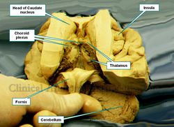
The term [thalamus] arises from the Latin (Thalamo], meaning “the wedding chamber”, "the inner chamber" or “the wedding bed”. It also has a similar meaning in Greek. Early anatomists thought that the enclosure of the lateral ventricles with the fornix as a roof formed a chamber. The lager nuclear mass known as the thalamus looked to some as a double bed in a chamber, hence the name.
It did help that the thalamus has a larger posterior protuberance akin to a cushion or a pillow, the pulvinar.
The thalamus is a paramedian structure which forms the lateral walls of the third ventricle of the brain, as well as part of the floor of the lateral ventricles of the brain. It can sometimes (30%) present a midline mass which communicates both thalami across the third ventricle. It is separated from the mesencephalon (midbrain) by the subthalamic region.
It is an important relay system, as all the sensory information to the brain (with the exception of the olfactory) has a synaptic stop at one or more of the thalamic nuclei before being sent to the cortex of the brain. The thalamus also receives important motor collaterals, as well as cerebellar and subthalamic information. This makes the thalamus an important entity in the regulation of fine motor control. Thalamic dysfunction can lead to sleep disorders and coma.
The thalamus also serves as an important relay between sensory, motor, limbic, and hypothalamic activity, making it critical to the relation of emotions, visceral responses, and prefrontal cortical activity. It also plays an important role in wakefulness, and consciousness.
In the accompanying image the thalamus is found covered by the choroid plexuses of the lateral ventricle.
Note: The links to Google Translate include an icon that will allow you to hear the pronunciation of the word. Image property of CAA, Inc. Photographer: E. Klein
- Details
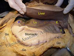
The adjectival term [epiploic] arises from the Greek term [επίπλουν] (pronounced “epiploun”) which is synonymous with the Latin term [omentum], referring to two abdominal peritoneal membranes, the lesser omentum and the greater omentum. For more information on the word [omentum] click here.
The word itself is used in Greek in the expression [επιπλέουν πάνω] (epiploun pano) which means “to float upon”, referring to the fact that the fatty omental apron “floats” or “drapes” upon the abdominal viscera. Hippocrates of Cos (460 BC - 370 BC) referred to the greater omentum as epiploon. This anatomical name evolved towards the Latin version, which is used today. In spite of this there are other languages where the Greek root is still used. As examples, in Spanish the terms are “epiplón mayor” and “epiplón menor”, and in French they are “grand epiploon” and “petit epiploon”.
Because of the presence of fat in the greater omentum, the medical adjectival term [epiploic] has also evolved to mean “fatty”, such as in the case of the epiploic appendages, a series of fatty appendages found in the colon.
Images property of:CAA.Inc.. Photographer:D.M. Klein
- Details
Aaron Ruhalter, MD, FACS
I am sad to let everybody know that Dr. Ruhalter passed on yesterday January 25th, 2016. A good friend, guide, and mentor, Dr. Ruhalter was always reminding me to keep on using drawings and sketches to teach human anatomy, a subject he loved, and at which he excelled. I will miss him dearly. May he rest in peace. Dr. Miranda
Dr. Aaron Ruhalter was for many years the Executive Director of Medical Education at the Johnson & Johnson Endo-Surgery Institute in Cincinnati. Dr. Ruhalter is a Professor of Anatomy and a former Professor of Surgery at the Robert Wood Johnson Memorial Hospital in New Jersey. He was also one of the founding members of the American Association of Clinical Anatomists (AACA).
His review on the surgical anatomy of the parotid gland, submandibular triangle, and floor of the mouth is outstanding. This review was published in 1997 in the book "Mastery of Surgery", third edition, by Drs. Lloyd M. Nyhus, Robert J. Baker, and Josef E. Fischer.
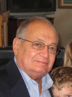
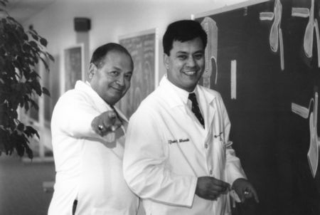
Dr. Miranda worked with Dr. Ruhalter for several years, both at the Endo-Surgery Institute and at the University of Cincinnati College of Medicine. The picture above shows both of them preparing an anatomy blackboard session at the Institute, back in 1994.
- Details
Refers to the internal lining of an artery or a vein. The tunica (layer) intima is composed of an inner layer known as endothelium (inner fabric or inner layer) and a subendothelial layer.
Also known as the [tunica intima vasorum], Latin for intimal (inner) layer of the vessels, the tunica inima is also known as "Bichat's tunic"
- Details
The term [bruxism] arises from the Greek [βρυγμός] (vrygm?s), meaning “a gnashing of the teeth”, or “bruxism”.
Bruxism is usually a subconscious problem, presenting most of the times at night while the individual is asleep, and does not continue into the waking hours. In some people bruxism does continue into the day and the person is constantly gnashing the teeth, but not conscious of the activity. Awake bruxism is sometimes called [bruxomania].
Bruxism is usually idiopathic (of unknown origin) or it can be secondary to other medical conditions.
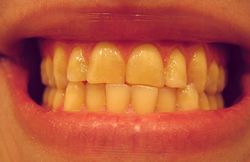
Patients with bruxism usually present with damage to the teeth by the constant attrition of the teeth by laterally grinding of the teeth of the maxilla against the teeth of the jaw. This can lead to severe tooth damage with exposure of the dentin through and absent, fractured, or damaged enamel. A secondary problem is misalignment of the teeth. The accompanying image shows misalignment and tooth damage.
Note: The links to Google Translate include an icon that will allow you to hear the pronunciation of the word.
Image by DRosenbach (en:Image:Deviated midline.JPG) [Public domain], Wikimedia Commons


