Medical Terminology Daily (MTD) is a blog sponsored by Clinical Anatomy Associates, Inc. as a service to the medical community. We post anatomical, medical or surgical terms, their meaning and usage, as well as biographical notes on anatomists, surgeons, and researchers through the ages. Be warned that some of the images used depict human anatomical specimens.
You are welcome to submit questions and suggestions using our "Contact Us" form. The information on this blog follows the terms on our "Privacy and Security Statement" and cannot be construed as medical guidance or instructions for treatment.
We have 754 guests online

Jean George Bachmann
(1877 – 1959)
French physician–physiologist whose experimental work in the early twentieth century provided the first clear functional description of a preferential interatrial conduction pathway. This structure, eponymically named “Bachmann’s bundle”, plays a central role in normal atrial activation and in the pathophysiology of interatrial block and atrial arrhythmias.
As a young man, Bachmann served as a merchant sailor, crossing the Atlantic multiple times. He emigrated to the United States in 1902 and earned his medical degree at the top of his class from Jefferson Medical College in Philadelphia in 1907. He stayed at this Medical College as a demonstrator and physiologist. In 1910, he joined Emory University in Atlanta. Between 1917 -1918 he served as a medical officer in the US Army. He retired from Emory in 1947 and continued his private medical practice until his death in 1959.
On the personal side, Bachmann was a man of many talents: a polyglot, he was fluent in German, French, Spanish and English. He was a chef in his own right and occasionally worked as a chef in international hotels. In fact, he paid his tuition at Jefferson Medical College, working both as a chef and as a language tutor.
The intrinsic cardiac conduction system was a major focus of cardiovascular research in the late nineteenth and early twentieth centuries. The atrioventricular (AV) node was discovered and described by Sunao Tawara and Karl Albert Aschoff in 1906, and the sinoatrial node by Arthur Keith and Martin Flack in 1907.
While the connections that distribute the electrical impulse from the AV node to the ventricles were known through the works of Wilhelm His Jr, in 1893 and Jan Evangelista Purkinje in 1839, the mechanism by which electrical impulses spread between the atria remained uncertain.
In 1916 Bachmann published a paper titled “The Inter-Auricular Time Interval” in the American Journal of Physiology. Bachmann measured activation times between the right and left atria and demonstrated that interruption of a distinct anterior interatrial muscular band resulted in delayed left atrial activation. He concluded that this band constituted the principal route for rapid interatrial conduction.
Subsequent anatomical and electrophysiological studies confirmed the importance of the structure described by Bachmann, which came to bear his name. Bachmann’s bundle is now recognized as a key determinant of atrial activation patterns, and its dysfunction is associated with interatrial block, atrial fibrillation, and abnormal P-wave morphology. His work remains foundational in both basic cardiac anatomy and clinical electrophysiology.
Sources and references
1. Bachmann G. “The inter-auricular time interval”. Am J Physiol. 1916;41:309–320.
2. Hurst JW. “Profiles in Cardiology: Jean George Bachmann (1877–1959)”. Clin Cardiol. 1987;10:185–187.
3. Lemery R, Guiraudon G, Veinot JP. “Anatomic description of Bachmann’s bundle and its relation to the atrial septum”. Am J Cardiol. 2003;91:148–152.
4. "Remembering the canonical discoverers of the core components of the mammalian cardiac conduction system: Keith and Flack, Aschoff and Tawara, His, and Purkinje" Icilio Cavero and Henry Holzgrefe Advances in Physiology Education 2022 46:4, 549-579.
5. Knol WG, de Vos CB, Crijns HJGM, et al. “The Bachmann bundle and interatrial conduction” Heart Rhythm. 2019;16:127–133.
6. “Iatrogenic biatrial flutter. The role of the Bachmann’s bundle” Constán E.; García F., Linde, A.. Complejo Hospitalario de Jaén, Jaén. Spain
7. Keith A, Flack M. The form and nature of the muscular connections between the primary divisions of the vertebrate heart. J Anat Physiol 41: 172–189, 1907.
"Clinical Anatomy Associates, Inc., and the contributors of "Medical Terminology Daily" wish to thank all individuals who donate their bodies and tissues for the advancement of education and research”.
Click here for more information
- Details
The term [suctorial] is based on the Latin [sugere] meaning “sucking” and evolves to modern Latin as the term [suctorius]. It is the base for the English verb “to suck”, the Spanish term [succionar], and the French [succion].
It is used to describe the small fat pad that covers the external and posterior aspect of the buccinator muscle. Also known as Bichat’s fat pads, these pads are said to help the suction activity of the baby. As they are not needed later in life, they tend to reduce in size.
Sources:
1. “Anatomy of the buccal fat pad and its clinical significance” Jackson, IT Plastic and Reconstructive Surgery, 06/1999, Volume 103, Issue 7
2. “Gray’s Anatomy” Henry Gray, 1918
3. “Understanding Anatomical Terms” Mehta, LA, et al. Clin Anat 9:330-336 (1996)
- Details
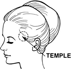
The etymology (origin) of the term [temporal] is Latin and derives from [tempus and temporis] meaning "time". It is said that the name was given to the area of the head where the hair initially starts to become gray and whitish, marking the "passage of time". Many philosophists and scholars have said that "life is only temporal".
Anatomically, the [temple] is the area of the temporal region. The word "temple" as used in anatomy has a separate etymology from the word temple, used as "place of worship". Both come from Latin, but the word for the place of worship comes from [templum] meaning a "shrine" or a "place of worship".
Use of the term [temporal] is found in:
• Temporal bone
• Temporal muscle
• Temporal fascia
Image: By Pearson Scott Foresman [Public domain], via Wikimedia Commons
Click here for the link to the original image
- Details
This article is part of the series "A Moment in History" where we honor those who have contributed to the growth of medical knowledge in the areas of anatomy, medicine, surgery, and medical research.

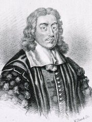
Thomas Willis
UPDATED: Thomas Willis (1621-1675). An English physician and anatomist, Willis was born on his parents' farm in Great Bedwyn, Wiltshire, where his father held the stewardship of the Manor. He was a kinsman of the Willys baronets of Fen Ditton, Cambridgeshire. He graduated M.A. from Christ Church, Oxford in 1642. In the Civil War years he was a royalist, and was dispossessed of the family farm at North Hinksey by Parliamentary forces. In the 1640's Willis was one of the royal physicians to Charles I of England. He obtained his medical degree in 1646.
Thomas Willis might well be one of the greatest physicians of the 17th century.He is one of the founders of the Royal Society of London. He is remembered by his many publications, especially "Cerebri Anatome: Cui accessit Nervorum Descriptio et Usu", where he describes the arterial anastomoses at the base of the brain. This work is also the first detailed description of the vasculature of the brain. Willis described nine cranial nerves.
He is considered as the father of Neurology as a discipline. He used the term "neurology" for the first time in 1664. He described several neurological conditions
The Arterial Circle of Willis is a famous eponymous structure found at the base of the brain. It represents an anastomotic roundabout that connects the right and left sides as well as the carotid and vertebral arterial territories that supply the brain. Named after Thomas Willis, this structure was known well before him, but it was Willis who described its function. You will be redirected to a detailed description of this structure if you click here.
Sources:
1. "The legendary contributions of Thomas Willis (1621-1675): the arterial circle and beyond" Rengachary SS et al J Neurosurg. 2008 Oct;109(4):765-75
2. "Thomas Willis, a pioneer in translational research in anatomy (on the 350th anniversary of Cerebri anatome)" Arraez-AybarJournal of Anatomy, 03/2015, Volume 226, Issue 3
3. " The naming of the cranial nerves: A historical review" Davis, M Clinical Anatomy, 01/2014, Volume 27, Issue 1
4. "Observations on the history of the circle of Willis". Meyer A, Hieros, R.Med Hist 6:119–130, 1962
Original image in the public domain courtesy of the National Library of Medicine.
- Details
- Written by: Prof. Claudio R. Molina, MSc
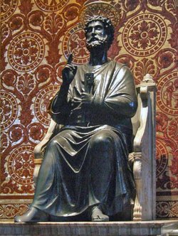
Also referred to as “benediction hand”, “Preacher`s hand”, “Papal Hand”, “Pope Blessing hand”, etc. This sign is linked to an ulnar nerve neuropathy, and there is a long time controversy about the name of this sign as well as its etiology, where some authors contend that there may be involvement of the median nerve.
Scholars, books and Internet sites are not clear about it. If there is an ulnar nerve neuropathy the person attempting to open his hand would be left with the second and third digits in extension, while the fourth and fifth digits would be flexed at the interphalangeal joints but extended at the metacarpophalangeal joints due the loss of the function of the interossei muscles and lumbrical muscles of the fourth and fifth digits.
The key for this sign is based in on the blessing act which is performed with an open hand and not with a fist. This is the reason why an ulnar neuropathy and not a median nerve neuropathy is the undelaying cause of the Papal Benediction Sign.
Presumably this term was the result of an injury of Saint Peter’s (the first Pope) ulnar nerve which caused him to bless using the “Preacher’s hand”. It stands to reason that everyone copied him as we can see in the lustrations and the art of the Catholic Church.
Article written by: Prof. Claudio R. Molina, MsC.
Sources:
1. Futterman, B. (2015). Analysis of the Papal Benediction Sign: The ulnar neuropathy of St. Peter. Clinical Anatomy (New York, N.Y.), 28(6), 696–701. Click here for the article
2. "From Vulcan Salute To Papal Blessing, Ulnar Nerve Damage Caused Original Benediction Sign | Box | NYIT". Nyit.edu. N.p., 2017. Web. 14 Jan. 2017
Image by By Mattana [Public domain], via Wikimedia Common: Click here for the link to the original image
- Details
We would like to welcome Professor Claudio R. Molina. MsC. as a contributor to Medical Terminology Daily.
Prof. Molina s a Physical Therapist, has a Masters degree in Biomedical Sciences, and is a Professor in the Human Anatomy Department of the Medical School at the Finis Terrae University in Santiago, Chile.
He is also a Postgraduate teacher for students of the “Anatomical Bases of Normal Imaging” diploma program at the Medical school of the same university.
Clinical Anatomy Associates, Inc is proud to have Dr. Cortés as a contributor to "Medical Terminology Daily" and as a consultant to our team.
- Details

Intended initially as a humorous view of anatomy, the "Anatomical Laws of Miranda" have a very serious objective. They support the fact that in every interventional case the operator should be extremely aware of the potential anatomical variations present. They also point to the fact that in real life, human anatomy does not look exactly like the anatomy books, models or prosections, and practice in the art of dissection and constant study are needed to ensure the proper identification of anatomical structures in surgery.
These laws are not original, they have been partially expressed at one time or another by several anatomists and surgeons, including Dr. Aaron Ruhalter and Dr. Robert Acland. What I have done is put them officially together and create the corollaries.
The anatomical laws of Miranda
- 1. The only constant in anatomy is variation
- 2. Nothing in the human body is really colored... or labeled
- 3. No anatomical structure has the moral obligation to be where they are supposed to be
There are corollaries to these laws and visitors to this web site are invited to provide us with their thoughts and addenda to the anatomical laws.
Corollaries:
1a. In the case of the so-called "anatomical constants"...law number 1 also applies
2.a. Black and white anatomy books are sometimes better to study than color atlases
2.b. Arteries are not red, nerves are not yellow, and lymphatic vessels are definitely not green!
2c. Nothing looks exactly like the anatomy books, computer simulations, or models. Food for thought for those medical schools that are eliminating dissection from their medical curricula.
3.a. This leads to that dreadful "Oooops!" sometimes heard in surgery
3.b. This also leads to the comment "It HAS to be around here!", which is dreadful if the "here" is a patient in surgery.


