
Medical Terminology Daily (MTD) is a blog sponsored by Clinical Anatomy Associates, Inc. as a service to the medical community. We post anatomical, medical or surgical terms, their meaning and usage, as well as biographical notes on anatomists, surgeons, and researchers through the ages. Be warned that some of the images used depict human anatomical specimens.
You are welcome to submit questions and suggestions using our "Contact Us" form. The information on this blog follows the terms on our "Privacy and Security Statement" and cannot be construed as medical guidance or instructions for treatment.
We have 667 guests online
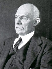
William S. Halsted, MD
(1852 – 1922)
American anatomist, teacher, and surgeon, William Stewart Halsted was born in New York City, USA to a wealthy family of English origin. His father was involved in charitable work and Governor and trustee to a city hospital. Not a brilliant student initially, Halsted took an undergraduate in Liberal Arts in Yale, CT., after which he entered the Medical College of Physicians at the Columbia College, where he excelled.
As a second-year medical student Halsted applied and obtained a position in surgery at a local hospital. In here he learned about Lister’s antiseptic technique and became an adamant proponent of it to reduce infection. In 1877 Halsted obtained his MD. After a short time as House Physician at the New York Hospital, Halsted traveled to Europe to further his education studying for two years at the Universities of Vienna, Leipzig, and W?rzburg.
Besides being at the forefront of surgical and antiseptic techniques (introducing the use of rubber gloves in surgery), Halsted was extremely concerned with the way medical students were taught in the US. He pioneered bedside clinical round discussions with the medical students after two years of basic sciences studies. Halsted developed the idea of a patient chart; he also developed the residency program for medical students in use today.
Halsted is probably the most influential researcher and surgeon at the turn of the century. He dedicated time to the study of intestinal anastomoses and the use of silk as a suture material. His experimental work in 1887 proved that the inclusion of the submucosa layer in an anastomosis was mandatory, as well that a single layered anastomosis was enough to attain closure. Perhaps Halsted’s most important contribution was the application and use of the scientific method to surgical questions. Halsted’s principles, also known as "Halsted's Rules of Surgery", set the standards used today in surgical suturing and surgical stapling.
He also pioneered the development and surgical techniques for radical mastectomy as a treatment for breast cancer.
As a side effect of this studied in anesthesia and the use of cocaine for anesthesia, Halsted became addicted to this substance, a problem that followed him through the years. Without impairing his capacity as a researcher and a surgeon, Halsted eventually recovered. He died in Baltimore in 1922 as a complication to surgery.
Sources:
1. Dubay, A. D., & Franz, G. M. (2003). Acute Wound Healing: The Biology of Acute Wound Failure. Surg Clin NA, 83, 463-481.
2. Halsted, W. S. (1887). Circular Suture of the Intestine - An Experimental Study. Am J Med Sci, 436-461.
3. “William Stewart Halsted: his life and contributions to surgery” Osborne, P. Lancet Oncol 2007; 8: 256–65
4. “William Stewart Halsted: Surgical pioneer” Burress, P Endoc Today (2010), 8: (2) 22
5. “William Stewart Halsted (1852–1922) Neurological stamp” Haas, LF J Neurol Neurosurg Psych 2000;69:641
Original image courtesy of "Images from the History of Medicine" at www.nih.gov

"Clinical Anatomy Associates, Inc., and the contributors of "Medical Terminology Daily" wish to thank all individuals who donate their bodies and tissues for the advancement of education and research”.
Click here for more information


- Details

Click for a larger image
Kernicterus is a disorder where excess bilirubin accumulates in the deep neural tissues of the brain and can cause brain damage in the newborn.
It is characterized by jaundice and a limpness of the newborn, devoid of energy. Can present with seizures, convulsions, and muscle spasms.
This condition is treatable and requires awareness from the parents if yellowness of the skin (jaundice) is detected along with the above-mentioned signs in the early days post-partum. There are other signs not mentioned in this article
The word [kernicterus] comes from the German word [kern], meaning “nucleus” or “core”. In this particular word the term kern refers to the fact that one of the most importantly affected brain structures in kernicterus are the basal nuclei of the brain (wrongly called the "basal ganglia"). It also includes the word [icterus] from the Greek word [ικτερός] pronounced (ikterós). The word [icterus] in Greek was originally used to denote a yellow bird, and is now used to denote the yellow color of jaundice.
We would like to thank diseasepictures.com for the image in this article. For additional information on neonatal jaundice, click here.
Sources:
1. Clayman, L. "The AMA Encyclopedia of Medicine" 1989. Random House, NY
2. “The Origin of Medical Terms” Skinner HA 1970 Hafner Publishing Co.
Thanks to Jackie Miranda-Klein for her contribution suggesting this word.
- Details
The term [parenchyma] is a Greek term (παράένχέω). Its origin and meaning have little relation to the medical use of the term. The word means "that what is poured" or to "pour in". The actual definition of the term is "the proper mass of a solid organ". If someone refers to the "liver parenchyma", they are referring to the hepatic tissue, so it is with any other solid organ.
The etymology of the word is obscure and reflects ancient physiological theories and history. Vesalius mentions that the word was introduced by Erasistratus circa 300BC. He thought that the blood was "poured" into the organ and then this poured fluid would congeal to form the organ's proper mass. With time this concept was abandoned, but the word persisted to its modern meaning.
Interesting, there are many which accentuate the word wrongly. The accent or stress should be on the letter "e" and not on the letter 'y", so it should be pronounced "parénchyma"
- Details

Layers of the GI tract
The gastrointestinal (GI) tract is formed, with a few exceptions, by four concentric layers of tissue. These are, from deep to superficial, the mucosa, submucosa, muscular (or muscularis) and the serosa layers. This is the simplified version. The fact is that there are more sublayers.
The mucosa layer is characterized by the presence of intestinal villi, which in the stomach and small intestine contribute to absorption of the digested food. The mucosa has a thin layer of connective called the "lamina propia" and external to it a thin layer of smooth muscle, the muscularis mucosae.
The submucosa layer is formed by irregular connective tissue and contains on its most external region a plexus of nerves and neurons, the "submucosal plexus of Meissner", which provides parasympathetic innervation to glands and the muscularis mucosae.
The muscular layer, also known as the "muscularis" is composed of two sublayers of smooth muscle. The deep layer contains circular fibers and is known either as the "circular muscle layer" or the "muscularis interna", the superficial layer contains longitudinal smooth muscle fibers and is known as the "longitudinal muscle layer" or the muscularis externa. Between both muscle layers lies the "myenteric plexus of Auerbach", a layer of sympathetic and parasympathetic nerves and neurons that provides nerve supply to the muscular layer. The combined action of this plexus on the muscular layer is responsible for peristalsis.
The serosa layer is the outer or external layer and is formed by a layer of peritoneum. As such, this layer can also be called "visceral peritoneum".
There are variations from GI organ to GI organ in the arrangement, content, glands, thickness of the layers, etc. The most important differences can be found in the thoracic esophagus and most of the rectum which are devoid of a serosa layer, and in the stomach, where there is a third muscular layer, deep to the circular layer, called the "oblique layer" that contributes fibers to the lower esophageal sphincter found at the esophagogastric junction.
An important point to make is the presence of two interconnected ganglionated plexuses that are represented in the GI tract by the submucosal plexus of Meissner and the myenteric plexus of Auerbach which form the GI intrinsic autonomic nervous component . These two plexuses extend from the esophagus to the rectum and allow for the GI tract to operate almost independently from the extrinsic autonomic nervous system which moderates their activity. Ganglionated plexuses are present in organs that have rhythmic activity, such as peristalsis. Ganglionated plexuses are also present in the heart.
Sources:
1. "The bowel and beyond: the enteric nervous system in neurological disorders" Rao, M & Gershon, M. Nat Rev Gastroenterol Hepatol. 2016 Sep; 13(9): 517–528.
2. "Advances in Enteric Neurobiology: The “Brain” in the Gut in Health and Disease" Kulkami, S et al. Journal of Neuroscience 31 October 2018, 38 (44) 9346-9354
3. "The Brain-Gut Connection" John Hopklins Health
4. "Think Twice: How the Gut's "Second Brain" Influences Mood and Well-Being" Hadhazy, B. Scientific American February 2010
Images property of:CAA.Inc.Artist:Dr. E. Miranda
- Details

Click for a larger image
UPDATED: The word [manubrium] is Latin and mean "handle", referring to the area where a person holds an instrument or device. To exemplify this, in Spanish the vernacular use of the word [manubrio] refers to the handles of bicycle or even the steering wheel of a car.
In anatomy, the term is used with the same meaning. In the malleus, a hammer-like ossicle of the middle ear, the manubrium is the handle-like extension of the bone that attaches to the tympanic membrane.
In the case of the sternum, the [manubrium sterni] is the superior portion bound by the sternal angle (of Louis) inferiorly. The use of the word manubrium can be explained because in early anatomy, the sternum was known by the Latin term [gladius] referring to the similarity of the sternum to the short sword of the gladiators. The area where you hold the sword is the handle, ergo, manubrium.
The manubrium has a superior and median notch called the "suprasternal notch" or the "jugular notch". It is important because in the case of a mediastinoscopy, the incision is made just superior to this landmark. The manubrium articulates superolaterally with the clavicle and inferolaterally with the superior aspect of the cartilage of the second rib. The rest of the rib cartilage articulates with the body of the sternum.
Image property of:CAA.Inc.. Artist: Mark J. Zuptich
- Details
The word [induration] arises from the Latin words induratio, meaning "thick or hard" and indurare, meaning "hardening".
It refers to a pathological hardening of tissues caused by tumoration or edema, increase of fibrous or connective tissue, or other causes. It is a good, descriptive term when stating a patient's symptoms. The term has been in use in English since the 14th century.
Note: The links to Google Translate include an icon that will allow you to hear the pronunciation of the word.
- Details
At the beginning of 1998, Clinical Anatomy Associates, Inc. was formed as an Ohio Corporation. Our mission is to deliver industry relevant, cutting-edge Training, Marketing, and R&D services that will enable our clients to gain a competitive advantage. Over the past two decades, Clinical Anatomy Associates, Inc. has become the go-to R&D resource for feasibility studies that require cadaver studies and anatomical research. We are also a preferred training solution for Sales Representatives, Distributors, Engineers, Clinicians, and Marketing Managers in the areas of Medical Terminology, Clinical Anatomy, and Surgical Procedures. Our expertise allows us to deliver training in a variety of medical and anatomical topics.
In 2012 Dr. Efrain A. Miranda, CEO of Clinical Anatomy Associates started "Medical Terminology Daily" (MTD), a website/blog as a service to the medical community, medical students, and the medical industry. MTD posts medical or surgical terms, its meaning and usage, as well as biographical notes on anatomists, surgeons, and researchers through the ages. These posts are also shared on Facebook to a group of followers.
20 year anniversary for Clinical Anatomy Associates, Inc. and 6 years for Medical Terminology Daily! Help us congratulate our staff and specially the contributors and friends of Medical Terminology Daily.
Our thanks to all our customers, friends, and contributors for an amazing 20 years!!! Looking forward to more!!