Medical Terminology Daily (MTD) is a blog sponsored by Clinical Anatomy Associates, Inc. as a service to the medical community. We post anatomical, medical or surgical terms, their meaning and usage, as well as biographical notes on anatomists, surgeons, and researchers through the ages. Be warned that some of the images used depict human anatomical specimens.
You are welcome to submit questions and suggestions using our "Contact Us" form. The information on this blog follows the terms on our "Privacy and Security Statement" and cannot be construed as medical guidance or instructions for treatment.
We have 305 guests online

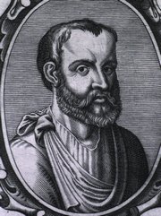
Georg Eduard Von Rindfleisch
(1836 – 1908)
German pathologist and histologist of Bavarian nobility ancestry. Rindfleisch studied medicine in Würzburg, Berlin, and Heidelberg, earning his MD in 1859 with the thesis “De Vasorum Genesi” (on the generation of vessels) under the tutelage of Rudolf Virchow (1821 - 1902). He then continued as a assistant to Virchow in a newly founded institute in Berlin. He then moved to Breslau in 1861 as an assistant to Rudolf Heidenhain (1834–1897), becoming a professor of pathological anatomy. In 1865 he became full professor in Bonn and in 1874 in Würzburg, where a new pathological institute was built according to his design (completed in 1878), where he worked until his retirement in 1906.
He was the first to describe the inflammatory background of multiple sclerosis in 1863, when he noted that demyelinated lesions have in their center small vessels that are surrounded by a leukocyte inflammatory infiltrate.
After extensive investigations, he suspected an infectious origin of tuberculosis - even before Robert Koch's detection of the tuberculosis bacillus in 1892. Rindfleisch 's special achievement is the description of the morphologically conspicuous macrophages in typhoid inflammation. His distinction between myocardial infarction and myocarditis in 1890 is also of lasting importance.
Associated eponyms
"Rindfleisch's folds": Usually a single semilunar fold of the serous surface of the pericardium around the origin of the aorta. Also known as the plica semilunaris aortæ.
"Rindfleisch's cells": Historical (and obsolete) name for eosinophilic leukocytes.
Personal note: G. Rindfleisch’s book “Traité D' Histologie Pathologique” 2nd edition (1873) is now part of my library. This book was translated from German to French by Dr. Frédéric Gross (1844-1927) , Associate Professor of the Medicine Faculty in Nancy, France. The book is dedicated to Dr. Theodore Billroth (1829-1894), an important surgeon whose pioneering work on subtotal gastrectomies paved the way for today’s robotic bariatric surgery. Dr. Miranda.
Sources:
1. "Stedmans Medical Eponyms" Forbis, P.; Bartolucci, SL; 1998 Williams and Wilkins
2. "Rindfleisch, Georg Eduard von (bayerischer Adel?)" Deutsche Biographie
3. "The pathology of multiple sclerosis and its evolution" Lassmann H. (1999) Philos Trans R Soc Lond B Biol Sci. 354 (1390): 1635–40.
4. “Traité D' Histologie Pathologique” G.E.
Rindfleisch 2nd Ed (1873) Ballieres et Fils. Paris, Translated by F Gross
"Clinical Anatomy Associates, Inc., and the contributors of "Medical Terminology Daily" wish to thank all individuals who donate their bodies and tissues for the advancement of education and research”.
Click here for more information
- Details
Kabourophobia is the fear of crabs and lobsters.
The etymology of the word [kabourophobia] comes from the Greek word [καβουρης] (pronounced “kavouris”), meaning [crab], and the suffix [-phobia], also from the Greek, arises from the word [φοβία] (pronounced “fovía”)
Kabourophobia is an extremely rare phobia, but it was brought to the public’s attention when a modern pop singer stated that she was afraid of crabs. Also, a prank (maybe acted) was shown on video on the internet with a man surrounded by lobsters.
Kabourophobia is very specific, and it can also be a part of a wider phobia called ostraconophobia, which is the fear of crustaceans, adding shrimp, oysters, clams, crabs, lobsters, etc.
An interesting point is that the word [crab] in Greek has another acception, that is the word [Καρκίνος] (pronounced “karkinos”), which is the root for the medical term [cancer].
We thank Jackie Miranda-Klein for her contribution suggesting this word.
- Details

Galen of Pergamon
(129AD - 200AD
The word sympathetic is the adjectival form of sympathy. This word arises from the Greek [συμπάθεια]and is composed of [syn/sym] meaning “together” and [pathos], a word which has been used to mean “disease”. In reality “pathos” has to do more with the “feeling of self”. Based on this, the word sympathy means “together in feeling”, which is what we use today.
How the term got to be used to denote a component of the so-called autonomic nervous system is part of the history of Medicine and Anatomy.
Galen of Pergamon (129AD-200AD), whose teachings on Medicine and Anatomy lasted as indisputable for almost 1,500 years, postulated that nerves were hollow and allowed for “animal spirits” to travel between organs and allowed the coordinated action of one with the other, in “sympathy” with one another. As the knowledge of the components of the nervous system grew, this concept of “sympathy” stayed, becoming a staple of early physiological theories on the action of the nervous system.
Jacobus Benignus Winslow (1669-1760) named three “sympathetic nerves” one of them was the facial nerve (the small sympathetic), the other the vagus nerve, which he called the “middle sympathetic”, and the last was what was known then as the “intercostalis nerve of Willis” or “large sympathetic", today’s sympathetic chain. Other nerves that worked coordinated with this “sympathetics” were considered to work in parallel with it. It is from this concept that the term “parasympathetic” arises.
Interestingly, the ganglia on the sympathetic chain were for years known as “small brains” and it was postulated that there was a separate multi-brain system coordinating the action of the thoracic and abdominopelvic viscera. The coordination between this “autonomous nervous system” and the rest of the body was made by way of the white and gray rami communicantes.
Today we know that there is only one brain and only one nervous system with an autonomic component which has a “sympathetic” component that is mostly in charge of the “fight or flight” reaction and a “parasympathetic” component that has a “slow down” or “depressor” function. Both work coordinated, so I guess Galen was not "off the mark" after all.
So, we still use the terms “sympathetic” and “parasympathetic”, but the origin of these terms has been blurred by history.
Sources:
1. "Claudius Galenus of Pergamum: Surgeon of Gladiators. Father of Experimental Physiology" Toledo-Pereyra, LH; Journal of Investigative Surgery, 15:299-301, 2002
2. "The Origin of Medical Terms" Skinner, HA 1970 Hafner Publishing Co.
3. "Medical Meanings:A Glossary of Word Origins" Haubrish, WS American College of Physicians Philadelphia, 1997
4. "The History of the Discovery of the Vegetative (Autonomic) Nervous System" Ackerknecht, EH Medical History, 1974 Vol 18.
Original image courtesy of Images from the History of Medicine at nih.gov
Note: The links to Google Translate include an icon that will allow you to hear the pronunciation of the word.
- Details
The medical term [epistaxis] refers to a “nose bleed”.
It is considered to be a Modern Latin term that originates from the Greek word [επίσταξη] (epístaxí). The word is composed of [επί] [epi-] meaning "on", "upon", or "above", and [στάζει] (stázei), meaning "in drops", "dripping".
The term was first used by Hippocrates, but only as [στάζει] , to denote dripping of the nose, and was later changed to [επίσταξη] to denote “dripping upon”. The term itself does not include or denote that the blood loss is from the nose, but its meaning has been implied and accepted for centuries. The plural form for epistaxis is epistaxes.
Skinner (1970) says that the term was first used in English in a letter by Thomas Beddoes (1760-1808) in a letter to Robert W. Darwin (1766-1848) in 1793. Robert Darwin was an English physician, father or Charles Darwin (1809-1882) author of “The Origin of the Species”.
Sources:
1. "The Origin of Medical Terms" Skinner, HA 1970 Hafner Publishing Co.
2. "Medical Meanings - A Glossary of Word Origins" Haubrich, WD. ACP Philadelphia
Note: The links to Google Translate include an icon that will allow you to hear the pronunciation of the word.
- Details
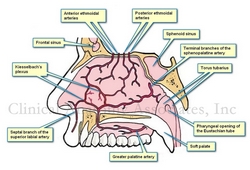
Kiesselbach's plexus is named after Dr. Wilhelm Kiesselbach (1839 – 1902), a German otolaryngologist. It is an area in the anteroinferior aspect of the nasal septum where several arteries from different origins meet and anastomose.
This arterial plexus is also known as the "locus Kiesselbachii, Kiesselbach's triangle, or Little's plexus, or Little's area. This area of the anteroinferior nasal septum has a propensity for epistaxis or nasal bleeding. In fact, close to 90% of nose bleeds (epistaxes) happen in this area.
There is a secondary area where epistaxis may happen, but this is a venous nose bleed. This is Woodruff's plexus, a venous plexus found in the posterior aspect of inferior turbinate on the lateral wall of the nose.
Thanks to Jackie Miranda-Klein for suggesting this post.
- Details
- Written by: Efrain A. Miranda, Ph.D.
This article is part of the series "A Moment in History" where we honor those who have contributed to the growth of medical knowledge in the areas of anatomy, medicine, surgery, and medical research.

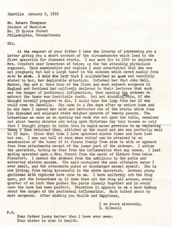
Letter from Ephraim McDowell
to Robert Thompson
As some of you may know by now, I am a collector of antique medical books and books that relate to the history of anatomy and medicine.
As important as the books themselves are, there are details beyond the book itself that can take hours of my time doing research. The first one is the bookplates (also known as Ex-Libris). The have been used for centuries by book owners and collectors to identify the books in their collections, a tradition that seems to be falling in disuse. Not me, I have one that you can see here. Some of these can lead to places that you cannot imagine initially. One of these bookplates took me to research a potential resident ghost in a library!
Provenance is also important. Where was it printed? Who owned it? Who was the illustrator? etc. I recently acquired a second copy of the book “EPHRAIM MCDOWELL, FATHER OF OVARIOTOMY AND FOUNDER OF ABDOMINAL SURGERY. With an Appendix on JANE TODD CRAWFORD”. By AUGUST SCHACHNER, M.D. Cloth, 8vo.A p. 33I. Philadelphia, J. B. Lippincott CO., I921. Dr. McDowell has also been featured in this blog in the series "A Moment in History".
This second copy is most valuable because of the papers found within the book. There is a series of notes, newspaper clippings, and copies of letters! Here is a detail of what I have found:
The book seems to have belonged to Cecil Stryker, MD.,a physician in Cincinnati, and one of the founders of the American Diabetes Association (ADA). There is a copy of a letter by Dr. Ephraim McDowell to Dr. Robert Thompson (Sr.) dated January 2nd, 1829, a year before Dr. McDowell's death. The letter is shown in the image attached. In this letter Dr. McDowell describes in his own words the ovariotomy he performed on Jane Todd. He also describes other ovariotomies he performed and his opinion on "peritoneal inflammation".
There is a note from Dr. Cecil Striker to "Bob" dated 6/3/73 when he gifted a copy of this book. In the note Dr. Striker explains that he bought several copies of the book and he is sending this copy to him. There is also a copy of Dr. McDowell's prayer (costs 25 cents), and a page of the Kentucky Advocate newspaper published in Danville, KY and dated Sunday April 15, 1973 on the restoration of Dr. Alban Goldsmith home, a surgeon who assisted Dr. McDowell in his first ovariotomy (first in the world, that is).
Last, there is a note dated September 16, 1974 from the wife of Dr. West T. Hill, Chairman of the Dramatic Arts Department at Centre College in Danville, Kentucky. In this handwritten note she mentions the McDowell family reunion that took place on June 15 and 16, 1974 in Danville. With the note comes the program and registration form for the festivities! Dr. West T. Hill was one of the many responsible for the restoration of the MacDowell Home and Museum. Today Danville has the West T. Hill community theatre that honors his name.
All of this in one book, as I always say "You know where you are going to start reading it, but you never know where are you going to end in researching it". This book will be a great addition to my library catalog. Dr. Miranda.
- Details
Rudi Coninx, MD is a physician and Chief a.i. Humanitarian policy and Guidance at the World Health Organization (WHO), based in Geneva. He obtained his MD from the University of KU Leuven Belgium, a Doctorate in Tropical Medicine from the Prins Leopold Instituut voor Tropische Geneeskunde, and an MPH from the John Hopkins University School of Medicine.
His CV shows more than twenty five years of national and international experience in policy and strategy development and analysis, policy dialogue, technical advice and program management support to countries and WHO country offices. Considerable experience in strengthening WHO country offices and in working with partners and networks at the global as well as filed level. Coordinated the WHO Country Focus Policy for more than five years and worked as a member in various strategic planning, decentralization, and global and regional partnership groups, including national and international committees, taskforces. Published several articles on policy analysis, management and health and development in regional and international journals.
He is also an Associate Faculty, Bloomberg School of Public Health, Johns Hopkins University, USA.
He has held a series of positions with the International Committee of the Red Cross, and also within the World Health Organization. His LinkedIn profile can be found here.
Thanks to Dr. Coninx for taking time of his busy schedule and collaborating with "Medical Terminology Daily" with the article "Did Andreas Vesalius really die from scurvy?" which he co-authored with Theo Dirix. We look forward to his future writings in this blog.


