Medical Terminology Daily (MTD) is a blog sponsored by Clinical Anatomy Associates, Inc. as a service to the medical community. We post anatomical, medical or surgical terms, their meaning and usage, as well as biographical notes on anatomists, surgeons, and researchers through the ages. Be warned that some of the images used depict human anatomical specimens.
You are welcome to submit questions and suggestions using our "Contact Us" form. The information on this blog follows the terms on our "Privacy and Security Statement" and cannot be construed as medical guidance or instructions for treatment.
We have 467 guests online

Jean George Bachmann
(1877 – 1959)
French physician–physiologist whose experimental work in the early twentieth century provided the first clear functional description of a preferential interatrial conduction pathway. This structure, eponymically named “Bachmann’s bundle”, plays a central role in normal atrial activation and in the pathophysiology of interatrial block and atrial arrhythmias.
As a young man, Bachmann served as a merchant sailor, crossing the Atlantic multiple times. He emigrated to the United States in 1902 and earned his medical degree at the top of his class from Jefferson Medical College in Philadelphia in 1907. He stayed at this Medical College as a demonstrator and physiologist. In 1910, he joined Emory University in Atlanta. Between 1917 -1918 he served as a medical officer in the US Army. He retired from Emory in 1947 and continued his private medical practice until his death in 1959.
On the personal side, Bachmann was a man of many talents: a polyglot, he was fluent in German, French, Spanish and English. He was a chef in his own right and occasionally worked as a chef in international hotels. In fact, he paid his tuition at Jefferson Medical College, working both as a chef and as a language tutor.
The intrinsic cardiac conduction system was a major focus of cardiovascular research in the late nineteenth and early twentieth centuries. The atrioventricular (AV) node was discovered and described by Sunao Tawara and Karl Albert Aschoff in 1906, and the sinoatrial node by Arthur Keith and Martin Flack in 1907.
While the connections that distribute the electrical impulse from the AV node to the ventricles were known through the works of Wilhelm His Jr, in 1893 and Jan Evangelista Purkinje in 1839, the mechanism by which electrical impulses spread between the atria remained uncertain.
In 1916 Bachmann published a paper titled “The Inter-Auricular Time Interval” in the American Journal of Physiology. Bachmann measured activation times between the right and left atria and demonstrated that interruption of a distinct anterior interatrial muscular band resulted in delayed left atrial activation. He concluded that this band constituted the principal route for rapid interatrial conduction.
Subsequent anatomical and electrophysiological studies confirmed the importance of the structure described by Bachmann, which came to bear his name. Bachmann’s bundle is now recognized as a key determinant of atrial activation patterns, and its dysfunction is associated with interatrial block, atrial fibrillation, and abnormal P-wave morphology. His work remains foundational in both basic cardiac anatomy and clinical electrophysiology.
Sources and references
1. Bachmann G. “The inter-auricular time interval”. Am J Physiol. 1916;41:309–320.
2. Hurst JW. “Profiles in Cardiology: Jean George Bachmann (1877–1959)”. Clin Cardiol. 1987;10:185–187.
3. Lemery R, Guiraudon G, Veinot JP. “Anatomic description of Bachmann’s bundle and its relation to the atrial septum”. Am J Cardiol. 2003;91:148–152.
4. "Remembering the canonical discoverers of the core components of the mammalian cardiac conduction system: Keith and Flack, Aschoff and Tawara, His, and Purkinje" Icilio Cavero and Henry Holzgrefe Advances in Physiology Education 2022 46:4, 549-579.
5. Knol WG, de Vos CB, Crijns HJGM, et al. “The Bachmann bundle and interatrial conduction” Heart Rhythm. 2019;16:127–133.
6. “Iatrogenic biatrial flutter. The role of the Bachmann’s bundle” Constán E.; García F., Linde, A.. Complejo Hospitalario de Jaén, Jaén. Spain
7. Keith A, Flack M. The form and nature of the muscular connections between the primary divisions of the vertebrate heart. J Anat Physiol 41: 172–189, 1907.
"Clinical Anatomy Associates, Inc., and the contributors of "Medical Terminology Daily" wish to thank all individuals who donate their bodies and tissues for the advancement of education and research”.
Click here for more information
- Details

Surface anatomy art by Danny Quirk - with permission
UPDATED: Surface anatomy is a subset of human anatomy that studies the relationship of external anatomical landmarks and deeply situated structures. These landmarks are created by cartilage, bones, tendons and muscles. In some cases they may be caused by a hypertrophic or diseased organ. Palpation is and art and my favorite book on the subject is the "Trail Guide to the Body" series by Andrew Biel
Surface anatomy is a core clinical skill for physicians, physical therapists and other health care professionals (HCP), as palpation in specific locations can lead to rapid diagnosis in certain cases. It is also used in surgery to determine the appropriate place for incisions or insertion of a trocar in the case of minimally invasive surgery.
It is true that individual habitus and anatomical variations must make the HCP wary of potential mistakes, but in the majority of cases these anatomical landmarks and diagnoses are correct.
Many book chapters have been written on the topic, but unfortunately, the need to dedicate time to modern discoveries have reduced the time spent on this wonderful learning tool to the point that modern books of anatomy barely touch upon the subject. Many feel today that the need for this tool has been made irrelevant by the bed-side availability of ultrasound imaging. I have to agree with Standring (2012) when she states “I am not convinced that surface anatomy is under growing threat from modern imaging technology…” There is and always will be the need for applied surface anatomy, although maybe not in the way and depth it was used in the past when imaging technology was scarce and expensive.
In order to make the importance of surface anatomy relevant in medical schools, some have added body painting as a tool in their anatomical curriculum with great results. Will it be used everywhere? I doubt it, but here again is a link between human anatomy and art, which our contributor Pascale Pollier presents through her art in "Artem Medicalis".
The artwork in this article is by Danny Quirk. Click on the image for a larger depiction. The video links are for the RMIT University Muscle Man and the Skeletal Man body paintings from the class by Dr. Claudia Diaz.
Personal note: One of the best book chapters on Surface Anatomy was written in "The Anatomical Basis of Clinical Practice" by Becker, Wilson and Gehweiler (1971). Unfortunately, although the content is very good, the images used, the tongue-in-cheek humor, and writing style used by the authors, plus the times at which the book was published forced the publisher stop the sales of this book. It was banned from use in medical colleges through the country. Known to many as the "green book" because of its cover, it is today considered a collector's curiosity. In the "sources" section of this article you can find a links to a journal article on the subject as well as images of the book. Dr. Miranda.
Sources:
1. “Evidence-Based Surface Anatomy” Standrig, S J Clin Anat (2012) 25:813–815
2. “Giving Color to a New Curriculum: Body paint as a Tool in Medical Education” Den Akker, JW. Et al J Clin Anat (2002) 15:356–362
3. “Body-Painting: A Tool Which Can Be Used to Teach Surface Anatomy” Nanjundaiah, K. J Clin Diag Res (2012) October, Vol-6(8): 1405-1408 Copy of the article here
4. “RMIT students paint anatomical man into human textbook” RMIT Dr. Diaz, C. Copy of the article here
5. “Should We Use Body Painting to Teach Anatomy?” Gambino, M article at Smithsonian.com
6. “The role of Fresh Tissue Dissection and Anatomic Body Painting in Anatomy Education” Bennet, C. PPT presentation on PDF here
7. " The anatomical Basis of Medical Practice" Becker, RF. Wilson, JW, Gehweiler, JA 1971, Baltimore, Williams & Wilkins
8. "The Pornographic Anatomy Book? The Curious Tale of The Anatomical Basis of Medical Practice" Halperin EC, Acad Med (2009) 84:2; 278-283 Article here
9. "The Objectification of Female Surface Anatomy" Ruiz, V. Internet article. Click here
- Details
UPDATED: In surface anatomy the abdomen can be divided into nine regions by named lines (or planes): transpyloric, transtubercular, and midclavicular. These regions have specific visceral content.
• Hypochondriac regions (right and left): [Hypo]="below"; [chondr]="cartilage"; [iac]=”pertaining to”. In this context, the term means “below or deep to the cartilage (of the ribs)". The right hypochondriac region contains the liver, gallbladder, portal vein, and the right colic flexure. The left hypochondriac region contains the stomach, spleen, tail of the pancreas, and left colic flexure. For a detail on how the name of this region relates to a mental disorder, click here.
• Epigastric region: [Epi]="above"; [gastr]="stomach”; [ic]=”pertaining to". The term means “above the stomach”. This region contains mostly stomach and abdominal esophagus
• Lumbar regions (right and left): Right and left lumbar regions. The term [lumbar] refers to the area of the loins. The right lumbar region contains the ascending colon and part of the right kidney. The left lumbar region contains the descending colon and part of the left kidney
• Umbilical region: Centered around the umbilicus, this region contains mostly small intestine, abdominal aorta, and greater omentum
• Inguinal regions (right and left): From the Latin [inguen]=”groin". Gaius Plinius Secundus aka “Pliny” (23-79 AD) first used this term naming after a plant (inguinalis) which he used to treat hernias of the groin. The right inguinal region contains the cecum and small intestine. The left inguinal region contains the sigmoid colon. In older days, as shown in the sketch, these regions were called the "iliac regions".
• Hypogastric region: [Hypo]="below"; [gastr]="stomach”; “ic”=”pertaining to. The term means “below the stomach”. The hypogastric region contains mostly small intestine and greater omentum. This region used to be called the "pubic region"
A clinical importance of these abdominal regions is that a ventral hernia is usually named by the anatomical region where it protrudes. Based on the image, you will see then why a hernia can be umbilical, inguinal, lumbar, epigastric, or hypogastric.
Sources:
1. "Clinical Anatomy" Brantigan, OC 1963 McGraw Hill
2. "Tratado de Anatomia Humana" Testut et Latarjet 8th Ed. 1931 Salvat Editores, Spain
3. Davis, Gwilym G. "Applied Anatomy: The Construction of the Human Body Considered in Relation to Its Functions, Diseases, and Injuries"; Philadelphia: J.B. Lippincott Co., 1910
Image modified from the original Davis, 1910
- Details
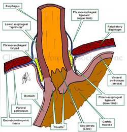
UPDATED: The [phrenoesophageal ligament] or phrenoesophageal membrane is part of a complex system that closes off the esophageal hiatus, one of the seven hiatuses in the respiratory diaphragm, preventing the herniation of abdominal structures into the thoracic mediastinum.
The connective tissue layer called the endoabdominopelvic fascia, which lines the inner aspect of the abdominopelvic cavity, is found as a "glue" between the respiratory diaphragm and the parietal peritoneum. At this point the endoabdominopelvic fascia is called the "infradiaphragmatic fascia".
When the infradiaphragmatic fasia gets to the edge of the esophageal hiatus, it splits into ascending and a descending components or limbs. These are the superior and inferior phrenoesophageal ligaments or phrenoesophageal membranes. These phrenoesophageal ligaments create a circular disc-like plug between the abdomen and the thorax. This "plug" is reinforced by a infradiaphragmatic fat pad found internal to the phrenoesophageal ligament.
The phrenoesophageal ligaments are reinforced externally. On their thoracic aspect by the endothoracic fascia, and on the abdominal side, by parietal peritoneum.
The ascending limb fuses superiorly with the esophageal fascia, which lines the external aspect of the longitudinal muscle of the esophagus, as the thoracic esophagus does not have a serosa layer. The descending limb fuses inferiorly with the esophageal fascial covering of the longitudinal ligament as it is covered by the peritoneum . Failure of the phrenoesophageal ligaments can predispose to esophageal hiatus hernia.
- Details
The following article is embedded from our Facebook page https://www.facebook.com/CAAInc.
This year the 2018 meeting of the American Association of Clinical Anatomists is being held in Atlanta, GA., at the Grand Hyatt Buckhead Hotel and Conference Center. The program is full of interesting topics and is already a hit with all the attendees. Looking forward to the program.
- Details
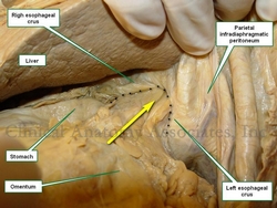
Esophageal hiatus hernia in situ.
The arrow points to stomach and greater
omentum herniating into the thorax
UPDATED: An esophageal hiatus hernia (also known as a hiatal hernia) is caused by a dilation of the esophageal hiatus and its component structures, the phrenoesophageal membranes (ligaments).
Since the intraabdominal pressure is higher than the intrathoracic pressure, abdominal contents -usually stomach and greater omentum- can herniate through the dilated esophageal hiatus into the mediastinum, the central region of the thoracic cavity. This presents as a hernia sac whose walls are formed by endothoracic fascia, phrenoesophageal membranes and parietal peritoneum.
There are two main types of esophageal hiatus hernias. Type I is known as a "sliding hiatal hernia" and is characterized by a complete ascension of the esophagogastric junction and abdominal esophagus into the thoracic hernia sac. This is usually accompanied by a typical "hourglass image" in a radiographic assessment, and also presents with gastroesophageal reflux disease (GERD). Type I esophageal hiatus hernias are more common.
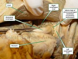
Esophageal hiatus hernia, reduced.
The dotted line shows the edge of
the enlarged esophageal hiatus
Type II esophageal hiatus hernia is known as a "paraesophageal hernia" and represent about 5 - 15% of esophageal hiatus hernias. In this case, the esophagogastric junction maintains its anatomical position inferior to the respiratory diaphragm, but the fundus and body of the stomach, along with some greater omentum herniate alongside the esophagus into the mediastinal region of the thoracic cavity. Although there can be GERD, this type of hernia usually presents with little symptomatology, and when it does, symptoms are related to ischemia or partial to complete obstruction. There are variations of type II hernia, which are classified as Type III and IV. Type IV, although rare, will include other viscera in the hernia sac, including colon, spleen, or even small intestine.
The accompanying images above depict a Type I esophageal hiatus hernia. The superior image shows the hernia in situ where the stomach and greater omentum are still in the hernia sac. The inferior image shows the contents reduced and the abdominal esophagus being pulled into the abdominal cavity. The dotted line shows the dilated esophageal hiatus that needs to be repaired to prevent recurrence of the pathology.
Click on this link for additional information on esophageal hiatus hernia surgery.
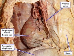
The image below answers a question by Victoria Guy Ratcliffe, who asked via Facebook "What would it be if it feels like you've got a blockage right at the level of the heart? That's too high for a hiatal hernia, isn't it?" The image answers the question. It shows a dissection of the left side of the thorax. The anterior thoracic wall and the left lung have been removed. The heart is immediately superior and anterior to the esophageal hiatus, and the hernia sac of a Type I esophageal hiatus hernia is seen immediately posterior and in contact with the heart. Whether this means that you will "feel" the hernia, it is up for debate, as all these structures have visceral innervation. Most probably, a well-developed Type II esophageal hiatus hernia might interfere with swallowing at this level, causing the sensation she mentions. Thanks for the question, Tori.
For additional information:
"Approaches to the Diagnosis and Grading of Hiatal Hernia" Kahrilas et al Best Pract Res Clin Gastroenterol. 2008 ; 22(4): 601–616.
- Details
The term [hiatus] derives from the Latin word [hiare], meaning to "gape" or to "yawn". In human anatomy this term is used to mean an "opening" or a "defect". It must be pointed out that in anatomy (and surgery) the term "defect" does not necessarily mean "defective". In most cases a "defect" is a normal opening in a structure, such as the esophageal hiatus. The plural form is either [hiatus] or [hiatuses].
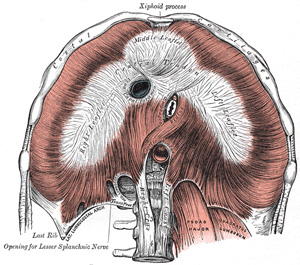
In the case of the respiratory diaphragm, there are seven such openings, seven normal hiatuses. On top of this, you can find an abnormal opening caused by incomplete congenital closure of the dome of the diaphragm, a congenital diaphragmatic hernia (CDH), also known as Bochdalek's hernia, found in the posterior aspect of the respiratory diaphragm.
The seven hiatuses of the respiratory diaphragm are:
• Aortic hiatus
• Inferior vena cava hiatus
• Hiatuses (2) for the superior epigastric vessels, which are the inferior continuation of the internal thoracic (mammary) vessels. Also known as the hiatuses of Morgagni. A hernia in a newborn through this hiatus is also considered a CDH.
• Hiatuses (2) for the splanchnic nerves
Based on the above it is wrong (maybe not wrong, but incomplete) to say that a patient has a "hiatal hernia", as the term does not include which hiatus is involved. In fact the hernia of Morgagni is also a "hiatal hernia" as the hernia passes through a normal defect in the respiratory diaphragm. Come to think of it, it could also be a hernia in a hiatus somewhere else in the body, such as a hernia of Schwalbe, a type or pelvic diaphragm hernia.
Note: Thanks to DHREAMS of the Columbia University Medical Center for the link on CDH.
Sources:
1. "The Origin of Medical Terms" Skinner, HA 1970 Hafner Publishing Co.
2. "Medical Meanings - A Glossary of Word Origins" Haubrich, WD. ACP Philadelphia
3 "Tratado de Anatomia Humana" Testut et Latarjet 8 Ed. 1931 Salvat Editores, Spain
4. "Anatomy of the Human Body" Henry Gray 1918. Philadelphia: Lea & Febiger Image modified by CAA, Inc. Original image by Henry Vandyke Carter, MD., courtesy of bartleby.com


