Medical Terminology Daily (MTD) is a blog sponsored by Clinical Anatomy Associates, Inc. as a service to the medical community. We post anatomical, medical or surgical terms, their meaning and usage, as well as biographical notes on anatomists, surgeons, and researchers through the ages. Be warned that some of the images used depict human anatomical specimens.
You are welcome to submit questions and suggestions using our "Contact Us" form. The information on this blog follows the terms on our "Privacy and Security Statement" and cannot be construed as medical guidance or instructions for treatment.
We have 496 guests online

Jean George Bachmann
(1877 – 1959)
French physician–physiologist whose experimental work in the early twentieth century provided the first clear functional description of a preferential interatrial conduction pathway. This structure, eponymically named “Bachmann’s bundle”, plays a central role in normal atrial activation and in the pathophysiology of interatrial block and atrial arrhythmias.
As a young man, Bachmann served as a merchant sailor, crossing the Atlantic multiple times. He emigrated to the United States in 1902 and earned his medical degree at the top of his class from Jefferson Medical College in Philadelphia in 1907. He stayed at this Medical College as a demonstrator and physiologist. In 1910, he joined Emory University in Atlanta. Between 1917 -1918 he served as a medical officer in the US Army. He retired from Emory in 1947 and continued his private medical practice until his death in 1959.
On the personal side, Bachmann was a man of many talents: a polyglot, he was fluent in German, French, Spanish and English. He was a chef in his own right and occasionally worked as a chef in international hotels. In fact, he paid his tuition at Jefferson Medical College, working both as a chef and as a language tutor.
The intrinsic cardiac conduction system was a major focus of cardiovascular research in the late nineteenth and early twentieth centuries. The atrioventricular (AV) node was discovered and described by Sunao Tawara and Karl Albert Aschoff in 1906, and the sinoatrial node by Arthur Keith and Martin Flack in 1907.
While the connections that distribute the electrical impulse from the AV node to the ventricles were known through the works of Wilhelm His Jr, in 1893 and Jan Evangelista Purkinje in 1839, the mechanism by which electrical impulses spread between the atria remained uncertain.
In 1916 Bachmann published a paper titled “The Inter-Auricular Time Interval” in the American Journal of Physiology. Bachmann measured activation times between the right and left atria and demonstrated that interruption of a distinct anterior interatrial muscular band resulted in delayed left atrial activation. He concluded that this band constituted the principal route for rapid interatrial conduction.
Subsequent anatomical and electrophysiological studies confirmed the importance of the structure described by Bachmann, which came to bear his name. Bachmann’s bundle is now recognized as a key determinant of atrial activation patterns, and its dysfunction is associated with interatrial block, atrial fibrillation, and abnormal P-wave morphology. His work remains foundational in both basic cardiac anatomy and clinical electrophysiology.
Sources and references
1. Bachmann G. “The inter-auricular time interval”. Am J Physiol. 1916;41:309–320.
2. Hurst JW. “Profiles in Cardiology: Jean George Bachmann (1877–1959)”. Clin Cardiol. 1987;10:185–187.
3. Lemery R, Guiraudon G, Veinot JP. “Anatomic description of Bachmann’s bundle and its relation to the atrial septum”. Am J Cardiol. 2003;91:148–152.
4. "Remembering the canonical discoverers of the core components of the mammalian cardiac conduction system: Keith and Flack, Aschoff and Tawara, His, and Purkinje" Icilio Cavero and Henry Holzgrefe Advances in Physiology Education 2022 46:4, 549-579.
5. Knol WG, de Vos CB, Crijns HJGM, et al. “The Bachmann bundle and interatrial conduction” Heart Rhythm. 2019;16:127–133.
6. “Iatrogenic biatrial flutter. The role of the Bachmann’s bundle” Constán E.; García F., Linde, A.. Complejo Hospitalario de Jaén, Jaén. Spain
7. Keith A, Flack M. The form and nature of the muscular connections between the primary divisions of the vertebrate heart. J Anat Physiol 41: 172–189, 1907.
"Clinical Anatomy Associates, Inc., and the contributors of "Medical Terminology Daily" wish to thank all individuals who donate their bodies and tissues for the advancement of education and research”.
Click here for more information
- Details
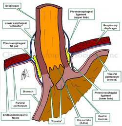
UPDATED: The esophageal hiatus is one of the seven hiatuses found in the respiratory diaphragm allowing passage of structures between the thorax and abdomen. As it name implies, the esophageal hiatus is the passageway for the esophagus. It also allows passage of the anterior and posterior vagus nerves, (CN X).
The hiatus is bound by two muscular crura, both of which arise from the right tendinous aortic crus. Since the intraabdominal pressure is higher than the intrathoracic pressure, there is a series of structures at the phrenoesophagogastric junction to close the esophageal hiatus.
The infradiaphragmatic parietal peritoneum reflects off the diaphragm towards the stomach to form its serosa layer (visceral peritoneum). At the same time the infradiaphragmatic fascia, also known as the endoabdominopelvic fascia, splits into two components or limbs. These are the superior and inferior phrenoesophageal ligaments or phrenoesophageal membranes. (the root [-phren-] means "diaphragm"). These phrenoesophageal ligaments create a disc-like plug between the abdomen and the thorax. This "plug" is reinforced by a circular infradiaphragmatic fat pad. The phrenoesophageal ligaments are reinforced on their thoracic aspect by the endothoracic fascia.
The lower esophagus has a dilation (evident in the image) called the "esophageal ampulla", in relation to this dilation the circular muscle layer of the esophagus slightly thickens creating the so-called "lower esophageal sphincter". This area is not a true anatomical sphincter, but rather is a functional sphincter.
The esophagogastric mucosal junction shows a marked transition in the shape of a wavy line. This is called the Z-line or the ora serrata. Extensions of the gastric mucosa and submucosa inferior to the ora serrata create a valve-like flap called the "gastroesophageal flap valve". When viewing this mucosal flap through and endoscope, it looks corrugated and flower-like, hence it is also called the "rosette".
The congenital or pathological dilation of the esophageal hiatus can predispose to esophageal hiatus hernia.
Sources:
1 "Tratado de Anatomia Humana" Testut et Latarjet 8 Ed. 1931 Salvat Editores, Spain
2. "Anatomy of the Human Body" Henry Gray 1918. Philadelphia: Lea & Febiger
Original image by Dr. E. Miranda
- Details
The esophagogastric junction is a complex anatomical region found at the esophageal hiatus of the respiratory diaphragm. It allows passage of he esophagus from the thorax into the abdomen.
The hiatus is bound by two muscular crura, both of which arise from the right tendinous aortic crus. Since the intraabdominal pressure is higher than the intrathoracic pressure, there is a series of structures at the phrenoesophagogastric junction to close the esophageal hiatus.
The infradiaphragmatic parietal peritoneum reflects off the diaphragm towards the stomach to form its serosa layer (visceral peritoneum). At the same time the infradiaphragmatic fascia, also known as the endoabdominopelvic fascia, splits into two components or limbs. These are the superior and inferior phrenoesophageal ligaments or phrenoesophageal membranes. (the root [-phren-] means "diaphragm"). These phrenoesophageal ligaments create a disc-like plug between the abdomen and the thorax. This "plug" is reinforced by a circular infradiaphragmatic fat pad. The phrenoesophageal ligaments are reinforced on their thoracic aspect by the endothoracic fascia.
The lower esophagus has a dilation (evident in the image) called the "esophageal ampulla", in relation to this dilation the circular muscle layer of the esophagus slightly thickens creating the so-called "lower esophageal sphincter". This area is not a true anatomical sphincter, but rather is a functional sphincter.
The esophagogastric mucosal junction shows a marked transition in the shape of a wavy line. This is called the Z-line or the ora serrata. Extensions of the gastric mucosa and submucosa inferior to the ora serrata create a valve-like flap called the "gastroesophageal flap valve". When viewing this mucosal flap through and endoscope, it looks corrugated and flower-like, hence it is also called the "rosette".
The congenital or pathological dilation of the esophageal hiatus can predispose to esophageal hiatus hernia.
Sources:
1 "Tratado de Anatomia Humana" Testut et Latarjet 8 Ed. 1931 Salvat Editores, Spain
2. "Anatomy of the Human Body" Henry Gray 1918. Philadelphia: Lea & Febiger
Original image by Dr. E. Miranda
- Details
The word acetabulum is formed by the combination of the Latin root [acetum], meaning "vinegar", and the Latin suffix [-abulum] a diminutive of [abrum], meaning a "cup", "holder", or "receptacle". Thus formed, the word acetabulum means "a small vinegar cup".
Roman soldiers liked to drink their water mixed with a small quantity of vinegar, so as to reduce the sensation of thirst. This mix was called "Posca". An acetabulum was used to add specific quantities of vinegar to the water, so over time the acetabula (plural form of acetabulum) were considered measuring devices. It is said that they measured one cup, or 2 1/2 oz. of wine.
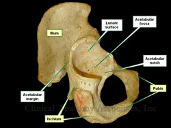
The anatomical acetabula are bilateral cup-like depressions in the os coxae which serve as a component of the coxofemoral joint (hip joint). They are found at the intersection of the three bony components of the os coxae, the ilium, ischium, and pubic bone and look anteroinferiorly.
The acetabulum has several components:
• Acetabular margin: An incomplete circular bony edge or border that marks the edge of the acetabulum
• Acetabular notch: The area where the acetabular margin is incomplete
• Acetabular labrum: Labrum (Lat. :lip). The acetabular labrum is a complete circular ring of fibrocartilage found on the acetabular margin that helps maintain the head of the femur in place. It is not shown in the accompanying image
• Lunate surface: A smooth, half-moon shaped area on the floor of the acetabulum. It is covered with hyaline cartilage and allows for articulation with the head of the femur
• Acetabular fossa: The non-articular region of the floor of the acetabulum. It contains fat, vessels, and the ligament of the head of the femur
Interesting fact: You may find that in older English anatomy books the acetabulum is referred to as the cotyloid cavity. The word cotyloid arises from the Greek [κοτυλοειδές] and means "similar to a cup". This separation in terms still exists when studying anatomy in other languages. For example, in Spanish the acetabulum is called "cavidad cotiloídea" or "cotilo", and in French it is called "cavité cotyloïde" or "cotyle". I guess the Greek soldiers did not drink vinegar with their water...
Image property of: CAA, Inc. Photographer: David M. Klein
- Details
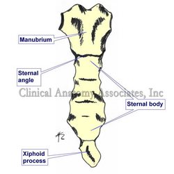
UPDATED:The sternal angle is the term used to denote the angulation at the joint between the manubrium and the body of the sternum. This transverse joint is called the "manubriosternal joint" and is a secondary cartilaginous joint of a type known as a symphysis. The angle varies between 160 and 169 degrees.
It is know eponymously as the "angle of Louis" named after Antoine Louis1 (1723-1792), a French physician. The importance of the sternal angle is that of an anatomical superficial landmark, which forms a horizontal plane which indicates a series of anatomical occurrences, as follows:
• Location of the cartilages of the second rib
• Beginning and end of the aortic arch
• Boundary between the inferior and superior mediastinum
• Location of the bifurcation of the trachea
• Posteriorly, the plane of the sternal angle passes trough the T4-T5 intervertebral disc (sometimes a little lower, through the superior aspect of T5)
• Highest point of the pericardial sac.
• It is the point where the right and left pleurae meet in the midline. They touch, but their pleural spaces do not communicate (usually).
1. Some authors contest the eponym, adjudicating it to Pierre Charles Alexander Louis (1787-1872), another French physician.
Image property of: CAA.Inc.. Artist: David M. Klein
Thoracic anatomy, pathology and surgery, are some of the many lecture topics developed and presented by Clinical Anatomy Associates, Inc. For more information Contact Us.
- Details
- Written by: Pascale Pollier

Cover of the book by Theo Dirix
My partner in crime and fellow traveler, Theo Dirix, has just published a new account of our common quest for the lost grave of Andreas Vesalius. Until the scientific results of our latest mission in Zakynthos in September 2017, will become public, this collection of articles published since 2014 represents a detailed and complete status quaestionis of a search that will never be the same anymore.
I'm proud and grateful to be part of a team he describes a most tenacious.
Following is a remarkable quote from the book: "The beast you have in your hands may appear as aged and stubborn: indeed, the texts collected here are not new and they regularly echo each other. The beast barks and growls: these words do not intend to examine or research but were meant to sell a project to potential sponsors. I feel the taste of the creature’s spit in my face, but pleading not guilty to any accusation of self-glorification, I do hope I managed to teach it a few tricks you will enjoy. While continuing to write about Vesalius’s death and his grave, black dogs may still be scratching at my hermitage. When I will finally throw open the doors to the beauty beyond, here’s hoping the encounter with the female spider will taste as fresh as a first kiss and be the beginning of something else."
No surprise some have described the book as: "a truly captivating story (a Live Adventure!) written in a fascinating, passionate and inspiring way. Theo Dirix, with his unique style is describing facts from his adventure to locate the grave of Vesalius and he is mentioning with great respect all his collaborators, the friends of Vesalius and those who share the same passion for Anatomy and Art." (Vasia Hatzi on Med in Art).
The book can be ordered here: https://www.shopmybooks.com/US/en/book/theo-dirix-32/in-search-of-andreas-vesalius. (English version of the website). More information about the author on his website www.theodirix.com. or here.
Personal note: Thanks to Pascale Pollier, a contributor to this website, for allowing us to publish this article, originally published on Vesalius Continuum.
I received a personalized copy from the author, Theo Dirix; Thank you very much for the recognition and the use of this website as reference in some of your comments. It is a great read for anyone even mildly interested in the life and specially the death and disappearance of the grave of Andreas Vesalius. There are several passages in the book that I will have to research and transform in articles for this blog.
For those who collaborated in the GoFundMe campaign because or our article entitled Do you want your name in a book? The Quest for the Lost Grave.... this is the book and the name of all the contributors are listed in it!
The quest continues... Dr. Miranda
- Details
- Written by: Randall K. Wolf, MD.
If you arrived to this article looking for information on Atrial Fibrillation, you will find some in this article. If you need to contact Dr. Wolf, please click here.
 Dr. Randall K. Wolf
HOUSTON AFib PATIENT EXPERIENCE SEMINAR
Saturday, April 21st, 2018 9am – 4pm
Westin at Memorial City, 945 Gesner Rd.
Houston, TX 77024
877-900-AFIB (2342)
This seminar is free and open to the public. To attend, please call the telephone number to register.
WELCOME MESSAGE FROM DR. RANDALL WOLF
In my experience over the last 18 years as a physician who specializes in the treatment of Atrial fibrillation (AFib), I have learned AFib sufferers want two things: Hope and a chance to feel better.
The first step to hope and to feeling better is to self educate. Learn about the latest medications, techniques and devices to treat AFib. Ask questions. Get a second opinion. Take charge of your health.
The purpose of the Houston AFib Patient Experience Seminar is to help AFib sufferers like you take charge of your health.
About 30 million people worldwide carry an AFib diagnosis. Today seems everyone either has AFib or knows someone that has AFib. When I first held an Afib seminar in Beijing, China, over 1200 people with AFib signed up for the seminar. It was standing room only!
Despite the common occurrence of AFib around the world, a recent study found that in patients who were diagnosed with AFib, 40-50% of patients with an elevated risk of stroke were not treated with the best therapy, and the rate of stroke over the next five years was 10%.
Here in Houston, we can do better! Learn more about AFib right here today, and I guarantee you will have hope and be more likely to reach your goal of feeling better.
Towards an AFib free healthy life,
Randall K. Wolf, MD.
ABOUT THE HOUSTON AFIB PATIENT EXPERIENCE SEMINAR
The University of Texas McGovern Medical School, Cardiothoracic and Vascular Surgery Department in Houston, is proud to host the inaugural Houston AFIB Patient Experience Seminar. The purpose is to educate the public in an interactive format allowing the audience to engage in conversation in a question/answer format with leading medical professionals. Our list of panel members and guest presentations include surgeons, cardiologists, neurologists, pulmonologists as well as testimonials from AFib patients. We are honored to be able to bring awareness to the resources and options available to patients suffering from AFIB.
NOTE: If you cannot attend the seminar, there is more information on Atrial Fibrillation at this website; click here.


