![]()
Medical Terminology Daily (MTD) is a blog sponsored by Clinical Anatomy Associates, Inc. as a service to the medical community. We post anatomical, medical or surgical terms, their meaning and usage, as well as biographical notes on anatomists, surgeons, and researchers through the ages. Be warned that some of the images used depict human anatomical specimens.
You are welcome to submit questions and suggestions using our "Contact Us" form. The information on this blog follows the terms on our "Privacy and Security Statement" and cannot be construed as medical guidance or instructions for treatment.
We have 1034 guests and no members online
")
Marcia Crocker Noyes
(1869 – 1946)
Further to my comment on old books and research that started with an interesting bookplate (Ex-Libris). I continued my research and found that the person in charge of the Osler library bookplate was a fascinating individual that today maybe a ghost in the MedChi library and building in Baltimore... This is certainly an article that can be called "A Moment in History"
Marcia Crocker Noyes was the librarian at The Maryland State Medical Society from 1896 to 1946 and was a founding member of the Medical Library Association.[1][2][3]
Sir William Osler, MD. a famous Johns Hopkins surgeon was a noted bibliophile and had a large personal collection of books on various topics. When he became the President of MedChi in 1896, he was dismayed at the condition of the library and knew that with the right person and some stewardship, it could become a significant collection. Sir William asked his friend, Dr. Bernard Steiner, a physician and President of the Enoch Pratt Free Library in Baltimore for suggestions of a librarian, and Dr. Steiner recommended Marcia Crocker Noyes. A native of New York, and a graduate of Hunter College, Marcia had moved to Baltimore for a lengthy visit with her sister, and took a “temporary” position at the Pratt Library, which turned into three years. Although she had no medical experience or background, she was enthusiastic, and most importantly, she was willing to move into the apartment provided for the librarian, who needed to be available 24 hours a day.
The image in this article is Ms. Noyes on her first year on the job. Marcia developed a book classification system for medical books, based on the Index Medicus, and called it the Classification for Medical Literature. The system uses the alphabet with capital letters for the major divisions of medicine and lower-case ones for the sub-sections. The system was used for many years, but it's now dated and the Faculty's original shelving scheme was never changed. The card catalogs still reflect her classification and many of the cards are written in Marcia's back-slanting handwriting.
Marcia knew enough to ask the Faculty's members about medical questions, terminology and literature. She gradually won over the predominantly male membership and they became her greatest allies; Sir William at the start, and then for nearly 40 years, Dr. John Ruhräh, a wealthy pediatrician with no immediate family of his own. She made a point of attending almost every Faculty function, and in 1904, under guidelines from the American Medical Association, Marcia was made the Faculty Secretary. For much of her first 10 years, she was the Faculty's only full-time employee, only being assisted by Mr. Caution, the Faculty's janitor. Later in life Marcia would say that she hired him because of his name!
Within ten years, the library had outgrown its space, and plans, spearheaded by Marcia and Sir William before his move to Oxford, were made to build a headquarters building, mainly to house the library's growing collection of medical books and journals.
Marcia was instrumental in the design and building of the new headquarters. She travelled to Philadelphia, New York and Boston to look at their medical society buildings, and eventually, the Philadelphia architectural firm, Ellicott & Emmart was selected to design and build the new Faculty building. Every detail of the building held her imprimatur, from the graceful staircase, to the light-filled reading room, and all of the myriad details of the millwork, marble tesserae, and most of all, the four-story cast iron stacks. She was on-site, climbing up unfinished staircases, checking out the progress of the building, which was built in less than one year at a cost of $90,000.
Among the features of the new building was a fourth-floor apartment for her. She referred to it as the "first penthouse in Baltimore" and it had a garden and rooftop terrace. The library collection eventually grew to more than 65,000 volumes from medical and specialty societies around the world. Journals were traded back and forth, and physicians eagerly anticipated the arrival of each new issue. At the same time, Marcia was involved in the Medical Library Association as one of eight founding members. The MLA promotes medical libraries and the exchange of information. One of the earliest mandates of the MLA was the Exchange, a distribution and trade service for those who had duplicates or little-used books in their collections. Initially, the Exchange was run out of the Philadelphia medical society, but in 1900 it was moved to Baltimore and Marcia oversaw it. Several hundred periodicals and journals were received and sent each month, a huge amount of work for a tiny staff. In 1904, the Faculty had run out of room to manage the Exchange, so it was moved to the Medical Society of the Kings County (Brooklyn). But without Marcia's excellent administrative skills, it floundered and in 1908, the MLA asked Marcia to take charge once again.
In 1909, when the new Faculty building opened, there was enough room to run the Exchange and with the help of MLA Treasurer, noted bibliophile and close friend, Dr. John Ruhräh, it once again became successful. Additionally, Marcia and Dr. Ruhräh combined forces to revive the MLA's bulletin, which had all but ceased publication in 1908, taking the Exchange with it. This duo maintained editorial control from 1911 until 1926. In 1934, around the time of Dr. Ruhräh's death, Marcia became the first “unmedicated” professional to head the MLA. During her tenure, the MLA incorporated, the first seal was adopted, and the annual meeting was held in Baltimore. Marcia wanted to write the history of the MLA once she retired from full-time work at the Faculty, but her health was beginning to fail. She had back problems and had suffered a serious burn on her shoulder as a young woman, possibly from her time running a summer camp, Camp Seyon, for young ladies in the Adirondack Mountains. In 1946, a celebration was planned to honor Marcia's 50 years at the Faculty. But she was adamant that the physicians wait until November, the actual date of her 50 years. However, they knew she was gravely ill, and might not make it until then, so a huge party was held in April. More than 250 physicians attended the celebration, but the ones she was closest to in the early years, were long gone. She was presented with a suitcase, a sum of money to use for travelling, and her favorite painting of Dr. John Philip Smith, a founder of the Medical College in Winchester, Virginia. It was painted by Edward Caledon Smith, a Virginia painter who had been a student of the painter Thomas Sully.[4] She adored this painting and vowed, jokingly, to take it with her wherever she went.
The painting was not to stay with her for very long, for she died in November 1946, and left it to the Faculty in her will. Her funeral was held in the Faculty's Osler Hall, named for her dear friend. More than 60 physicians served as her pallbearers, and she was buried at Baltimore's Green Mount Cemetery. In 1948, the MLA decided to establish an award in the name of Marcia Crocker Noyes. It was for outstanding achievement in medical library field and was to be awarded every two years, or when a truly worthy candidate was submitted. In 2014, the Faculty began giving a bouquet of flowers to the winner of the award in Marcia's name, and in honor of her work. Much evidence exists for this tradition, as we know that the physicians, especially Drs. Osler and Ruhräh, frequently gave her bouquets of flowers. Marcia also cultivated flower gardens at the Faculty and decorated the rooms with her work.
Today, the MedChi building is open for tours and if the rumors are to be believed Ms. Marcia Crocker Noyes is still at work in her beloved library as the "resident ghost" [1][5]

NOTE: This article has been modified from the original Wikipedia article on Marcia Crocker Noyes. The article itself is well-written with interesting images of the subject. I would encourage you to visit it. The second insert is from book 00736 in my personal library and shows in pencil, the incredibly small handwriting of Marsha C. Noyes.
Sources:
1. "Marcia, Marcia, Marcia" MedChi Archives blog.
2. "Marcia C. Noyes, Medical Librarian" (PDF). Bulletin of the Medical Library Association. 35 (1): 108–109. 1947. PMC 194645
3. Smith, Bernie Todd (1974). "Marcia Crocker Noyes, Medical Librarian: The Shaping of a Career" (PDF). Bulletin of the Medical Library Association. 62 (3): 314–324. PMC 198800Freely accessible. PMID 4619344.
4. Edward Caledon BRUCE (1825-1901)"
5. Behind the scenes tour MedChiBuilding
"Clinical Anatomy Associates, Inc., and the contributors of "Medical Terminology Daily" wish to thank all individuals who donate their bodies and tissues for the advancement of education and research”.
Click here for more information


- Details
- Written by: Pavlos Plessas
NOTE: In 2014 Pavlos Plessas presented the compelling theory that Andreas Vesalius died in 1564 from scurvy on the island of Zakynthos. With his permission his original article entitled "Powerful indications that Vesalius died from scurvy" was published in this blog in 2016.
His theory was later challenged by Theo Dirix and Dr. Rudi Coninx in this same blog with the article "Did Andreas Vesalius really died from scurvy?". Pavlos Plessas' rebuttal to the latter article is published here from a letter to Theo Dirix.
...continued from: An answer regarding the death of Andreas Vesalius (2). For the initial article, click here.

Pavlos Plessas
Click on the image for author information
16. The study’s sample is small, not representative of a general population, based on answering a questionnaire and not observation
Clinical studies of this nature face obvious limitations. If the authors are prepared to disbelieve the testimony of people who lived in a confined space with many victims of scurvy until the final fatal outcome, it is no wonder they complain about the study. Doubting is their prerogative, however, the way to discredit the study is to either find an error in the data or its interpretation, or conduct their own study and come up with different results.
17. The quoted personality changes peak on day 107. Certainly not an early symptom
Do the authors consider a symptom only when it peaks? Is pain not pain until it becomes unbearable?
18. Elevation of this triad is also found in prolonged semi starvation, and deficiencies of B-complex vitamins
The study at this point refers to Brozek and indirectly to the Minnesota Starvation Experiment of 1944-45, meaning semi-starvation over many months. It is clearly not relevant here as Vesalius did not face food shortages for more than a few weeks. As for vitamins of the B complex if the authors are ready to suggest that Vesalius may have died from beriberi or pellagra I am ready and happy to argue.
19. These changes are characteristic of individuals who are physically ill, as the subjects were
Yes, but in the beginning they did not know that they were ill (no clinical signs). Besides, there is another study which indicates that “behavioural change in human scurvy patients has a physiological rather than a solely psychological basis” (6). And there is a suggested biological explanation. “Vitamin C is a cofactor in the biosynthesis of catecholamines, most notably in the conversion of dopamine to norepinephrine, which may explain the behavior and mood disorders associated with vitamin C deficiency” (7). “Acutely hospitalized patients experience emotional distress for many reasons; therefore, it may seem unexpected that simple correction of their vitamin C deficiency could account for such rapid and dramatic improvements in psychological well-being. There are several reasons why this possibility merits serious consideration. First, the result is biologically plausible. Psychological dysfunction is known to occur in vitamin C deficiency, presumably because of the involvement of ascorbate in neuronal transmission and in brain neurotransmitter and fuel metabolism.” (8)
20. Vesalius’ reaction was normal in the view of many passengers getting sick, dying and being thrown overboard
Did all the passengers start begging the crew not to throw their dead bodies overboard? Did Boucherus do it? Why did he then only report Vesalius doing it? Was it normal behaviour for a distressed 16th century aristocrat to beg lowly-born sailors?
21. The only known symptoms of Vesalius are consistent with many other diseases
I think the best way for someone to debunk the scurvy theory is to make a list of diseases that could have killed Vesalius. It appears that the authors do not consider it too great a challenge. But these diseases have to be compatible with the circumstances and the authors will have to do considerably better than the suggestion of food poisoning.
22. Vesalius died from exhaustion combined by illness
All sources agree that Vesalius died from an illness. He had gone through an ordeal, he was probably malnourished and dehydrated to some extent but there is no evidence of exhaustion. He was sick, not tired. He was a passenger on a ship, not a galley slave. The crew, who worked around the clock, were fine. The only exhaustion was that of his reserves of Vitamin C.
23. In order to have a definite diagnosis, it will be important to locate the grave of Andreas Vesalius
Amen. I have one more reason to wish it than most of the many people who wish Theo and Pascale good luck in the search for the grave. I expect to be vindicated. And I hope that maybe my work will contribute in the beyond doubt identification of Vesalius’ remains..
Sources:
1. "Voyages and Travels in the Levant in the Years 1749, 50, 51, 52" London 1766, p. 147
2. "Medicina Nautica: an Essay on the Diseases of Seamen" Volume III, London 1803, p. 387
3. "De magnis Hippocratis" Lienibus Libellus, Antwerp 1564, pp. 26a – 31b
4. A voyage round the world in the years MDCCXL, I, II, III, IV, 5th edition, London 1749, p. 101.
5. Robert A. Kinsman and James Hood, Some behavioral effects of ascorbic acid deficiency, The American Journal of Clinical Nutrition, April 1971.
6. Fiona E. Harrison, Behavioural and neurochemical effects of scurvy in gulo knockout mice, Journal for Maritime Research, Volume 15, Issue 1, 2013.
7. Olivier Fain, Musculoskeletal manifestations of scurvy, Joint Bone Spine 72, 2005.
8. Wang et al, Effects of vitamin C and vitamin D administration on mood and distress in acutely hospitalized patients, the American Journal of Clinical Nutrition, 2013.
PERSONAL NOTE: My thanks to all the authors who are part of this ongoing discussion and who are also friends and contributors to this blog. Everybody is correct in the fact that the only way to find the truth of the cause of death of Andreas Vesalius is to find his grave. The quest is ongoing and hopefully we are closer every day to this objective. Dr. Miranda
- Details
- Written by: Pavlos Plessas
NOTE: In 2014 Pavlos Plessas presented the compelling theory that Andreas Vesalius died in 1564 from scurvy on the island of Zakynthos. With his permission his original article entitled "Powerful indications that Vesalius died from scurvy" was published in this blog in 2016.
His theory was later challenged by Theo Dirix and Dr. Rudi Coninx in this same blog with the article "Did Andreas Vesalius really died from scurvy?". Pavlos Plessas' rebuttal to the latter article is published here from a letter to Theo Dirix.
...continued from: An answer regarding the death of Andreas Vesalius (1)...
Pavlos Plessas
Click on the image for author information
8. Echtius’ treatise was only published after his death in 1556
This is incorrect. Echtius was alive almost a decade after that and heard Boucherus describe Vesalius’ death with his own ears. The treatise was first published in 1564, the year of Vesalius’ death, albeit was wrongly attributed to Wierus.
9. Echtius believed scurvy was caused by a blocked spleen, leading to an excess of black bile
Echtius believed that an excess of black bile (melancholic humour) caused scurvy. He wrote that in addition to eating preserved foods and mouldy biscuit, and drinking foul water, the following conditions led to an excess of black bile: warm air, lack of sleep, hard manual work, anxiety and fevers. Each one individually could cause scurvy even if the diet was good . The reasons for Vesalius’ illness as indicated by the sources are: eating rotten biscuit, drinking corrupt water, hot weather and extreme worrying. Please compare with the list of causes given by Echtius in his treatise.
10. It is claimed that extreme fear and irrational behaviour … are well known early symptoms [Plessas]. This is not the case
I quote Rev. Richard Walter of the Anson expedition, who saw many of his shipmates die from scurvy: This disease is likewise usually attended with a strange dejection of spirits; and with shiverings, tremblings, and a disposition to be seized with the most dreadful terrors on the slightest accident (4).
11. This (absence of extreme fear and irrational behaviour) is also the observation of one of the authors (RC) having observed scurvy patients in Ethiopian prisons
Did any of Dr. Coninx’s patients see other inmates die from scurvy? Did he ever observe one of his patients witness an accident? Were his patients evaluated by a psychiatrist? I would hazard the guess that the answer to all the questions is no. I choose to believe Rev. Richard Walter.
12. The Italian Pietro Bizzari based his account on what he had been told by an anonymous Venetian goldsmith
Bizzari’s account is not credible. It clashes with the accounts of three different people, who saw Vesalius’ grave in Santa Maria delle Grazie.
13. Metellus describes the symptoms of Vesalius’ illness
No, he does no such thing.
14. The possibility of rotten food as a cause of death on the ship is plausible
I hope this is not a suggestion that a great physician like Vesalius could not recognise the symptoms of food poisoning in others. If he did, it would have been possible to identify the particular source and he would not have fallen ill himself. Even if that was not possible they could have resorted to sharing the supplies of the crew, who had suffered no cases of illness. Surely it is best to be malnourished than risk death from food poisoning. In addition, had the cause of death been food poisoning, the sources would not have blamed Vesalius’ worrying for his illness. Finally, since the authors seem to agree that Metellus’ version of events is the most reliable, how is it possible for a man on the verge of death from food poisoning to be walking on the seashore of Zakynthos? Food poisoning as a cause of death is not plausible even though the sources claim the disease was somehow related to food and water shortage.
15. A 1971 study by Kinsman and Hood which allegedly claims that personality changes are amongst the first symptoms of scurvy
Why allegedly? I quote from the study: The personality changes occurred at an earlier stage of depletion than the psychomotor changes, which did not appear until obvious clinical scurvy was present (5). So the study does claim that personality changes are amongst the first symptoms of scurvy, the very first as a matter of fact. According to figure 3 of the study the MMPI T-scores started increasing when the level of Vitamin C in the body was at the equivalent of 761 mg. Clinical signs of scurvy became apparent only when the level went down to 300 mg.
Article continued here: An answer regarding the death of Andreas Vesalius (3)
Sources:
1. "Voyages and Travels in the Levant in the Years 1749, 50, 51, 52" London 1766, p. 147
2. "Medicina Nautica: an Essay on the Diseases of Seamen" Volume III, London 1803, p. 387
3. "De magnis Hippocratis" Lienibus Libellus, Antwerp 1564, pp. 26a – 31b
4. A voyage round the world in the years MDCCXL, I, II, III, IV, 5th edition, London 1749, p. 101.
5. Robert A. Kinsman and James Hood, Some behavioral effects of ascorbic acid deficiency, The American Journal of Clinical Nutrition, April 1971.
6. Fiona E. Harrison, Behavioural and neurochemical effects of scurvy in gulo knockout mice, Journal for Maritime Research, Volume 15, Issue 1, 2013.
7. Olivier Fain, Musculoskeletal manifestations of scurvy, Joint Bone Spine 72, 2005.
8. Wang et al, Effects of vitamin C and vitamin D administration on mood and distress in acutely hospitalized patients, the American Journal of Clinical Nutrition, 2013.
- Details
- Written by: Pavlos Plessas
NOTE: In 2014 Pavlos Plessas presented the compelling theory that Andreas Vesalius died in 1564 from scurvy on the island of Zakynthos. With his permission his original article entitled "Powerful indications that Vesalius died from scurvy" was published in this blog in 2016.
His theory was later challenged by Theo Dirix and Dr. Rudi Coninx in this same blog with the article "Did Andreas Vesalius really died from scurvy?". Pavlos Plessas' rebuttal to the latter article is published here from a letter to Theo Dirix.
Pavlos Plessas
Click on the image for author information
An answer to Theo (and Rudi) regarding the death of Vesalius
Three years after my 2014 speech at the Vesalius Continuum meeting, where I suggested that scurvy appeared as the only possible explanation to Vesalius’ death, Dr. Rudi Coninx and Theo Dirix published a refutation. Theo is a dear friend and Dr. Coninx has dealt with scorbutic patients in Ethiopia. In addition, Theo is – along with another good friend, Pascale Pollier – the driving force behind the search for Vesalius’ remains. Arguments are always welcome, since they bring us closer to the truth, and are particularly important amongst people with common goals. Here is my reaction.
Quotes have been altered or truncated for brevity.
1. Vesalius must have eaten well in the Holy Land because he was a nobleman
The point I made was that at that time of year there was extreme scarcity of foods containing Vitamin C in that area, not that Vesalius was devoid of means or that he was not well looked after by the monks who hosted him.
2. Vesalius ate the food that protected monks from scurvy
It did not protect them. There is at least one recorded outbreak of scurvy in a Holy Land monastery (1). The circumstances were not unusual so scurvy in those monasteries was probably not uncommon.
3. Scurvy was not uncommon in the area but people were not dying in large numbers
If they had been suddenly forced to migrate en masse, through desert and sea for three months in Vesalius’ footsteps, there would have been a very large number of deaths.
4. Liver and kidneys are also sources of Vitamin C
True, but they are not prime cuts. How many times a month would a nobleman be offered offal? I would say probably none.
5. Forty days at sea is not long enough to develop scurvy. Symptoms appear after 3 months at least
This is true for a previously healthy-eating subject of a study. An 18th century British warship could easily have a dozen dead and another 50 sick in less than 2 months (2) . There are many factors such as activity, infections, temperature, stress, smoking and possibly others, like age, gender, weight and genetic make-up. The most important though is the amount of Vitamin C in the body when the period of deprivation starts. Some historians seem to believe that West European aristocrats would not have fared very well in this field because of their diet. There is an intriguing study by Susan Maclean Kybett that suggests Henry VIII of England, a contemporary of Vesalius, died from scurvy. And he was neither in the Middle East nor at sea. Vesalius had been travelling for seven months prior to his death. Of those, more than two he spent at sea and more than three in arid conditions during the summer. This is how and where he spent the last five months of his life. He could have been affected by scurvy and died even earlier than he did.
6. “Clinical description is typical for scurvy”. This is simply not true
What is not true is that I ever made the above statement. I am not a doctor and I did not make a diagnosis. I presented a theory based on historical research. What I said is that every single thing we know about this case, not only the few symptoms known to us, either points to scurvy or is compatible with scurvy. At the same time no other illness fits the picture. I also made two more observations. First that the sources, which do not name the disease – scurvy had no universally accepted name yet – and do not consciously describe its symptoms, do give a number of causes for it, all of which feature in a list of causes of scurvy in the treatise of Johannes Echtius. Second that the fact Echtius took the trouble to meet Georgius Boucherus – the man who travelled with Vesalius and paid for his burial – and hear for himself the details of what happened is probably not coincidental.
7. Vesalius would have recognized the symptoms of scurvy and described them
We have no description of the disease or its symptoms by Vesalius. There is no reason to believe that Vesalius would have described symptoms of scurvy but not of the plague or of cholera for example. All sources are unfortunately silent on the symptoms. His mental state and his sudden death were only mentioned because they were unusual and impressive events, and in the case of the former also as a factor that contributed to his illness. I am the one who considers them as symptoms of an illness.
Article continued here: An answer regarding the death of Andreas Vesalius (2)
Sources:
1. "Voyages and Travels in the Levant in the Years 1749, 50, 51, 52" London 1766, p. 147
2. "Medicina Nautica: an Essay on the Diseases of Seamen" Volume III, London 1803, p. 387
3. "De magnis Hippocratis" Lienibus Libellus, Antwerp 1564, pp. 26a – 31b
4. A voyage round the world in the years MDCCXL, I, II, III, IV, 5th edition, London 1749, p. 101.
5. Robert A. Kinsman and James Hood, Some behavioral effects of ascorbic acid deficiency, The American Journal of Clinical Nutrition, April 1971.
6. Fiona E. Harrison, Behavioural and neurochemical effects of scurvy in gulo knockout mice, Journal for Maritime Research, Volume 15, Issue 1, 2013.
7. Olivier Fain, Musculoskeletal manifestations of scurvy, Joint Bone Spine 72, 2005.
8. Wang et al, Effects of vitamin C and vitamin D administration on mood and distress in acutely hospitalized patients, the American Journal of Clinical Nutrition, 2013.
- Details

Click for a larger image
The "Canal of Nuck" is the patent embryological remnant of the processus vaginalis in the female. The processus vaginalis is an evaginated extension of the peritoneum that forms to the side of the gubernaculum, a small fibrous cord that is attached to the lower pole of the gonad in the embryo. On the other end, the gubernaculum attaches to the inner aspect of the labioscrotal fold, an embryonic structure that will become the scrotum in the male and the labia majora in the female.
In the male, the processus vaginalis accompanies the gubernaculum and the testicle, on its descent towards the scrotum. In the female, the gonad (ovary) stays in the pelvis and the embryological remnants of the gubernaculum become the proper ovarian ligament (uteroovarian ligament) and the round ligament of the uterus which enters the inguinal canal, splits into multiple small fibers that disappear in the tissues of the labium majus.

Click for a larger image
In the male (and female) the walls of the processus vaginalis normally fuse, closing the communication between the scrotum (and the labia majora) and the main peritoneal cavity. If they remain open, the name is different, although the pathological consequences are similar (hernia, cysts or hydrocele). In the male, it is called a “patent processus vaginalis”, and in the female it is called the “Canal of Nuck”, which is found patent in 10-20% of the cases, although its presence does not per se imply the presence of pathology.
It was first described by Anton Nuck, a Dutch surgeon and anatomist (1650-1692) in his book "Adenographia Curiosa & uteri foeminei anatome nova" published in 1691. In this book he questions why do some females present with inguinal hernias: "Haecce , praeter alias herniarium species , in utroque sexu obvias auditoribus meis anno fuperiori demonftrandi , difficile vifum fuit explicare , qui Hernia foeminarum inguinales orirentur?" Why when it is easy to see (the canal) in other species it is so difficult to explain to those listening why only some women have inguinal hernias?

Click for a larger image
In figure XL of the same book he proceeds to show the open processus vaginalis which was from then on known as the eponymic "Canal of Nuck"
The images in this article are from “Case Report: Infected Hydrocele of the Canal of Nuck” by Mandahan, P and Batthi, K. (see sources) Figure 1 shows the superficial hydrocele herniation; figure 2 shows the infected hydrocele; and figure 3 shows the excised opened hydrocele. Read the full article here.
http://dx.doi.org/10.1155/2013/275257
My personal thanks to Dr. Sanford Osher who suggested this article. Dr. Miranda
- Details
- Written by: Coninx, R; Dirix, T
Did Andreas Vesalius really die from scurvy on the island of Zakynthos in 1564?
Evidence does not support this theory.
NOTE: The following article authored by Theo Dirix, and Dr. Rudi Coninx, is a rebuttal of Pavlos Plessas' theory that Andreas Vesalius indeed died from scurvy. The original article entitled "Powerful indications that Vesalius died from scurvy" by Pavlos Plessas was presented in a meeting at the island of Zakynthos in 2014.

Dr. Rudi Coninx, and Theo Dirix
For the first segment of this article, click here.
...continued...
4. The sudden death
Vesalius did indeed die upon arrival in Zakynthos. And sudden death is indeed associated with scurvy.
We know this from two accounts. Both are second hand accounts:
The Italian Pietro Bizzari based his account on what he had been told by an anonymous Venetian goldsmith. The goldsmith claimed that he had happened upon the sick Vesalius by chance on a deserted beach and, in spite of the opposition he had faced from the Zakynthians, had tried to assist him in his final hours and had buried him with his own hands in a plot he had purchased for that purpose. This account suggests the Vesalius’ death was not sudden, but was prolonged process, lasting at least hours, if not days. None of the typical scurvy signs like the bleeding and joint pain, the foul mouth, are mentioned in the account.
The second written account has come to us via Johannes Metellus, the Latinised name of a Frenchman named Jean Matal. Metellus claims to have received this information from a German traveler from Nuremberg named Georg Boucher. Boucher travelled on the same ship with Vesalius to Zakynthos.
Boucher tells us that Vesalius was poorly supplied with food, fell ill, initially with worry over the breakout of the disease and his own fate, and soon after disembarking dropped dead. Boucher arranged for a stone to be put on his grave. Boucher’s account is generally considered more reliable
Metellus describes the symptoms of Vesalius’ illness and they do not match the description of a scorbutic patient. The disease described by Metellus includes symptoms of extreme fear and irrational behaviour, immediately prior to his illness. These are not well known scurvy symptoms, as sometimes is claimed.
Metellus attributes the illness that broke out on Vesalius’ ship, at least partly, to food and water shortages. He is clear though in that the deaths were caused by illness and not directly by starvation or dehydration. Not everyone of the ship was affected. Boucher, for example, does not appear to have fallen ill at any point, despite having had to share the same food as other passengers. The possibility of rotten food as a cause of death on the ship is plausible.
The behaviour changes described by Metellus are sometimes given as proof of scurvy, and reference is made to a 1971 study by Kinsman and Hood [16] which allegedly claims that personality changes are amongst the first symptoms of scurvy.
However, this study was done on only five volunteer prisoners. Not a big sample and certainly not a sample representative of a general population, especially when making conclusions about personality disorders which, moreover are based on answering a questionnaire, not based on observation. In addition, the quoted personality changes appear related to the body pool of ascorbic acid, but the peak appears on day 107. Certainly not an early symptom. And finally, the authors admit that these signs are not specific for vitamin C deficiency: “elevation of this triad is also found in prolonged semi starvation, and deficiencies of B-complex vitamins. These changes are characteristic of individuals who are physically ill, as the subjects were”.
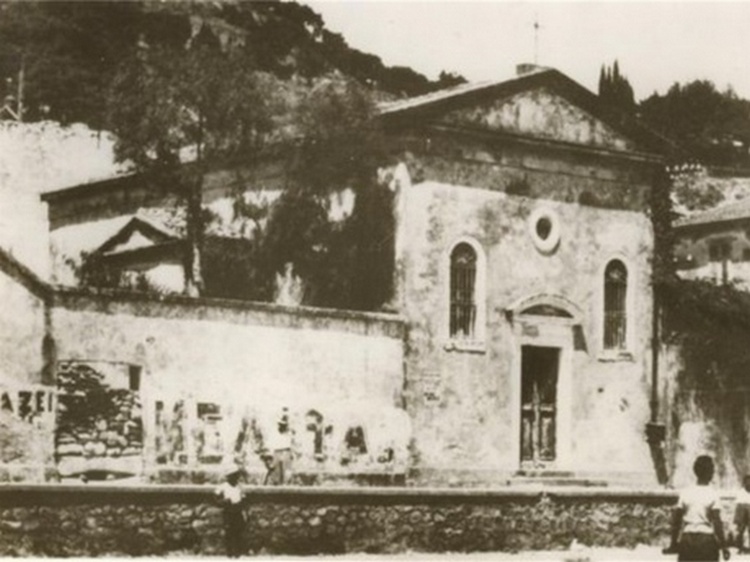
Church of Santa Maria delle Grazie
Click for a larger image
Vesalius’ immense worry and fear of falling ill, when he had not yet showed symptoms, would not point to an abnormal hypochondriac reaction, as alleged, but rather to a normal reaction. In the view of many passengers getting sick, die and being thrown overboard. Vesalius was known to be introvert. [quotation]
5. Burial on Zakynthos.
We know that Vesalius died in Zakynthos and was buried there as his tombstone has been described by several contemperous travellers. The German traveller Christoph Fürer Von Haimendorff visited the island in 1565, just a year after Vesalius’ death, and described Vesalius’ grave in the Franciscan monastery of Santa Maria delle Grazie. Von Haimendorff provided us with the details of the burial inscription. In 1586 the tombstone had disappeared, looted in the Turkish attack of 1571, according to Giovanni Zuallardo (Johannes Schwallart), a compatriot of Vesalius, who visited Zakynthos in 1586.
Conclusion
We conclude that the death from scurvy theory, while not entirely implausible, is not backed up by facts. The symptoms attributed to Andreas Vesalius prior to his death simply do not correspond to scurvy. While his altered mental state and his sudden death are compatible with scurvy, the absence of typical tell-tale signs of scurvy (gingival bleeding, joint pain, ecchymoses,) which were known at the time, would certainly have attracted the attention of Vesalius, one of the most prominent doctors of his era. The two remaining signs -altered mental state and his sudden death- are non-specific and are consistent with many other diseases. We therefore agree with the conclusion recently advanced by three eminent Vesalius experts (M. Biesbrouck, Th. Goddeeris and O. Steeno) that Vesalius died from exhaustion combined by illness [17].
In order to have a definite diagnosis, it will be important to locate the grave of Andreas Vesalius, find the remaining bones and test them for signs of scurvy. X-ray changes to the long bones are typical for scurvy [18] and even in adults one should be able to find signs of osteopenia, pathological fractures or others.
This would provide a definitive diagnosis of scurvy, even 500 years after the death of the great anatomist.
Note: My personal thanks to Theo Dirix and Dr. Rudi Coninx for contributing this article to this blog. The search for the grave of Andreas Vesalius continues on and you can find more information on "The Quest for the Lost Grave" GoFundMe page. Dr. Miranda
Note: Pavlos Plessas has published a counterargument to this article in a separate document entitled "An answer regarding the death of Andreas Vesalius", also found in this blog.
Sources:
1. https://circulatingnow.nlm.nih.gov/2014/10/15/the-death-of-andreas-vesalius/ accessed 27.12.2016
2. Matheson Cullen, G. Vesalius and the inquisition myth. Lancet, January 14, 1928, p 105-6.
3. Dirix Th. In search of Andreas Vesalius. The quest for the lost grave. Lannoo, 2014.
4. https://en.wikipedia.org/wiki/Andreas_Vesalius accessed on January 21, 2016
5. Biesbrouck M, Goddeeris Th, Steeno O. The last months of Andreas Vesalius. A coda. In Vesalius, Acta Internationalia Historiae Medicinae. 2012, 18 (No 2), 70-75.
6. Plessas P. http://www.parathemata.com/2014/09/pavlos-plessas-powerful-indications.html 2014. Accessed January 21, 2016.
7. https://www.clinicalanatomy.com/andreas-vesalius
Aa https://ods.od.nih.gov/factsheets/VitaminC-HealthProfessional/
8. Fain, O. La Revue de Médecine Interne, 2004; vol 25, Issue 12, 872-880.
9. Hodges RE, Hood J, Canham HE, Sauberlich HE, Baker EM. Clinical manifestations of ascorbic acid deficiency in man. Am J Clin Nutr 1971;24:432-43.
x. Hirschman JV, Raugi GJ. Adult scurvy. Journal of American Academy of Dermatology, 1999, 41; No 6, 895-909.
xx. Bartley W, Krebs HA, O’Brien JRP. Vitamin C requirement of human adults. Medical Research Council Special Report Series No 280. London: Her Majesty’s Stationary Office; 1953. P 1-179. Quoted in Hirschmann et al.
xxx Hodges RE, Baker EM, Hood J, Saueberlich HE, March SC. Experimental scurvy in man. American Journal of Clinical Nutrition. 1969;22:535-48.
10. Harrisons Principles of Internal Medicine, 1998; p 484-85
11. Leung FW, Guze PA: Adult scurvy. Annals of Emergency medicine; 1981; 10:652-655
12. Bennet M, Coninx R. The mystery of the wooden leg: vitamin C deficiency in East African prisons. Tropical Doctor, 2005; 35: 81-84.
13. Carpenter K. The history if scurvy and vitamin C.Cambridge University Press 1986, p29.
11. Bown S R. 4he Age of Scurvy. How a surgeon, a mariner and a gentleman helped Britain win the battle of Trafalgar. Summersdale, 2003, p 96-99.
15. Lind J. A treatise of the scurvy. Containing an inquiry into the nature, causes and cure of that disease. Together with a critical and chronological view of what has been published on the subject. Edinburgh: Sands, Murray and Cochran: 1753.
16. Kinsman RA, Hood J: Some behavioural effects of ascorbic acid deficiency. The American Journal of Clinical Nutrition, 1971, 455-464.
17. Biesbrouck M, Goddeeris Th, Steeno O: ‘Post Mortem’ Andreae Vesalii (1514 – 1564). Deel II. Het graf van Andreas Vesalius op Zakynthos. A. Vesalius, nr.4 December 2015. [in Dutch].
18. Bruce M Rothschild. Scurvy imaging. http://emedicine.medscape.com/article/413463-overview
- Details
- Written by: Coninx, R; Dirix, T
Did Andreas Vesalius really die from scurvy on the island of Zakynthos in 1564?
Evidence does not support this theory.
NOTE: The following article authored by Theo Dirix, and Dr. Rudi Coninx, is a rebuttal of Pavlos Plessas' theory that Andreas Vesalius indeed died from scurvy. The original article entitled "Powerful indications that Vesalius died from scurvy" by Pavlos Plessas was presented in a meeting at the island of Zakynthos in 2014.
Dr. Rudi Coninx, and Theo Dirix
For the first segment of this article, click here.
...continued...
We believe the arguments in favour of scurvy unfortunately are not sufficient to draw a conclusion. And for each of the argument in favour of the scurvy theory, there are strong counterarguments too.
We will examine the main arguments here
1. Travelers to the Holy Land did have not enough food to eat that contained vitamin C.
There is no evidence that Vesalius, a man of means with introduction letters from the King of Spain, was deprived of food during his travels in the Holy Land. He could certainly buy food, and probably stayed at monasteries, sharing the same food that protected the residing monks from developing scurvy. And while scurvy was not uncommon, residents did not die in large numbers because of lack of vitamin C. Dying from scurvy was, even then, was limited to a small segment of the population.
Minimal daily intake of vitamin C for an adult is 90 mg per day [aa]. Most of that intake comes from fruits and vegetables (oranges, grapefruits …) but liver and kidney are also sources of vitamin C. These are all foods that Vesalius would have consumed in Jerusalem.
2. The long sea voyage led to scurvy.
The sea voyage is too short (40 days) to develop scurvy. It takes about three months (120 days) for scurvy to develop [8]. In one study it took healthy volunteers four months to develop signs of scurvy when fed a vitamin C deprived diet [9] although some showed signs at day 29. The first sign to appear was petechial haemorrhage. In 1939, Johan Crandon, a surgical resident at Boston City Hospital, experimented on himself by eating a diet totally devoid of vitamin C [x]. Fatigue developed after 3 to 4 months, and the pathognomonic hyperkeratotic papules appeared on day 134 and the perifollicular haemorrhages on the legs on day 162. Symptoms appear after 3 to 4 months only, and there is no evidence of this in any description of Vesalius symptoms.
It has also been argued that the trip through the Sinai desert must have contributed to the presumed low intake of vitamin C.
Vesalius’s entire stay in the Holy Land, including the Sinai desert trip –presumably lasting a month and a half- did not last for more than 4 months: he arrived in Jerusalem in May 1564 and boarded ship in Alexandria in September 1564. We believe this is not long enough to deplete all vitamin C and cause symptoms, even adding a 40 day sea voyage. And it is unlikely that his stay in Jerusalem was entirely free of vitamin C, as he was a respected guest of high authorities. We know that there was “no hint of shortages before their departure from Egypt” [Plessas]
Most studies indicate the earliest detectable change occurs after 120 to 180 days [xx] although there are studies where early signs appear earlier [xxx]. But in this study, which was stopped after 3 months, no serious effects occurred.
3. “Clinical description is typical for scurvy”.
This is simply not true. The clinical description we have, from second hand accounts, are vague and non-specific. The clinical signs and symptoms of scurvy are well known and clearly described today [10]. The typical clinical signs for scurvy are absent from all accounts: gingival bleeding, an early and typical sign, or the “rotten mound” is never mentioned. Subcutaneous bleeding [11] – ecchymoses - or joint pains, leading to difficulties walking, are never mentioned. Gum bleeding are typical signs, and so are swollen and painful legs, also leading to difficulties in walking [12]. They are all absent from the accounts. Non-specific signs such as laziness are also typical, with muscle pain in the legs as an initial sign, but they always evolve into bleeding, gum problems, putrid smell and other signs that Vesalius would have described. As a doctor, Vesalius would have been well placed to recognize these signs and to describe them. None of them appear in any account.
Authors claiming typical signs were present make a lot of the so-called melancholy and refer to the writings of the 16th century expert Johannus Echthius who wrote a treatise about scurvy in Latin, as was customary in these days, in 1541, although it was only published after his death in 1556 [13] Despite being considered an expert on scurvy, Echtihius –and all “experts” of his day could recognize the disease – they knew little about the causes of scurvy, its link with vitamin C and the treatment. In fact Echthius and his 16th century colleagues were dead wrong about the causes of scurvy. Echthius obtained his knowledge entirely from studying Greek and Roman classics, Celsus, Galen and others, and came to the conclusion that scurvy was caused by a blocked spleen, leading to an excess of black bile. The treatment therefore consisted in hot and wet medicines like oil and vitriol. The only useful cure, citrus juice, was considered a “cold” medicine, therefore of no use [14]. Doctors at the time recommended avoiding fruits and vegetables in case of scurvy! Knowledge about the causes of scurvy would have to wait till the work of James Lind, an officer of the British Royal Navy who published his now famous treatise on scurvy in 1753 [15]
But Echthius did know how to recognize the symptoms of scurvy: stomachache, a complaint of the mouth, and sceletyrbe, a complaint of the legs. Vesalius showed none of these symptoms.
In another argument in favour of the scurvy theory, it is claimed that extreme fear and irrational behaviour … are well known early symptoms [Plessas ]. This is not the case. Lind was correct when he observed that “a listlessness to action … a lazy inactive disposition” that degenerates in “a universal lassitude” [Hisrchman] are typical signs. This is also the observation of one of the authors (RC) having observed scurvy patients in Ethiopian prisons.
Article continued here: Did Andreas Vesalius really die from scurvy? (3)
Sources:
1. https://circulatingnow.nlm.nih.gov/2014/10/15/the-death-of-andreas-vesalius/ accessed 27.12.2016
2. Matheson Cullen, G. Vesalius and the inquisition myth. Lancet, January 14, 1928, p 105-6.
3. Dirix Th. In search of Andreas Vesalius. The quest for the lost grave. Lannoo, 2014.
4. https://en.wikipedia.org/wiki/Andreas_Vesalius accessed on January 21, 2016
5. Biesbrouck M, Goddeeris Th, Steeno O. The last months of Andreas Vesalius. A coda. In Vesalius, Acta Internationalia Historiae Medicinae. 2012, 18 (No 2), 70-75.
6. Plessas P. http://www.parathemata.com/2014/09/pavlos-plessas-powerful-indications.html 2014. Accessed January 21, 2016.
7. https://www.clinicalanatomy.com/andreas-vesalius
Aa https://ods.od.nih.gov/factsheets/VitaminC-HealthProfessional/
8. Fain, O. La Revue de Médecine Interne, 2004; vol 25, Issue 12, 872-880.
9. Hodges RE, Hood J, Canham HE, Sauberlich HE, Baker EM. Clinical manifestations of ascorbic acid deficiency in man. Am J Clin Nutr 1971;24:432-43.
x. Hirschman JV, Raugi GJ. Adult scurvy. Journal of American Academy of Dermatology, 1999, 41; No 6, 895-909.
xx. Bartley W, Krebs HA, O’Brien JRP. Vitamin C requirement of human adults. Medical Research Council Special Report Series No 280. London: Her Majesty’s Stationary Office; 1953. P 1-179. Quoted in Hirschmann et al.
xxx Hodges RE, Baker EM, Hood J, Saueberlich HE, March SC. Experimental scurvy in man. American Journal of Clinical Nutrition. 1969;22:535-48.
10. Harrisons Principles of Internal Medicine, 1998; p 484-85
11. Leung FW, Guze PA: Adult scurvy. Annals of Emergency medicine; 1981; 10:652-655
12. Bennet M, Coninx R. The mystery of the wooden leg: vitamin C deficiency in East African prisons. Tropical Doctor, 2005; 35: 81-84.
13. Carpenter K. The history if scurvy and vitamin C.Cambridge University Press 1986, p29.
11. Bown S R. 4he Age of Scurvy. How a surgeon, a mariner and a gentleman helped Britain win the battle of Trafalgar. Summersdale, 2003, p 96-99.
15. Lind J. A treatise of the scurvy. Containing an inquiry into the nature, causes and cure of that disease. Together with a critical and chronological view of what has been published on the subject. Edinburgh: Sands, Murray and Cochran: 1753.
16. Kinsman RA, Hood J: Some behavioural effects of ascorbic acid deficiency. The American Journal of Clinical Nutrition, 1971, 455-464.
17. Biesbrouck M, Goddeeris Th, Steeno O: ‘Post Mortem’ Andreae Vesalii (1514 – 1564). Deel II. Het graf van Andreas Vesalius op Zakynthos. A. Vesalius, nr.4 December 2015. [in Dutch].
18. Bruce M Rothschild. Scurvy imaging. http://emedicine.medscape.com/article/413463-overview