![]()
Medical Terminology Daily (MTD) is a blog sponsored by Clinical Anatomy Associates, Inc. as a service to the medical community. We post anatomical, medical or surgical terms, their meaning and usage, as well as biographical notes on anatomists, surgeons, and researchers through the ages. Be warned that some of the images used depict human anatomical specimens.
You are welcome to submit questions and suggestions using our "Contact Us" form. The information on this blog follows the terms on our "Privacy and Security Statement" and cannot be construed as medical guidance or instructions for treatment.
We have 1047 guests and no members online
")
Marcia Crocker Noyes
(1869 – 1946)
Further to my comment on old books and research that started with an interesting bookplate (Ex-Libris). I continued my research and found that the person in charge of the Osler library bookplate was a fascinating individual that today maybe a ghost in the MedChi library and building in Baltimore... This is certainly an article that can be called "A Moment in History"
Marcia Crocker Noyes was the librarian at The Maryland State Medical Society from 1896 to 1946 and was a founding member of the Medical Library Association.[1][2][3]
Sir William Osler, MD. a famous Johns Hopkins surgeon was a noted bibliophile and had a large personal collection of books on various topics. When he became the President of MedChi in 1896, he was dismayed at the condition of the library and knew that with the right person and some stewardship, it could become a significant collection. Sir William asked his friend, Dr. Bernard Steiner, a physician and President of the Enoch Pratt Free Library in Baltimore for suggestions of a librarian, and Dr. Steiner recommended Marcia Crocker Noyes. A native of New York, and a graduate of Hunter College, Marcia had moved to Baltimore for a lengthy visit with her sister, and took a “temporary” position at the Pratt Library, which turned into three years. Although she had no medical experience or background, she was enthusiastic, and most importantly, she was willing to move into the apartment provided for the librarian, who needed to be available 24 hours a day.
The image in this article is Ms. Noyes on her first year on the job. Marcia developed a book classification system for medical books, based on the Index Medicus, and called it the Classification for Medical Literature. The system uses the alphabet with capital letters for the major divisions of medicine and lower-case ones for the sub-sections. The system was used for many years, but it's now dated and the Faculty's original shelving scheme was never changed. The card catalogs still reflect her classification and many of the cards are written in Marcia's back-slanting handwriting.
Marcia knew enough to ask the Faculty's members about medical questions, terminology and literature. She gradually won over the predominantly male membership and they became her greatest allies; Sir William at the start, and then for nearly 40 years, Dr. John Ruhräh, a wealthy pediatrician with no immediate family of his own. She made a point of attending almost every Faculty function, and in 1904, under guidelines from the American Medical Association, Marcia was made the Faculty Secretary. For much of her first 10 years, she was the Faculty's only full-time employee, only being assisted by Mr. Caution, the Faculty's janitor. Later in life Marcia would say that she hired him because of his name!
Within ten years, the library had outgrown its space, and plans, spearheaded by Marcia and Sir William before his move to Oxford, were made to build a headquarters building, mainly to house the library's growing collection of medical books and journals.
Marcia was instrumental in the design and building of the new headquarters. She travelled to Philadelphia, New York and Boston to look at their medical society buildings, and eventually, the Philadelphia architectural firm, Ellicott & Emmart was selected to design and build the new Faculty building. Every detail of the building held her imprimatur, from the graceful staircase, to the light-filled reading room, and all of the myriad details of the millwork, marble tesserae, and most of all, the four-story cast iron stacks. She was on-site, climbing up unfinished staircases, checking out the progress of the building, which was built in less than one year at a cost of $90,000.
Among the features of the new building was a fourth-floor apartment for her. She referred to it as the "first penthouse in Baltimore" and it had a garden and rooftop terrace. The library collection eventually grew to more than 65,000 volumes from medical and specialty societies around the world. Journals were traded back and forth, and physicians eagerly anticipated the arrival of each new issue. At the same time, Marcia was involved in the Medical Library Association as one of eight founding members. The MLA promotes medical libraries and the exchange of information. One of the earliest mandates of the MLA was the Exchange, a distribution and trade service for those who had duplicates or little-used books in their collections. Initially, the Exchange was run out of the Philadelphia medical society, but in 1900 it was moved to Baltimore and Marcia oversaw it. Several hundred periodicals and journals were received and sent each month, a huge amount of work for a tiny staff. In 1904, the Faculty had run out of room to manage the Exchange, so it was moved to the Medical Society of the Kings County (Brooklyn). But without Marcia's excellent administrative skills, it floundered and in 1908, the MLA asked Marcia to take charge once again.
In 1909, when the new Faculty building opened, there was enough room to run the Exchange and with the help of MLA Treasurer, noted bibliophile and close friend, Dr. John Ruhräh, it once again became successful. Additionally, Marcia and Dr. Ruhräh combined forces to revive the MLA's bulletin, which had all but ceased publication in 1908, taking the Exchange with it. This duo maintained editorial control from 1911 until 1926. In 1934, around the time of Dr. Ruhräh's death, Marcia became the first “unmedicated” professional to head the MLA. During her tenure, the MLA incorporated, the first seal was adopted, and the annual meeting was held in Baltimore. Marcia wanted to write the history of the MLA once she retired from full-time work at the Faculty, but her health was beginning to fail. She had back problems and had suffered a serious burn on her shoulder as a young woman, possibly from her time running a summer camp, Camp Seyon, for young ladies in the Adirondack Mountains. In 1946, a celebration was planned to honor Marcia's 50 years at the Faculty. But she was adamant that the physicians wait until November, the actual date of her 50 years. However, they knew she was gravely ill, and might not make it until then, so a huge party was held in April. More than 250 physicians attended the celebration, but the ones she was closest to in the early years, were long gone. She was presented with a suitcase, a sum of money to use for travelling, and her favorite painting of Dr. John Philip Smith, a founder of the Medical College in Winchester, Virginia. It was painted by Edward Caledon Smith, a Virginia painter who had been a student of the painter Thomas Sully.[4] She adored this painting and vowed, jokingly, to take it with her wherever she went.
The painting was not to stay with her for very long, for she died in November 1946, and left it to the Faculty in her will. Her funeral was held in the Faculty's Osler Hall, named for her dear friend. More than 60 physicians served as her pallbearers, and she was buried at Baltimore's Green Mount Cemetery. In 1948, the MLA decided to establish an award in the name of Marcia Crocker Noyes. It was for outstanding achievement in medical library field and was to be awarded every two years, or when a truly worthy candidate was submitted. In 2014, the Faculty began giving a bouquet of flowers to the winner of the award in Marcia's name, and in honor of her work. Much evidence exists for this tradition, as we know that the physicians, especially Drs. Osler and Ruhräh, frequently gave her bouquets of flowers. Marcia also cultivated flower gardens at the Faculty and decorated the rooms with her work.
Today, the MedChi building is open for tours and if the rumors are to be believed Ms. Marcia Crocker Noyes is still at work in her beloved library as the "resident ghost" [1][5]

NOTE: This article has been modified from the original Wikipedia article on Marcia Crocker Noyes. The article itself is well-written with interesting images of the subject. I would encourage you to visit it. The second insert is from book 00736 in my personal library and shows in pencil, the incredibly small handwriting of Marsha C. Noyes.
Sources:
1. "Marcia, Marcia, Marcia" MedChi Archives blog.
2. "Marcia C. Noyes, Medical Librarian" (PDF). Bulletin of the Medical Library Association. 35 (1): 108–109. 1947. PMC 194645
3. Smith, Bernie Todd (1974). "Marcia Crocker Noyes, Medical Librarian: The Shaping of a Career" (PDF). Bulletin of the Medical Library Association. 62 (3): 314–324. PMC 198800Freely accessible. PMID 4619344.
4. Edward Caledon BRUCE (1825-1901)"
5. Behind the scenes tour MedChiBuilding
"Clinical Anatomy Associates, Inc., and the contributors of "Medical Terminology Daily" wish to thank all individuals who donate their bodies and tissues for the advancement of education and research”.
Click here for more information


- Details
This article is part of the series "A Moment in History" where we honor those who have contributed to the growth of medical knowledge in the areas of anatomy, medicine, surgery, and medical research.
 Hippocrates
Hippocrates of Cos (460 BC - 370 BC). A Greek physician, Hippocrates was born on the Greek island of Cos (Kos) c. 460BC. Considered the "Father of Medicine" he removed Medicine from the realms of superstition and magic. He was the first to record medical writings and is considered the first one to use and maintain proper medical terminology. There are many writing attributed to Hippocrates, but there is no assurance that these were actually written by Hippocrates himself. Hippocrates changed the art of medical diagnosis by replacing supernatural precepts with observation-based methodology. Natural, rather than supernatural causes, would from here on explain all disease processes, what was known as Rational Medicine.
He is known for having set the oath that governs medical principles, the Hippocratic Oath, although there are many authors that contend that this oath was written long time after he died.
Sources:
1. "Hippocrates himself" JAMA. 1968;204(12):1138-1139
2. "Hippocrates: father of medicine" Tan, S Y (01/01/2002). Singapore medical journal(0037-5675), 43(1), p.5.
- Details
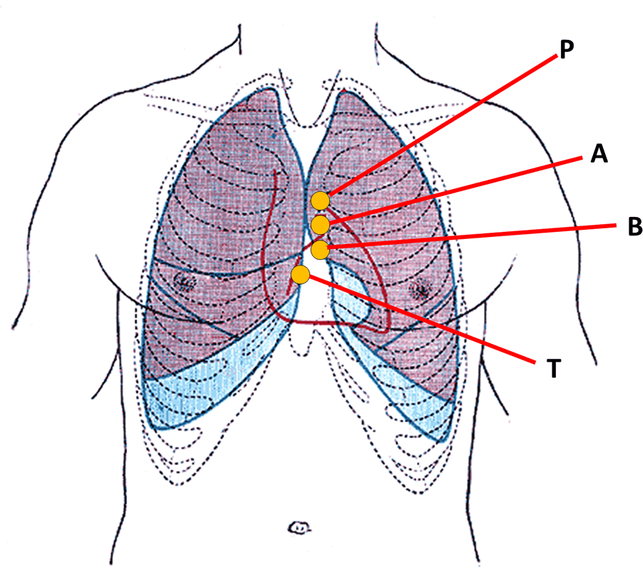
Anterior view of the thorax, showing surface
relations of bones, lungs (purple), pleura (blue),
and heart (red outline).
P. Pulmonary valve. A. Aortic valve.
B. Bicuspid valve. T. Tricuspid valve
This is a combined word arising from terms [atrium], [ventricle], and [sulcus]. For the etymology of each word, click on the corresponding link.
The atrioventricular sulcus, also know as the "coronary groove" or "coronary sulcus" is an evident incomplete groove between the atria and ventricles of the heart. It is complete posteriorly and is separated anterosuperiorly by the roots of the aorta and the pulmonary trunk. It contains the right coronary artery on the right side, and the circumflex artery on the left side, hence the name "coronary groove". These coronary arteries are not visible as they are usually covered by the epicardium and subepicardial fat.
The atrioventricular sulcus (and the corresponding coronaries) are also in relation to the deeper situated atrioventricular (AV) valves, the tricuspid valve on the right; and the mitral or bicuspid valve on the left side. The accompanying image depicts the location of the AV valves, and therefore the location of the AV sulcus. The image is an anterior view of the thorax, showing surface relations of bones, lungs (purple), pleura (blue), and heart (red outline). P. Pulmonary valve. A. Aortic valve. B. Bicuspid valve. T. Tricuspid valve
Sources:
1. "The Origin of Medical Terms" Skinner, HA 1970 Hafner Publishing Co.
2. "Medical Meanings - A Glossary of Word Origins" Haubrich, WD. ACP Philadelphia
3 "Tratado de Anatomia Humana" Testut et Latarjet 8 Ed. 1931 Salvat Editores, Spain
4. "Anatomy of the Human Body" Henry Gray 1918. Philadelphia: Lea & Febiger
Image modified by CAA, Inc. Original image by Henry Vandyke Carter, MD., courtesy of bartleby.com
- Details
This article is part of the series "A Moment in History" where we honor those who have contributed to the growth of medical knowledge in the areas of anatomy, medicine, surgery, and medical research.

Galen of Pergamum
Galen of Pergamon (129AD - 200AD). A Roman physician of Greek origin, Galen is a seminal character in Medicine and Physiology for the ages. He has been known as Galen, Galenus, Aelius Galenus, Claudius Galenus, Claudius Clarissimus Galen, and Galen of Pergamus. He was born in 129 A.D. in a Roman-Greek community in Pergamum (today's Turkey). As a very young man, he studied Medicine at the Pergamum temple of Asclepius. After traveling for additional studies, Galen obtained the appointment of "physician to the gladiators" back at this hometown of Pergamum.
The post required of him to study and develop hygiene, preventive medicine, as well as dealing with the gladiator's injuries. The horrible wounds allowed him to observe and study human anatomy and develop incredible skills at treating battle wounds. Galen traveled to Rome, where he was appointed Physician to the Emperor Marcus Aurelius.
Galen performed human and animal anatomical dissections, writing over 300 medical, pharmaceutical, and philosophical treatises in Greek, many of which were translated into other languages, especially Latin and Arabic.
Even though most of the original books were lost, the translations and interpretations of Galen's work have survived until today. His teachings and dictums were considered undisputable for over 1,500 years. In fact, in Medieval times and early Renaissance doubting Galen's teachings was considered heresy!
Galen's name is preserved in the eponymical "Vein of Galen", the great central cerebral vein.
Sources:
1. "Claudius Galenus of Pergamum: Surgeon of Gladiators. Father of Experimental Physiology" Toledo-Pereyra, LH; Journal of Investigative Surgery, 15:299-301, 2002
2. "Galen: history’s most enduring medic" Tan, SY; Singapore Med J 2002:3 (43):116 –117
3. "Galen and His Anatomic Eponym: Vein of Galen" Ustun, C.; Clinical Anatomy 17:454–457 (2004)
Original image in the public domain, courtesy of the National Library of Medicine
- Details
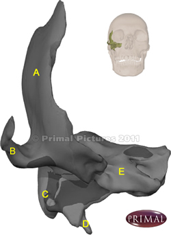
Click for a larger image
The temporal bone is a complex bone composed of several regions. The image shows an anterior view of the right temporal bone. To see the location of the bone, look at the inset that shows by transparency the location of the bone. Click on the image for a larger picture.
A. Squamous portion: From the Latin [squama], and meaning "scale-like", this portion of the bone is very thin, articulating with the parietal and sphenoid bones.
B. Zygomatic process: an anterior extension that articulates with the corresponding temporal process of the zygomatic bone.
C. Mastoid process: A Greek term from [-mast-] meaning breast, and the suffix [-oid] meaning "similar to".
D. Styloid process: Another Greek term from [stylos] meaning a "pillar", but also a "pen", therefore "shaped or similar to a pen". This is a slender and long inferior bony process. Close to the syloid process there are other processes, the pterygoid processes.
E. Petrous process: From the Latin [petrus] meaning "rock". The petrous process contains the components of the external auditory canal, the middle and inner ear, and a large canal through which passes the internal carotid artery.

First image modified from the original: "3D Human Anatomy: Regional Edition DVD-ROM." Courtesy of Primal Pictures.
Animation via Wikimedia Commons, public domain. Polygon data generated by Database Center for Life Science (DBCLS), CC BY-SA 2.1 JP.
- Details
This term has combined Greek components. The prefix [ect-] comes from [ectos], meaning "outside", and the root term [-top-] from [topos], meaning "place or location". The suffix [-ic] of course means "pertaining to". The word [ectopic] then means "outside its (normal) place or location".
The words has several uses. As an example, in atrial fibrillation, the atria of the heart will depolarize in abnormal or ectopic locations, causing a dysrhythmia. Another common use is in endometriosis, where there are abnormal or ectopic implantation sites of endometrium.
- Details
This article is part of the series "A Moment in History" where we honor those who have contributed to the growth of medical knowledge in the areas of anatomy, medicine, surgery, and medical research.

Oliver W. Holmes Sr.
Oliver W. Holmes Sr. (1809-1894). American physician, writer, and poet, Oliver Wendell Holmes Sr. was born in 1809 in Cambridge, MA. He started his studies in law, but soon turned to Medicine, studying part of his time in Paris. In 1843 he joined the fight against "puerperal fever", for which he was mocked, but he stood his ground on principle. A gifted writer, he published several books on essays, biography, and poetry. He was Dean of the Harvard Medical School. He received several honorary doctorates in Law and letters from Harvard and Cambridge. Little known is his contribution to Medicine by the coining of the terms "anesthesia" and "anesthetic", and that he was the father of a Supreme Court Judge, Justice Oliver Wendell Homes Jr.
The Journal of Clinical Anatomy published an article on Oliver W. Holmes Sr. profiling his many accomplishments.
Original image courtesy of www.nndb.com.
Sources:
1. "The Origin of Medical Terms" Skinner, H.A.(1970)
2. "Oliver Wendell Holmes, Sr. (1809–1894): Physician, jurist, poet, inventor, pioneer, and anatomist" Tubbs, RS et al, Clin Anat 25:8; 992-997 (2012)