![]()
Medical Terminology Daily (MTD) is a blog sponsored by Clinical Anatomy Associates, Inc. as a service to the medical community. We post anatomical, medical or surgical terms, their meaning and usage, as well as biographical notes on anatomists, surgeons, and researchers through the ages. Be warned that some of the images used depict human anatomical specimens.
You are welcome to submit questions and suggestions using our "Contact Us" form. The information on this blog follows the terms on our "Privacy and Security Statement" and cannot be construed as medical guidance or instructions for treatment.
We have 650 guests and no members online
")
Marcia Crocker Noyes
(1869 – 1946)
Further to my comment on old books and research that started with an interesting bookplate (Ex-Libris). I continued my research and found that the person in charge of the Osler library bookplate was a fascinating individual that today maybe a ghost in the MedChi library and building in Baltimore... This is certainly an article that can be called "A Moment in History"
Marcia Crocker Noyes was the librarian at The Maryland State Medical Society from 1896 to 1946 and was a founding member of the Medical Library Association.[1][2][3]
Sir William Osler, MD. a famous Johns Hopkins surgeon was a noted bibliophile and had a large personal collection of books on various topics. When he became the President of MedChi in 1896, he was dismayed at the condition of the library and knew that with the right person and some stewardship, it could become a significant collection. Sir William asked his friend, Dr. Bernard Steiner, a physician and President of the Enoch Pratt Free Library in Baltimore for suggestions of a librarian, and Dr. Steiner recommended Marcia Crocker Noyes. A native of New York, and a graduate of Hunter College, Marcia had moved to Baltimore for a lengthy visit with her sister, and took a “temporary” position at the Pratt Library, which turned into three years. Although she had no medical experience or background, she was enthusiastic, and most importantly, she was willing to move into the apartment provided for the librarian, who needed to be available 24 hours a day.
The image in this article is Ms. Noyes on her first year on the job. Marcia developed a book classification system for medical books, based on the Index Medicus, and called it the Classification for Medical Literature. The system uses the alphabet with capital letters for the major divisions of medicine and lower-case ones for the sub-sections. The system was used for many years, but it's now dated and the Faculty's original shelving scheme was never changed. The card catalogs still reflect her classification and many of the cards are written in Marcia's back-slanting handwriting.
Marcia knew enough to ask the Faculty's members about medical questions, terminology and literature. She gradually won over the predominantly male membership and they became her greatest allies; Sir William at the start, and then for nearly 40 years, Dr. John Ruhräh, a wealthy pediatrician with no immediate family of his own. She made a point of attending almost every Faculty function, and in 1904, under guidelines from the American Medical Association, Marcia was made the Faculty Secretary. For much of her first 10 years, she was the Faculty's only full-time employee, only being assisted by Mr. Caution, the Faculty's janitor. Later in life Marcia would say that she hired him because of his name!
Within ten years, the library had outgrown its space, and plans, spearheaded by Marcia and Sir William before his move to Oxford, were made to build a headquarters building, mainly to house the library's growing collection of medical books and journals.
Marcia was instrumental in the design and building of the new headquarters. She travelled to Philadelphia, New York and Boston to look at their medical society buildings, and eventually, the Philadelphia architectural firm, Ellicott & Emmart was selected to design and build the new Faculty building. Every detail of the building held her imprimatur, from the graceful staircase, to the light-filled reading room, and all of the myriad details of the millwork, marble tesserae, and most of all, the four-story cast iron stacks. She was on-site, climbing up unfinished staircases, checking out the progress of the building, which was built in less than one year at a cost of $90,000.
Among the features of the new building was a fourth-floor apartment for her. She referred to it as the "first penthouse in Baltimore" and it had a garden and rooftop terrace. The library collection eventually grew to more than 65,000 volumes from medical and specialty societies around the world. Journals were traded back and forth, and physicians eagerly anticipated the arrival of each new issue. At the same time, Marcia was involved in the Medical Library Association as one of eight founding members. The MLA promotes medical libraries and the exchange of information. One of the earliest mandates of the MLA was the Exchange, a distribution and trade service for those who had duplicates or little-used books in their collections. Initially, the Exchange was run out of the Philadelphia medical society, but in 1900 it was moved to Baltimore and Marcia oversaw it. Several hundred periodicals and journals were received and sent each month, a huge amount of work for a tiny staff. In 1904, the Faculty had run out of room to manage the Exchange, so it was moved to the Medical Society of the Kings County (Brooklyn). But without Marcia's excellent administrative skills, it floundered and in 1908, the MLA asked Marcia to take charge once again.
In 1909, when the new Faculty building opened, there was enough room to run the Exchange and with the help of MLA Treasurer, noted bibliophile and close friend, Dr. John Ruhräh, it once again became successful. Additionally, Marcia and Dr. Ruhräh combined forces to revive the MLA's bulletin, which had all but ceased publication in 1908, taking the Exchange with it. This duo maintained editorial control from 1911 until 1926. In 1934, around the time of Dr. Ruhräh's death, Marcia became the first “unmedicated” professional to head the MLA. During her tenure, the MLA incorporated, the first seal was adopted, and the annual meeting was held in Baltimore. Marcia wanted to write the history of the MLA once she retired from full-time work at the Faculty, but her health was beginning to fail. She had back problems and had suffered a serious burn on her shoulder as a young woman, possibly from her time running a summer camp, Camp Seyon, for young ladies in the Adirondack Mountains. In 1946, a celebration was planned to honor Marcia's 50 years at the Faculty. But she was adamant that the physicians wait until November, the actual date of her 50 years. However, they knew she was gravely ill, and might not make it until then, so a huge party was held in April. More than 250 physicians attended the celebration, but the ones she was closest to in the early years, were long gone. She was presented with a suitcase, a sum of money to use for travelling, and her favorite painting of Dr. John Philip Smith, a founder of the Medical College in Winchester, Virginia. It was painted by Edward Caledon Smith, a Virginia painter who had been a student of the painter Thomas Sully.[4] She adored this painting and vowed, jokingly, to take it with her wherever she went.
The painting was not to stay with her for very long, for she died in November 1946, and left it to the Faculty in her will. Her funeral was held in the Faculty's Osler Hall, named for her dear friend. More than 60 physicians served as her pallbearers, and she was buried at Baltimore's Green Mount Cemetery. In 1948, the MLA decided to establish an award in the name of Marcia Crocker Noyes. It was for outstanding achievement in medical library field and was to be awarded every two years, or when a truly worthy candidate was submitted. In 2014, the Faculty began giving a bouquet of flowers to the winner of the award in Marcia's name, and in honor of her work. Much evidence exists for this tradition, as we know that the physicians, especially Drs. Osler and Ruhräh, frequently gave her bouquets of flowers. Marcia also cultivated flower gardens at the Faculty and decorated the rooms with her work.
Today, the MedChi building is open for tours and if the rumors are to be believed Ms. Marcia Crocker Noyes is still at work in her beloved library as the "resident ghost" [1][5]

NOTE: This article has been modified from the original Wikipedia article on Marcia Crocker Noyes. The article itself is well-written with interesting images of the subject. I would encourage you to visit it. The second insert is from book 00736 in my personal library and shows in pencil, the incredibly small handwriting of Marsha C. Noyes.
Sources:
1. "Marcia, Marcia, Marcia" MedChi Archives blog.
2. "Marcia C. Noyes, Medical Librarian" (PDF). Bulletin of the Medical Library Association. 35 (1): 108–109. 1947. PMC 194645
3. Smith, Bernie Todd (1974). "Marcia Crocker Noyes, Medical Librarian: The Shaping of a Career" (PDF). Bulletin of the Medical Library Association. 62 (3): 314–324. PMC 198800Freely accessible. PMID 4619344.
4. Edward Caledon BRUCE (1825-1901)"
5. Behind the scenes tour MedChiBuilding
"Clinical Anatomy Associates, Inc., and the contributors of "Medical Terminology Daily" wish to thank all individuals who donate their bodies and tissues for the advancement of education and research”.
Click here for more information


- Details
This article is part of the series "A Moment in History" where we honor those who have contributed to the growth of medical knowledge in the areas of anatomy, medicine, surgery, and medical research.

Click for a larger image
Dr. Húmer Hültl (1868 – 1940) Hungarian surgeon, Húmer Hültl was born in 1868 in Felsobanya. Hültl studied in Budapest, earning his medical degree in 1891, and after surgical training he started to work as a surgeon in 1893.
By 1900, Dr. Hültl was the chief surgeon at the St. Stephen’s Hospital and later at the Sr. Rokus Hospital, and during WWI he was a commander of a Hungarian military hospital. Dr. Hültl’s attention to detail, careful asepsis (after Ignaz Semmelweis) and superb surgical technique earned him the moniker “The Paganini of the Knife”. Hültl was the first in his country to introduce the use of face masks, gloves, sterile cotton, and rubber gloves.
Dr. Hültl was very concerned about the consequences of spillage of gastrointestinal contents in the peritoneal cavity during surgery, covering all the walls of the cavity with sterile towels. At that time some surgical instruments had been invented to keep the edges of the intestines together while suturing. In 1907 Dr. Hültl envisioned a mechanical instrument that could place rows of staples transversely in the intestines thus avoiding spillage. With the aid of Victor Fisher, a German mechanical engineer, the first surgical stapler was constructed.
This original instrument was very bulky and heavy, weighing close to 11 pounds, and used a “bicycle-chain” type of mechanism to push a crankshaft that would push the staples into the anvil to form “B” shaped staples. It placed four rows or staggered staples. This device was first used in surgery on May 9th, 1908. A later, lighter variation of the instrument was later created, with a different crankshaft and weighing 8 pounds. Images of these instruments are available here.
Not many of these instruments were sold, but Dr. Hültl had set the stage for the development of the modern surgical stapler. Even today we still use the basic principles of his surgical stapler: “B" shaped staples, staggered rows of staples, and attention to the avoidance of leakage through the staple line. All of this makes Dr. Hültl an integral part of the history of surgical stapling.
Sources:
1. "Húmer Hültl: The Father of the Surgical Stapler" Robicsek, F.& Konstantinov, I. J Med Biogr February 2001 9: 16-19
2. “Current Practice of Surgical Stapling" Ravitch, MM; Steichen, FM, 1991.
Original image courtesy of "Surgical Stapler Museum" at www.surgicalstaplermuseum.com
- Details
The root term [-mur-] has its origin in the Latin word [murus] and means "wall". In medical terminology it is used mostly as [-mural] meaning "pertaining to a wall". It can be used in the following terms:
• Transmural: Through a wall
• Extramural: Outside a wall
• Intramural: Within a wall
Another term meaning "wall" is [parietal] from the Greek word [paries].
- Details
Click for a larger image
In medical terms a [sign] is an objective, observable, measurable expression of a pathology. To a physician, the combination of a patient's clinical and familial history (anamnesis), combined with the patient's symptoms and signs allows for a proper diagnosis. Some signs are so subtle and specific that they can only be observed and understood by a trained health care professional. Furthermore, some signs are pathognomonic, that is, by their presence they define a pathology. Furthermore
Contrary to symptoms (which are subjective), signs are not only objective, but comparable between individuals of the same species. Therefore we can compare the heart rate bmp (beats per minute) between normal and sick individuals allowing us to chart a range from normal to abnormal. The same is true for most signs such as body temperature, respiratory capacity, breathing rate, weight, height, etc.
Some signs are particular to a pathology, although they may not be pathognomonic. These specific signs are usually eponymic, such as:
• McMurray's sign: A click caused by the meniscus during manipulation of the knee; indicative of meniscal injury.
• Blumberg's sign: Sharp piercing pain on the abrupt release of steady pressure over the site of a suspected abdominal lesion, indicative of peritonitis. When used to diagnose appendicitis over McBurney's point it may be called Aaron's sign.
• Musset's sign: Rhythmical jerking of the head following the heart pulsations in aortic aneurysm and aortic insufficiency.
• Cardarelli's sign: An abnormal pulsation of the trachea that may be found in patients with an aneurysm of the aortic arch that causes left tracheal displacement.
• Caput medusae: A ring of dilated varicose veins radiating from the umbilicus, usually indicative of portal hypertension.
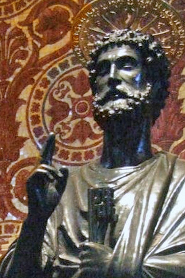
• Papal Benediction Sign: A contraction of the fourth and fifth digits, as in benediction (see image).
There are many more medical signs, this list is only an illustration of the concept
- Details
The medical term [symptom] has many definitions, as shown in this article. A good definition follows: A symptom is that what a patient refers, it is subjective, and cannot be measured or standardized (between patients). Examples of symptoms are thermal sensation ("I feel hot or feverish"), visceral sensations ("I feel a lump on my throat"), etc. In all cases the main characteristic of a symptom is that is subjective and patient-dependent. It is only relevant to a particular patient and cannot be compared from one patient to another.
Symptoms are part of the clinical study or history of a patient that help lead to a diagnosis.
• Merrian-Webster: "subjective evidence of disease or physical disturbance", "something that indicates the existence of something else"
• Memidex: "any sensation or change in bodily function that is experienced by a patient and is associated with a particular disease"
• Pandora World: "Feelings and perceptions reported by a patient indicative or that can be correlated with a disease process"
• Oxford Dictionaries: "A physical or mental feature that is regarded as indicating a condition of disease, particularly such a feature that is apparent to the patient"
Pain is a subjective entity, as it is characteristic to an individual. We all feel and respond to pain differently, as there are people with higher and lower thresholds to pain. Although I understand the need to have some type of standard, I dread the question posed by many..."From one to ten, can you tell me what your pain level is?. If the answer allows the physician or nurse to compare levels of pain within the same patient and see the evolution of a pathology, I am OK with that. But you cannot use that measurement to compare pain levels between patients!
The latest advances test for nerve activity following a noxa, but this just indicates that pain is being detected. Some say that the higher the recording the higher the pain. Possibly; but since pain is subjective we cannot use that measurement to compare pain levels between patients... at least that is my opinion. Dr. Miranda
- Details
The medical term [tenesmus] originates from the Greek [τεινεσμός], itself derivation from [τέντωμα] meaning "stretch", "distend", or to "strain". It refers to a symptom where the patient refers a constant urge to evacuate, with no or ineffective results. There are two types of tenesmus: rectal tenesmus and vesical tenesmus.
Because of the constant straining, tenesmus patients can have pain and cramping. Tenesmus can be one of the symptoms associated with the distal empty segment of colon or rectum found in a temporary or permanent diverting colostomy.
Note: The links to Google Translate in these articles include an icon that will allow you to hear the Greek or Latin pronunciation of the word.
- Details
This article is part of the series "A Moment in History" where we honor those who have contributed to the growth of medical knowledge in the areas of anatomy, medicine, surgery, and medical research.

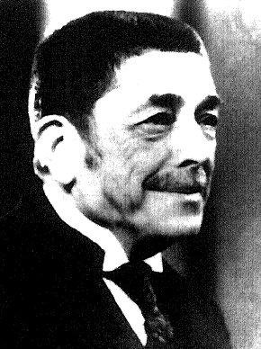
Mark M. Ravitch, MD
Mark M. Ravitch M.D.(1910-1989) American surgeon, historian, teacher, author, innovator, and visionary, Mark Mitchell Ravitch was born in 1910 in New York City. His parents were Russian immigrants, allowing to be fluent in Russian, which opened the doors to one of his many contributions to medicine and surgery: modern surgical stapling.
In 1934, Dr. Ravitch obtained his MD from Johns Hopkins, continuing in the same institution as a surgical intern, and later as a pediatrics resident, where he worked with Dr. Alfred Blalock, eventually becoming a professor of Surgery at Johns Hopkins, moving later to the Baltimore City Hospital. From 1943 to 1946 Dr. Ravitch was a surgeon in the Army.
He moved to the University of Chicago where from 1966 to 1969 he was professor of pediatric surgery and chief of pediatric surgery. His later and last move was to Pittsburgh, where he was professor of surgery at the University of Pittsburgh, and surgeon-in-chief at the Montefiore Hospital in Pittsburgh.
Dr. Ravitch is known for many important contributions to surgery, especially pediatric surgery, where he pioneered a procedure (the eponymic Ravitch procedure) to repair pectum excavatum in children, as well as research and develop a of nonoperative procedure to reduce intussuception using hydrostatic pressure reduction with barium enema. For this and many contributions he is known as one of the founding fathers of pediatric surgery.
A prolific writer and visionary, Dr. Ravitch authored 453 papers, 101 book chapters, 22 books, and served as editor of nearly 20 medical journals. In some of his writings he presented his vision for the development of surgery, even to the point of predicting heart transplantation surgery. Dr. Ravitch also was a surgical historian, with a wonderful library that was donated to the University of Pittsburgh upon his death.
In the medical industry Dr. Mark Ravitch is probably best know for bringing to the USA from the then USSR, the technology of surgical stapling. In 1958, while visiting Kiev, Dr. Ravitch and three other American physicians were shown the use in surgery of a bronchial surgical stapler developed in the USSR. They were able to procure one of these devices and bring it back to the USA. An American entrepreneur, Leon Hirsch, obtained the patents for these devices, founded the United States Surgical Corporation (now the Covidien Surgical Devices Division) and continued the development of the reusable and later the disposable surgical staplers. During the research Dr. Ravitch was joined by Dr. Felicien Steichen (1926 - 2011). Both Drs. Ravitch and Steichen were instrumental in the research and development of these modern surgical devices, making them part of the history of surgical stapling. Their work set the stage for the development of surgical stapling in minimally invasive procedures, so common today.
Dr. Ravitch died in 1989, still teaching students from his own hospital bed. His son Dr. Michael M. Ravitch (1943-2004) followed in his steps in medical education as an educational psychologist at Northwestern University's Feinberg School of Medicine.
Personal notes: I regret not having had the opportunity to meet Dr. Ravitch. In 2006 I spent several hours talking with Dr. Felicien Steichen about his trip to the USSR with Dr. Ravitch and the research and development that happened afterwards. When concluding my visit, Dr. Steichen presented me with a signed copy of his and Dr. Ravitch's book that reads:"Mark Ravitch would have enthusiastically applauded your efforts to teach the science of Anatomy that is the basis of the Art of Surgery". With the loss of both Drs. Ravitch and Steichen a wonderful chapter of the history of surgical stapling has closed.
A few years ago I was contacted by the Ravitch family. Knowing of my interest in Medical History, they donated a series of books signed by Dr. Ravitch. Recently the family also donated the personally typed diary of his trip tp Russia in 1958. They will be well cared and hopefully I will be able to have them printed as a book in the future. Dr. Miranda.
Sources:
1. "Naissance des sutures mecaniques modernes en chirurgie: petites et grandes histoires, en hommage a Mark Ravitch" Steichen,FM Chirurgie 1998,123 (6), 616.
2. "The Peaks of Excitement" Ann Surg 192: (1980) 3, 282 - 287
3. "A Century of Surgery, 1880-1980" Ravitch, Mark M.. Philadelphia
4. "Current Practice of Surgical Stapling" Ravitch, W & Steichen, F. 1991 Lea & Febiger USA
5. "Mark Ravitch (1910 - 1989) Editors, "Current Problems in Surgery" 1989
6. "All heart - Mark Ravitch" O'Donell B. J Ped Surg 25:1 (1990) 184
7. "Mark M. Ravitch: Historian and Innovator" Fingerete, AL, et al. J Surg Ed (2011) 155-158
8. "Reduction of intussusception by barium enema : A clinical and experimental study" Ravitch MM, McCune RM.Ann Surg. 1948;128:904-91
9. "The Surgical Curmudgeon" Pittmed, Spring 2013. 18-23