
Medical Terminology Daily (MTD) is a blog sponsored by Clinical Anatomy Associates, Inc. as a service to the medical community. We post anatomical, medical or surgical terms, their meaning and usage, as well as biographical notes on anatomists, surgeons, and researchers through the ages. Be warned that some of the images used depict human anatomical specimens.
You are welcome to submit questions and suggestions using our "Contact Us" form. The information on this blog follows the terms on our "Privacy and Security Statement" and cannot be construed as medical guidance or instructions for treatment.
We have 791 guests online
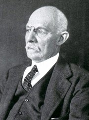
William S. Halsted, MD
(1852 – 1922)
American anatomist, teacher, and surgeon, William Stewart Halsted was born in New York City, USA to a wealthy family of English origin. His father was involved in charitable work and Governor and trustee to a city hospital. Not a brilliant student initially, Halsted took an undergraduate in Liberal Arts in Yale, CT., after which he entered the Medical College of Physicians at the Columbia College, where he excelled.
As a second-year medical student Halsted applied and obtained a position in surgery at a local hospital. In here he learned about Lister’s antiseptic technique and became an adamant proponent of it to reduce infection. In 1877 Halsted obtained his MD. After a short time as House Physician at the New York Hospital, Halsted traveled to Europe to further his education studying for two years at the Universities of Vienna, Leipzig, and W?rzburg.
Besides being at the forefront of surgical and antiseptic techniques (introducing the use of rubber gloves in surgery), Halsted was extremely concerned with the way medical students were taught in the US. He pioneered bedside clinical round discussions with the medical students after two years of basic sciences studies. Halsted developed the idea of a patient chart; he also developed the residency program for medical students in use today.
Halsted is probably the most influential researcher and surgeon at the turn of the century. He dedicated time to the study of intestinal anastomoses and the use of silk as a suture material. His experimental work in 1887 proved that the inclusion of the submucosa layer in an anastomosis was mandatory, as well that a single layered anastomosis was enough to attain closure. Perhaps Halsted’s most important contribution was the application and use of the scientific method to surgical questions. Halsted’s principles, also known as "Halsted's Rules of Surgery", set the standards used today in surgical suturing and surgical stapling.
He also pioneered the development and surgical techniques for radical mastectomy as a treatment for breast cancer.
As a side effect of this studied in anesthesia and the use of cocaine for anesthesia, Halsted became addicted to this substance, a problem that followed him through the years. Without impairing his capacity as a researcher and a surgeon, Halsted eventually recovered. He died in Baltimore in 1922 as a complication to surgery.
Sources:
1. Dubay, A. D., & Franz, G. M. (2003). Acute Wound Healing: The Biology of Acute Wound Failure. Surg Clin NA, 83, 463-481.
2. Halsted, W. S. (1887). Circular Suture of the Intestine - An Experimental Study. Am J Med Sci, 436-461.
3. “William Stewart Halsted: his life and contributions to surgery” Osborne, P. Lancet Oncol 2007; 8: 256–65
4. “William Stewart Halsted: Surgical pioneer” Burress, P Endoc Today (2010), 8: (2) 22
5. “William Stewart Halsted (1852–1922) Neurological stamp” Haas, LF J Neurol Neurosurg Psych 2000;69:641
Original image courtesy of "Images from the History of Medicine" at www.nih.gov

"Clinical Anatomy Associates, Inc., and the contributors of "Medical Terminology Daily" wish to thank all individuals who donate their bodies and tissues for the advancement of education and research”.
Click here for more information


- Details
These two terms are opposite, yet they need to be reviewed together. The word [antegrade] is Latin and means "to go before" or in a variation of the term, [anterograde] meaning "to go anterior or moving forward". The word [retrograde] is also Latin and means "to go or move behind (or posterior)". Based on their etymology, these words are of no use and confusing.
Now, if we use these words in the context of "proximal and distal" then the story changes. Based on our working definition of proximal and distal, the definition would be as follows:
Antegrade: Moving or in a relationship of proximal to distal. In the case of a hollow organ containing a moving fluid, it would mean "going with the flow"
Retrograde: Moving or in a relationship of distal to proximal. In the case of a hollow organ containing a moving fluid, it would mean "going against the flow"
The confusion caused by the lack of proper definition can be illustrated by this Letter to the Editor, published in The American Journal of Sports Medicine.
- Details
The prefix [-iso-] originates from the Greek [ίσος] meaning "equal". In medical terminology it is used to mean "same". Applications of this prefix include:
- Isometric: Same length. An isometric muscular contraction occurs when the muscle contracts, but the length of the muscle is the same, it does not change
- Isotonic: Equal or same tension. Refers to solutions that have the same osmotic pressure
- Isomorphous: Same shape or form
- Isosceles: Equal legs. An isosceles triangle is formed by three "legs" that have the same length
Note: The links to Google Translate in these articles include an icon that will allow you to hear the Greek or Latin pronunciation of the word.
- Details

Click for a larger image
The word [cingulum] is Latin and means "belt" or "girdle". The term was also used by Roman soldiers to denote a sword belt. The short sword of the Romans was called a "gladius" and this was the earlier name for the sternum. The plural Latin form for cingulum is "cingula".
The adjectival form for this word is [cingulate] and means "belted", "girdled", or "shaped like a belt"
In human anatomy the term [cingulum] or cingulate gyrus is used to describe a brain gyrus found deep in the medial aspect of the brain hemisphere, hugging the corpus callosum like a girdle. The cingulate gyrus (in green in the accompanying image) is part of the limbic system.
Interesting side notes: The Latin word cingulum evolved into the Spanish "cincha" used to refer to a strong leather belt, while in English it evolved into "cinch"... so when you are in "a cinch" you are basically "tied up" or "in a girdle". Also, the Greek equivalent term for cingulum is zoster, which explains why the pathology is called "herpes zoster", as the presentation of the pathology is in a tight, belt-like abdominothoracic rash.
- Details
This article is part of the series "A Moment in History" where we honor those who have contributed to the growth of medical knowledge in the areas of anatomy, medicine, surgery, and medical research.

Bernhard Siegfried Albinus
Bernhard Siegfried Albinus (1697-1770). German-Dutch physician, surgeon, and anatomist, Albinus was born in Frankfurt an der Oder, but lived most of his life in Leyden, in Holland, his adopted country. His real name was Bernhard Siegfried Weiss, which means “white” in German, the Latin version of which is “albus”, from where derives his Latinized name “Albinus”.
His father was also a physician, Bernard (or Bernhard) Weiss (1653 – 1721). He also took the last name Albinus, which makes following their history and genealogy a bit difficult.
Albinus moved to Leyden (Leiden) when he was only five years old, excelling at his studies and entering the University of Leyden at 12. He later moved to Paris, France to continue his studies on anatomy and surgery. He received his medical degree in 1719.
He began work at the University of Leyden as a Professor of Anatomy and Surgery, where he continued working until his death in 1770. He is considered one the most well-known anatomists of the 18th century.
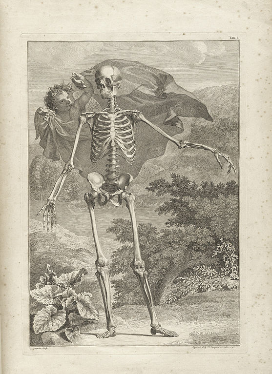
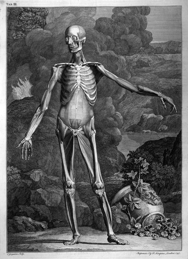
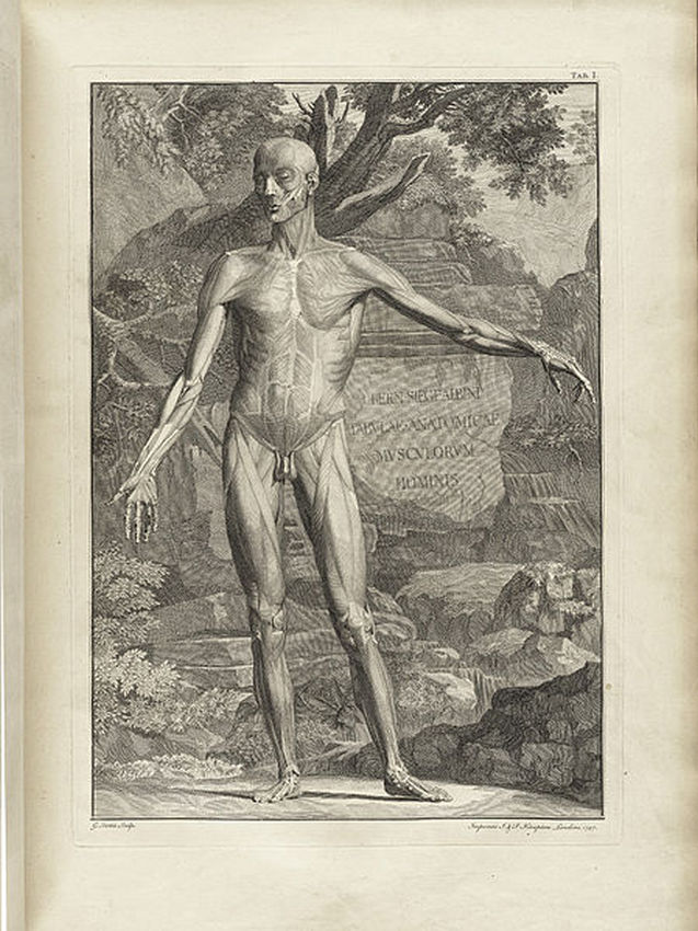
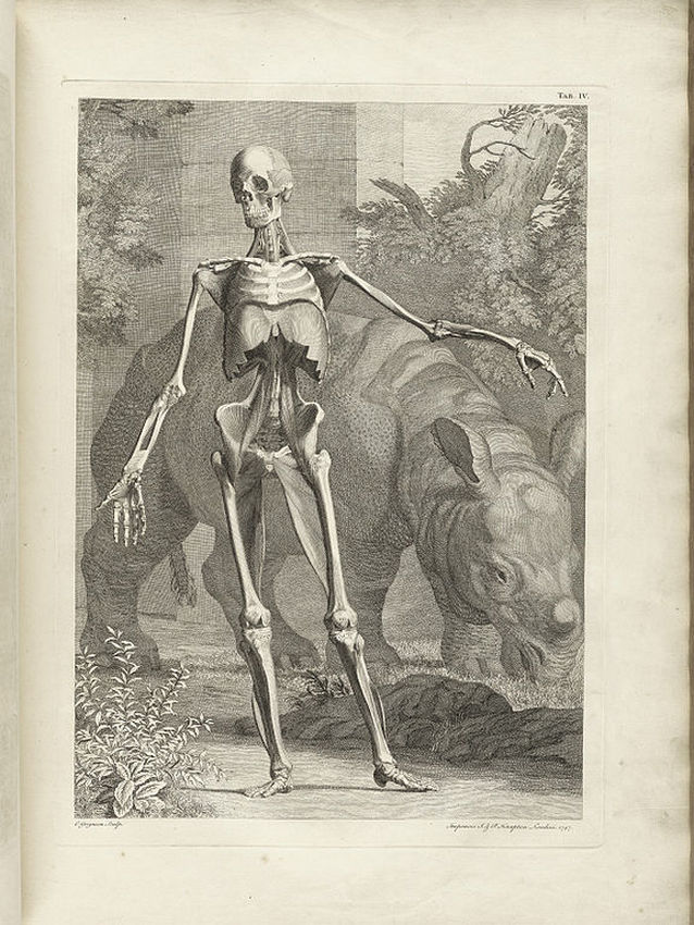
Because of his work with his colleague Herman Boerhaave (1668-1738), Albinus came in contact with Jan Wandelaar (1690-1759) an artist and engraver. It was this collaboration and the art of Jan Wandelaar which have made Albinus’ books and illustration famous. Albinus was criticized for the luscious, detailed, and sometimes strange backgrounds of his anatomical images, yet he defended Jan Wandelaar and his artistic expression. Albinus and Jan Wandelaar were dedicated to the faithful reproduction of anatomy in their publications, developing a grid system to reduce errors in production and printing.
During his tenure, Albinus was twice appointed Rector of the University and President of the College of Surgeons of Leyden. During this time, he became aware of the discovery of the copper plates created by Eustachius’ and lost for over a hundred years. In 1744 he published the plates in the book “Explanation of the Anatomical Tables of Eustachius “with his comments, stressing the fact that these images were better than those of Vesalius, published in 1543. This is no surprise, as Vesalius’ images were woodcuts, done before the technique of printing with copper plates became popular.
Although not well-known, Albinus’ name is eponymically attached to the risorius and scalenus minimus muscles. His famous publications include “Historia musculorum hominis” in 1734), “Icones ossium foetus humani” in 1737, “Tabulæ sceleti et musculorum corporis humani” in 1749, etc.
Some of Albinus and Jan Wandelaar images can be seen here. Click on the image to see a larger depiction:
Sources:
1. "Bernard Siegfried Albinus (1697-1770), German-Dutch anatomist" JAMA (1966), 196 (10): 910
2.“Bartholomeo Eustachio – The Third Man: Eustachius Published By Albinus” Fahrer, M. Ann Anat 187 (2005) 555—573
3. “Attic perfection in anatomy: Bernhard Siegfried Albinus (1697–1770) and Samuel Thomas Soemmerring (1755–1830)” Hildebrand, R. Ann Anat 187 (2005) 555—573
4. “Two Conceptions of the Human Form: Bernard Siegfried Albinus and Andreas Vesalius” Elkins, J. Artibus et Historiae, 7:14 (1986) 91-106
5. “Bartolomeo Eustachio: His Influence on Albinus and the Anatomical Models at La Specola, Florence” Hilloowala, R. J Hist Med All Sci (1986); 41 (4): 442 -462
Portrait and all other images in this article are in the public domain. Courtesy of Wikipedia. The links to read the books online courtesy of Archive.org.

- Details
Click for a larger image
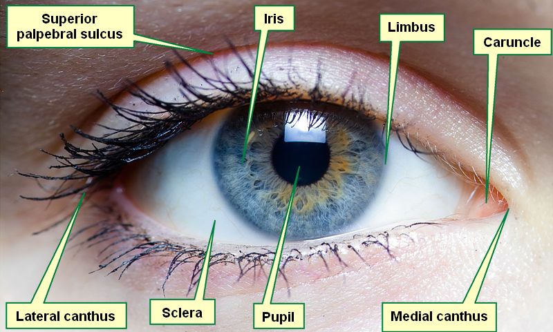
The word [canthus] arises from the Greek [kanthos] meaning "angle". It was later adopted into New Latin as [canthus]. The Latin plural form is [canthi].
In human anatomy it is used to describe the angles in the eye formed by the joining of the superior and inferior eyelids.
Each eye has a lateral and a medial canthus. At the medial canthus there is a small elevation of mucosa called the caruncle.
As an interesting note, the lateral canthal lines are a fancy way to refer to "crow's feet", the wrinkles that diverge from the lateral canthus.
Image modified from the original. Public domain. Courtesy of Wikipedia.
- Details
The word [fovea] is Latin, and means a "small pit" or a "small depression", a small round fossa. There are two plural forms in use. The first is the Latin form [foveae] and the second one is the English form [foveas]. In human anatomy foveae are usually round in shape.
There are several foveae described in the human body. One of them is the fovea centralis of the eye, a small round depression in the retina characterized by the presence of a large number of cone cells and responsible for highest visual acuity.