
Medical Terminology Daily (MTD) is a blog sponsored by Clinical Anatomy Associates, Inc. as a service to the medical community. We post anatomical, medical or surgical terms, their meaning and usage, as well as biographical notes on anatomists, surgeons, and researchers through the ages. Be warned that some of the images used depict human anatomical specimens.
You are welcome to submit questions and suggestions using our "Contact Us" form. The information on this blog follows the terms on our "Privacy and Security Statement" and cannot be construed as medical guidance or instructions for treatment.
We have 785 guests online
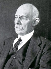
William S. Halsted, MD
(1852 – 1922)
American anatomist, teacher, and surgeon, William Stewart Halsted was born in New York City, USA to a wealthy family of English origin. His father was involved in charitable work and Governor and trustee to a city hospital. Not a brilliant student initially, Halsted took an undergraduate in Liberal Arts in Yale, CT., after which he entered the Medical College of Physicians at the Columbia College, where he excelled.
As a second-year medical student Halsted applied and obtained a position in surgery at a local hospital. In here he learned about Lister’s antiseptic technique and became an adamant proponent of it to reduce infection. In 1877 Halsted obtained his MD. After a short time as House Physician at the New York Hospital, Halsted traveled to Europe to further his education studying for two years at the Universities of Vienna, Leipzig, and W?rzburg.
Besides being at the forefront of surgical and antiseptic techniques (introducing the use of rubber gloves in surgery), Halsted was extremely concerned with the way medical students were taught in the US. He pioneered bedside clinical round discussions with the medical students after two years of basic sciences studies. Halsted developed the idea of a patient chart; he also developed the residency program for medical students in use today.
Halsted is probably the most influential researcher and surgeon at the turn of the century. He dedicated time to the study of intestinal anastomoses and the use of silk as a suture material. His experimental work in 1887 proved that the inclusion of the submucosa layer in an anastomosis was mandatory, as well that a single layered anastomosis was enough to attain closure. Perhaps Halsted’s most important contribution was the application and use of the scientific method to surgical questions. Halsted’s principles, also known as "Halsted's Rules of Surgery", set the standards used today in surgical suturing and surgical stapling.
He also pioneered the development and surgical techniques for radical mastectomy as a treatment for breast cancer.
As a side effect of this studied in anesthesia and the use of cocaine for anesthesia, Halsted became addicted to this substance, a problem that followed him through the years. Without impairing his capacity as a researcher and a surgeon, Halsted eventually recovered. He died in Baltimore in 1922 as a complication to surgery.
Sources:
1. Dubay, A. D., & Franz, G. M. (2003). Acute Wound Healing: The Biology of Acute Wound Failure. Surg Clin NA, 83, 463-481.
2. Halsted, W. S. (1887). Circular Suture of the Intestine - An Experimental Study. Am J Med Sci, 436-461.
3. “William Stewart Halsted: his life and contributions to surgery” Osborne, P. Lancet Oncol 2007; 8: 256–65
4. “William Stewart Halsted: Surgical pioneer” Burress, P Endoc Today (2010), 8: (2) 22
5. “William Stewart Halsted (1852–1922) Neurological stamp” Haas, LF J Neurol Neurosurg Psych 2000;69:641
Original image courtesy of "Images from the History of Medicine" at www.nih.gov

"Clinical Anatomy Associates, Inc., and the contributors of "Medical Terminology Daily" wish to thank all individuals who donate their bodies and tissues for the advancement of education and research”.
Click here for more information


- Details
Click for a larger image
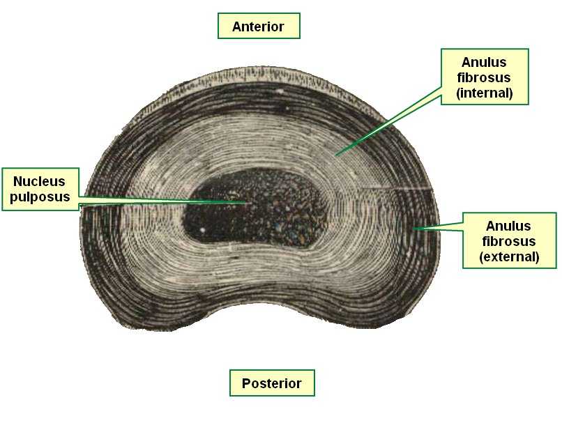
The word [lamella] is Latin and is the diminutive form of [lamina], which means "thin", or a "small plate". A lamella is a thin, wafer-like structure. The plural form is [lamellae]. The adjectival form [lamellar] refers to structures that resemble a stack of thin plates.
Many structures in the human body have a lamellar organization, such as compact bone, the pubic symphysis, and intervertebral discs. The accompanying image is a superior view of an intervertebral disc. Click on the image to see a larger depiction and the lamellar structure of the anulus fibrosus, the external component of the disc.
Image modified from the original from Testut and Latarjet, 1931
- Details
Click for a larger image
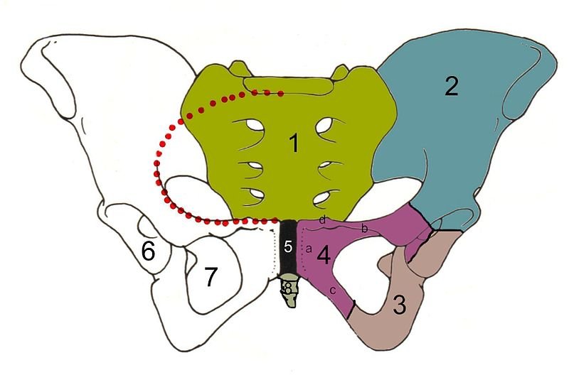
The pubic symphysis is a joint found between the pubic bones. It is composed by a layer of hyaline cartilage that covers the medial surface of each pubic bone (os pubis), and a fibrocartilaginous disc arranged in lamellae, somewhat akin to the structure of an intervertebral disc. It is classified as a fibrocartilaginous joint or a secondary cartilaginous joint. It generally presents with an internal midline cleft and is surrounded by strong ligaments. This joint has very little movement, limited to about one degree. The ligaments are:
• Superior pubic ligament: Found on the public crest, this ligament extends laterally as far as the pubic tubercles. It also serves as anchoring point for the tendons of the rectus abdominis muscles and the pyramidalis muscle.
• Inferior pubic ligament: Also known as the subpubic ligament this ligament forms an arch and attaches to the inferomedial aspect of the inferior pubic rami
• Anterior pubic ligament: Found anterior to the pubic symphysis, this ligament attaches to the periosteum of the anterior aspect of the body of the pubic bone
•Posterior pubic ligament: Found on the posterior aspect of the pubic bone, this ligament is the anterior boundary of the retropubic space of Retzius
Under the influence of hormones, the pubic symphysis becomes softer and more mobile in pregnant women. This is good for natural birth, but can be the cause of symphyseal pain in the later stages of pregnancy.
- Details
This medical word is formed by the prefix [-dys-] meaning “abnormal”, the root term [-kin-] meaning “movement”, and the suffix [-esia] meaning “condition” or “situation". Dyskinesia is “a condition of abnormal movement”.
Dyskinesia refers to a wide variety of involuntary movements that are either extraneous, or a fragmentation of normal movements of the face or limbs. The etiology for dyskinesia can be pharmacological, because of long-term use of antipsychotic medicine (tardive dyskinesia) or related to damage (trauma) or abnormal development of deep brain structures.
Dyskinetic movements are involuntary and classified as:
- Tremor: Small oscillating movements either at rest or during muscle activity, as in Parkinson’s disease
- Chorea: Irregular random and excessive movements
- Myoclonus: Rapid, fast and arrhythmic movements
- Tics: Brief and repetitive movements
- Dystonia: Postures that are involuntary and maintained for a brief time
Following are links to YouTube videos that show patients with different types of dyskinesia. These videos will open in a separate page
WARNING! These videos could be disturbing to some viewers. Videos courtesy of the JAMA Network.
Video 1: Demonstration of Kinesigenic Dyskinesia
Video 2: Choreathetotic Movement Attack in a Patient With Paroxysmal Nonkinesigenic Dyskinesia
Video 3: Orofacial Dyskinesia
- Details
This article is part of the series "A Moment in History" where we honor those who have contributed to the growth of medical knowledge in the areas of anatomy, medicine, surgery, and medical research.

Adrian Van Der Spigelius
Adriaan Van Der Spigelius (1578 - 1625). Sometimes knows as Adrianus Spigelius, was a Flemish surgeon, anatomist, and botanist born in Brussels. Just as Vesalius, he studied at the University of Louvain, and later in Padua, Italy. Having settled as a professor of anatomy in Venice, in 1616 he was offered and accepted the Chair of Anatomy at Padua. His posthumous work "De Humani Corporis Fabrica Libri X" was published in 1627.
Spigelius described the caudate lobe of the liver and the "linea semilunaris", the lateral border of the rectus abdominis muscle. Today, a ventral hernia that occurs in the linea semilunaris is said to be an "Spigelian hernia".
His name is also associated with the Spigelian fascia, formed by the combined aponeuroses of the external oblique muscle, the internal oblique muscle and the transversus abdominis muscle.
If you hover over Spigelius' image, you will see an anatomical drawing depicting the linea semilunaris (arrow).
Original image courtesy of Wikipedia. Hover image property of:CAA.Inc.. Artist:D.M. Klein
- Details
The root term [-kin-] and its variation [-kine-] originate from the Greek word [κινέω] (kineo) and means "to move", or "place in motion". Applications of this root term include:
- Kinesiology: Study of movement
- Dyskinesia: Abnormal movement
- Kinetic: Pertaining to movement
- Kinesis: Movement
- Cytokinesis: Cell movement
- Kinesthesia: Sensation of movement, etc.
The root terms [-cine-] and [-cinemat-] are a variation of [-kine-] and also mean "movement". They are the basis of the word "cinematography" which means "process of writing or recording movement" in other words "moving pictures". The shortened version gives us [cinema]. Medical use of these root terms gives us:
- Cineangiography: Recording of vessels in movement
- Cineangiocardiography: Recording of the heart vessels in movement
- Coronary cineangiography: Pertaining to movement of the coronary vessels
- Details

Click for a larger image
The [pectineal ligament], also known as [Cooper's ligament) is a thickening of the periosteum that overlays the pectineal line of the pubis which is reinforced by fibers that extend from the inguinal ligament (Poupart's) and the lacunar ligament (Gimbernat's).
The medial boundary of the pectineal ligament is at the pubic tubercle, where the ligament is at its thickest. From here the pectineal ligament extends laterally as part of the pectineal line of the pubis. As it moves laterally the pectineal ligament thins slowly. There is no clear lateral boundary to the lateral aspect of the pectineal ligament.
This is important in the repair of inguinofemoral hernias though laparoscopic procedures. The pectineal ligament is one of the targets used to place staples or bone tacks to attach a mesh that reinforces the hernia repair.
Do not confuse this ligament with others with the same name found in the breast.
Image property of: CAA.Inc. Artist: Mark Zuptitch