Medical Terminology Daily (MTD) is a blog sponsored by Clinical Anatomy Associates, Inc. as a service to the medical community. We post anatomical, medical or surgical terms, their meaning and usage, as well as biographical notes on anatomists, surgeons, and researchers through the ages. Be warned that some of the images used depict human anatomical specimens.
You are welcome to submit questions and suggestions using our "Contact Us" form. The information on this blog follows the terms on our "Privacy and Security Statement" and cannot be construed as medical guidance or instructions for treatment.
We have 440 guests online

Jean George Bachmann
(1877 – 1959)
French physician–physiologist whose experimental work in the early twentieth century provided the first clear functional description of a preferential interatrial conduction pathway. This structure, eponymically named “Bachmann’s bundle”, plays a central role in normal atrial activation and in the pathophysiology of interatrial block and atrial arrhythmias.
As a young man, Bachmann served as a merchant sailor, crossing the Atlantic multiple times. He emigrated to the United States in 1902 and earned his medical degree at the top of his class from Jefferson Medical College in Philadelphia in 1907. He stayed at this Medical College as a demonstrator and physiologist. In 1910, he joined Emory University in Atlanta. Between 1917 -1918 he served as a medical officer in the US Army. He retired from Emory in 1947 and continued his private medical practice until his death in 1959.
On the personal side, Bachmann was a man of many talents: a polyglot, he was fluent in German, French, Spanish and English. He was a chef in his own right and occasionally worked as a chef in international hotels. In fact, he paid his tuition at Jefferson Medical College, working both as a chef and as a language tutor.
The intrinsic cardiac conduction system was a major focus of cardiovascular research in the late nineteenth and early twentieth centuries. The atrioventricular (AV) node was discovered and described by Sunao Tawara and Karl Albert Aschoff in 1906, and the sinoatrial node by Arthur Keith and Martin Flack in 1907.
While the connections that distribute the electrical impulse from the AV node to the ventricles were known through the works of Wilhelm His Jr, in 1893 and Jan Evangelista Purkinje in 1839, the mechanism by which electrical impulses spread between the atria remained uncertain.
In 1916 Bachmann published a paper titled “The Inter-Auricular Time Interval” in the American Journal of Physiology. Bachmann measured activation times between the right and left atria and demonstrated that interruption of a distinct anterior interatrial muscular band resulted in delayed left atrial activation. He concluded that this band constituted the principal route for rapid interatrial conduction.
Subsequent anatomical and electrophysiological studies confirmed the importance of the structure described by Bachmann, which came to bear his name. Bachmann’s bundle is now recognized as a key determinant of atrial activation patterns, and its dysfunction is associated with interatrial block, atrial fibrillation, and abnormal P-wave morphology. His work remains foundational in both basic cardiac anatomy and clinical electrophysiology.
Sources and references
1. Bachmann G. “The inter-auricular time interval”. Am J Physiol. 1916;41:309–320.
2. Hurst JW. “Profiles in Cardiology: Jean George Bachmann (1877–1959)”. Clin Cardiol. 1987;10:185–187.
3. Lemery R, Guiraudon G, Veinot JP. “Anatomic description of Bachmann’s bundle and its relation to the atrial septum”. Am J Cardiol. 2003;91:148–152.
4. "Remembering the canonical discoverers of the core components of the mammalian cardiac conduction system: Keith and Flack, Aschoff and Tawara, His, and Purkinje" Icilio Cavero and Henry Holzgrefe Advances in Physiology Education 2022 46:4, 549-579.
5. Knol WG, de Vos CB, Crijns HJGM, et al. “The Bachmann bundle and interatrial conduction” Heart Rhythm. 2019;16:127–133.
6. “Iatrogenic biatrial flutter. The role of the Bachmann’s bundle” Constán E.; García F., Linde, A.. Complejo Hospitalario de Jaén, Jaén. Spain
7. Keith A, Flack M. The form and nature of the muscular connections between the primary divisions of the vertebrate heart. J Anat Physiol 41: 172–189, 1907.
"Clinical Anatomy Associates, Inc., and the contributors of "Medical Terminology Daily" wish to thank all individuals who donate their bodies and tissues for the advancement of education and research”.
Click here for more information
- Details
The word "hyaline" is a derivate of the Greek [υαλώδης] (yalódis) meaning "glassy". It refers to a glassy, transparent substance. Although it is usually associated with hyaline cartilage, this term can be used by itself in daily English.
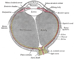
Galen of Pergamon used the term [hyaloid] (glassy, or similar to glass) to refer to the vitreous humor of the eye. As a result of this early anatomical term, today we have the following:
• Hyaloid membrane: also known as the vitreous membrane. It is a collagenous membrane separating the vitreous humor from the rest of the structures of the eye
• Hyaloid artery: a branch of the opthalmic artery which dissapears before birth
• Hyaloid canal: a small membranous canal in the vitrous humor extending between the lens and the optic disc. This can be seen in the accompanying image of a horizontal section of the eye.
Sources:
1. “Gray’s Anatomy” Henry Gray, 1918
2. "Tratado de Anatomia Humana" Testut et Latarjet 8th Ed. 1931 Salvat Editores, Spain
3. "Gray's Anatomy" 38th British Ed. Churchill Livingstone 1995
4. "The Origin of Medical Terms" Skinner, HA 1970 Hafner Publishing Co.
Image modified from the original by Henry VanDyke Carter, MD. in the book "Grays's Anatomy" by Henry Gray FRS. Public domain
Note: Google Translate includes the symbol (?). Clicking on it will allow you to hear the pronunciation of the word.
- Details
[Anular] means "ring" or "ring-shaped". The word [epiphysis] is composed of the preffix [epi-] meaning "outer" or "above", while [-physis] means "growth". The term [anular epiphysis] means the "outer ring-shaped growth".
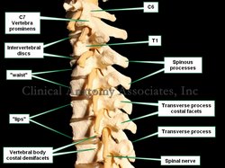
The [anular epiphysis], sometimes also called the [anular apophysis], is a bony ring found on the superior and inferior aspect of the vertebrae. This outer ring is formed by thickened cortical bone and leaves a central depression which has a more porous surface. In this central depression the vertebrae present with a thin layer of hyaline cartilage, which forms part of the vertebral endplate.
In a lateral view the presence of the anular epiphysis causes the superior and inferior borders of the vertebral body to protrude. This protrusion is called by some anatomists the "lips" of the vertebrae. To see these "lips" click here.
Image property of: CAA.Inc.Photographer: David M. Klein
- Details
The root term [-auricul-] arises from the Latin word [Auricula], which is a diminutive of [auris] meaning "ear". Auricula means "a little ear".
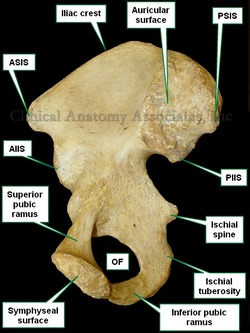
The adjectival form [auricular] is used in several places in human anatomy. One of them is the [auricular surface of the iliac bone] (see image) referring to a roughened area in the medial aspect of the iliac bone where it articulates with the sacrum, forming the sacroiliac joint. The Latin term for this articular surface is facies auricularis ossis ilii. A corresponding auricular surface is found in the sacral bone, the facies auricularis ossis sacri.
[Auricular] is also used in the term [auricular cartilage], referring to the fibrocartilage found in the ear or [pinna].
A shortened version is the root term [-aur(i)-], meaning the same. It is used in the word [auricle] referring to the atrial appendages of the heart. These appendages when seen from the outside of the heart really look like little "boxer's ears". This image (click here) shows the left atrial appendage (LAA) or left auricle.
By the way, did you know that one of the digits was called "digitus auricularis"? Read the story here.
Image property of: CAA.Inc.Photographer: David M. Klein
- Details
This article is part of the series "A Moment in History" where we honor those who have contributed to the growth of medical knowledge in the areas of anatomy, medicine, surgery, and medical research.
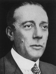
Enrique Finochietto, MD (1881 – 1948) Argentinian surgeon and researcher, Enrique Finochietto was born in 1881 in the city of Buenos Aires. He studied in both public and private schools, entering medical school at age 16. He received his medical degree from the University of Buenos Aires in 1904.
Shortly after graduation, he became an intern at the Rawson hospital of Buenos Aires. Dr. Finochietto would become part of the staff of this hospital and be part of its staff all his life. He traveled to Europe to study with surgeons such as Calot, Kocher, Roux, and others, visiting France, Switzerland, Austria, and Italy.
Upon his return he dedicated himself to surgery and endoscopy, and with the help of his brother Ricardo started research and invented many medical and surgical devices, including a motorized surgical table, endoscopic devices and surgical instruments, some of which are in use today throughout the world.
During World War I Dr. Finochietto worked for no fee (ad honorem) as surgeon-in-chief at the Argentine Hospital in Paris. For his dedication and work, France awarded him the Legion d’Honneur and the Red Cross. He came back to Argentina and then traveled to the USA where he visited with the Mayo Brothers and Harvey Cushing. One of Dr. Finochietto’s mottos was “Only the surgeon who goes beyond his obligations serves his duty”.
Although mostly remembered for the Finochietto rib spreader, Dr. Finochietto tackled most types of surgery during his career, including neurosurgery, gastrointestinal, thyroid, thoracic, and orthopedic.
Dr. Finochietto died on February 17, 1948. His legacy in Argentinian surgery lives on through his school of thought, research and disciples.
If you click on Dr. Finochietto’s image (courtesy of Wikipedia) you will see an image of his namesake rib spreader (courtesy of Surgiway, France ).
Sources:
1. “Memoir: Enrique Finochietto, MD (1881 – 1948)” DeBakey. ME. Ann Surg. 1948 Aug; 128(2): 319–320
2. “Enrique Finochietto” The Legacy of Surgery in Argentina” De La Fuente, SG. J Surg Ed (2007)64:2; 120-123
3. “El separador intercostal de Enrique Finochietto, el hospital Rawson, la escuela quir?rgica, y una an?cdota que los relaciona” Saadia, A. Rev Arg Cir Cardiovasc (2009) 3:3 149-150
4. “Rese?as hist?ricas: Enrique Finochietto” Parquet RA. Acta Gastroenterol Latinoam (2009); 39:18
5. “Surgery in Argentina” Beveraggi, EM. Arch Surg (1999) 134”438-444
6. “The Legends Behind Cardiothoracic Surgical Instruments” Ailawadi, G et al. Ann Thorac Surg (2010) 89:5; 1693-1700
- Details
The term [facet] is English and derives from the French word [facette] meaning "a small face". The French term is itself derived from the Latin [facies], meaning "face".
The term facet in anatomy is used to refer to a small smooth surface, usually an articular surface. Another use of the word, still meaning "a small face" can be found in the jeweler's term [facet] used to describe one of the many small surfaces in a diamond.
In spinal anatomy, the term “a facet joint” is most commonly used, but the term should be pronounced with the accent on the first syllable as in “fácet”! And just to be a bit more correct, the proper term for a so-called “facet joint” is “zygapophyseal joint”. This term is one of several medical terms that are used and/or pronounced incorrectly.
- Details
The [spinous process] is a single, median posterior bony process found in most vertebrae, with the exception of C1, and coccygeal vertebrae.
The spinous processes have regional variations according to the spinal region. Cervical vertebrae usually have a bifid spinous process, while C7 (vertebra prominens) has the longest spinous process in the cervical region
In the thoracic region the spinous processes are thin, slender and they overlap each other, while in the lumbar region these processes are thick and square. The spinous processes in the sacral region become progressively smaller.
Image property of: CAA.Inc.Photographer: David M. Klein


