Medical Terminology Daily (MTD) is a blog sponsored by Clinical Anatomy Associates, Inc. as a service to the medical community. We post anatomical, medical or surgical terms, their meaning and usage, as well as biographical notes on anatomists, surgeons, and researchers through the ages. Be warned that some of the images used depict human anatomical specimens.
You are welcome to submit questions and suggestions using our "Contact Us" form. The information on this blog follows the terms on our "Privacy and Security Statement" and cannot be construed as medical guidance or instructions for treatment.
We have 1118 guests online

Jean George Bachmann
(1877 – 1959)
French physician–physiologist whose experimental work in the early twentieth century provided the first clear functional description of a preferential interatrial conduction pathway. This structure, eponymically named “Bachmann’s bundle”, plays a central role in normal atrial activation and in the pathophysiology of interatrial block and atrial arrhythmias.
As a young man, Bachmann served as a merchant sailor, crossing the Atlantic multiple times. He emigrated to the United States in 1902 and earned his medical degree at the top of his class from Jefferson Medical College in Philadelphia in 1907. He stayed at this Medical College as a demonstrator and physiologist. In 1910, he joined Emory University in Atlanta. Between 1917 -1918 he served as a medical officer in the US Army. He retired from Emory in 1947 and continued his private medical practice until his death in 1959.
On the personal side, Bachmann was a man of many talents: a polyglot, he was fluent in German, French, Spanish and English. He was a chef in his own right and occasionally worked as a chef in international hotels. In fact, he paid his tuition at Jefferson Medical College, working both as a chef and as a language tutor.
The intrinsic cardiac conduction system was a major focus of cardiovascular research in the late nineteenth and early twentieth centuries. The atrioventricular (AV) node was discovered and described by Sunao Tawara and Karl Albert Aschoff in 1906, and the sinoatrial node by Arthur Keith and Martin Flack in 1907.
While the connections that distribute the electrical impulse from the AV node to the ventricles were known through the works of Wilhelm His Jr, in 1893 and Jan Evangelista Purkinje in 1839, the mechanism by which electrical impulses spread between the atria remained uncertain.
In 1916 Bachmann published a paper titled “The Inter-Auricular Time Interval” in the American Journal of Physiology. Bachmann measured activation times between the right and left atria and demonstrated that interruption of a distinct anterior interatrial muscular band resulted in delayed left atrial activation. He concluded that this band constituted the principal route for rapid interatrial conduction.
Subsequent anatomical and electrophysiological studies confirmed the importance of the structure described by Bachmann, which came to bear his name. Bachmann’s bundle is now recognized as a key determinant of atrial activation patterns, and its dysfunction is associated with interatrial block, atrial fibrillation, and abnormal P-wave morphology. His work remains foundational in both basic cardiac anatomy and clinical electrophysiology.
Sources and references
1. Bachmann G. “The inter-auricular time interval”. Am J Physiol. 1916;41:309–320.
2. Hurst JW. “Profiles in Cardiology: Jean George Bachmann (1877–1959)”. Clin Cardiol. 1987;10:185–187.
3. Lemery R, Guiraudon G, Veinot JP. “Anatomic description of Bachmann’s bundle and its relation to the atrial septum”. Am J Cardiol. 2003;91:148–152.
4. "Remembering the canonical discoverers of the core components of the mammalian cardiac conduction system: Keith and Flack, Aschoff and Tawara, His, and Purkinje" Icilio Cavero and Henry Holzgrefe Advances in Physiology Education 2022 46:4, 549-579.
5. Knol WG, de Vos CB, Crijns HJGM, et al. “The Bachmann bundle and interatrial conduction” Heart Rhythm. 2019;16:127–133.
6. “Iatrogenic biatrial flutter. The role of the Bachmann’s bundle” Constán E.; García F., Linde, A.. Complejo Hospitalario de Jaén, Jaén. Spain
7. Keith A, Flack M. The form and nature of the muscular connections between the primary divisions of the vertebrate heart. J Anat Physiol 41: 172–189, 1907.
"Clinical Anatomy Associates, Inc., and the contributors of "Medical Terminology Daily" wish to thank all individuals who donate their bodies and tissues for the advancement of education and research”.
Click here for more information
- Details
This article is part of the series "A Moment in History" where we honor those who have contributed to the growth of medical knowledge in the areas of anatomy, medicine, surgery, and medical research.

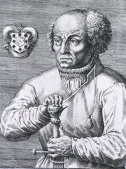
Paracelsus
Paracelsus (1493 – 1541). Swiss physician and alchemist , Phillipus Aureolus Theophrastus Bombastus Von Hohenheim was born in Einsiedeln in 1493 (one year after Columbus discovered America) in what is today is Switzerland. At an early age he became a migrant student, visiting several universities including Tubingen, Vienna, Wirttemberg, Heidelberg, and Cologne. There is discussion as to whether he received or not a medical degree, although most authors today agree he might had. In 1510 he moved to Ferrara where he attained (apparently) his medical degree in 1516. During his constant travels he started to understand that folk medical treatment based on actual observation was better than what was published and followed blindly by the physicians of the time.
He started to call himself “Paracelsus” which means “alongside Celsus”, seen as one of the greatest physicians in history. Paracelsus continued his travels, visiting Egypt and Jerusalem. It is at this time that he started delving into the world of alchemy, returning to his home circa 1524.
Paracelsus was appointed “Town Physician” of the city of Basle but created controversy when he started lecturing in German (not Latin) and invited the general public as well as his students to his lectures. In his presentations he introduced the concepts of direct observation of the patient and empirical treatment, based his statements on experiments and reasoning opposing the “classics” Galen, and Avicenna.
In 1527, during a demonstration he publicly burned the works of Avicenna to prove his point. This caused a backlash from the university and town authorities who expelled him in 1528. From this point on, Paracelsus’ life is constant wandering. He settles for a time and then travels again. In spite of his disdain for the works of the “medical greats”, he himself writes a large number of works, including medicine, surgery, theology, astronomy, magic, etc. Many of these works are not published until after his death as he is considered to be contradicting Galen. In 1530 he writes the best description of syphilis and recommended its treatment with mercury.
In 1541 he was appointed to a post on the staff of Duke Ernest of Bavaria, but he died in mysterious circumstances on September 24 of that year at the White Horse Inn in Salzburg.
Paracelsus is a controversial image, bound in legend. For many, Paracelsus was bombastic, quarrelsome, opinionated and a drunkard. For others he is a figure of his time, clashing with the classics and giving us a new way to look at the world and at diseases. He taught that wounds must be allowed to drain and not, as was common, packed with unhealthy materials. According to him, the human body primarily consists of salt, sulphur, and mercury, and it is the separation of these elements that causes illness. He introduced mineral baths and made opium, mercury, lead and other minerals part of his treatment, foreshadowing modern pharmacology with the use of chemical remedies, mercury for syphilis, laudanum and antimony. Paracelsus stated in 1538 that “Everything is a poison, the dose alone which makes a thing not a poison”.
I just discovered an interesting chain of events. For a time Paracelsus had a medical student that later decided not to continue his medical studies and instead dedicated himself to the new art of printing. His name was Johannes Oporinus and he was the printer that Andreas Vesalius selected to print his masterpiece, the "Fabrica".
Sources
1. “Paracelsus” Abbott.A. Nature 366: (1993) 98
2. “Paradigm lost: a celebration of Paracelsus on his quincentenary” Feder. G. Lancet. 341: (1993) 1396-1397
3. “Does Paracelsus deserve a place in the medical pantheon?” Bynum, B. 367: (2006) 29; 1389–1390
4. “Paracelsus: founder of medical chemistry” Endeavour 15:4 (1991) 147
5. “Paracelsus: the medical Luther” Leary B. 73: 3(1984) 131-133
6. “Paracelsus and the Philosopher’s Stone” TenHoor, W. Am J Surg (1935) 30:3 563-572
- Details
This word is of Greek origin composed of the root term [-arthr-], meaning "joint", as in an bony articulation, and the suffix [-(o)desis], meaning "to bind together".
An arthrodesis is the surgical immobilization of a joint by fusion or the application of surgical devices (rods, screws, etc) to the bony components of a joint.
- Details
This medical term is Greek and is composed of [γιατρός] (iatros) meaning "doctor", "physician", or "healer" and the suffix [-(o)genic], meaning "creation", "born of", or "beggining". An iatrogenic condition is that which is caused or created by the doctor or the hospital.
This is an expensive word, as iatrogenic conditions may lead to a lawsuit!
- Details
The word [leiomyoma] is of Greek origin with combined root terms. The term [-lei(o)-] arises from the Greek [λείος] meaning "smooth", the other root is [μυς] (mys) meaning "muscle". The suffix [-oma] means "tumor" or "mass". A [leiomyoma] is a "smooth muscle tumor". The medical plural form is [leiomyomata], or it can be [leiomyomas].
Since smooth muscle is involuntary muscle, leiomyomata are usually found in viscera. The most common leiomyomata are found in the uterus (see image), in the muscular or submucosal layer of the digestive system, mostly jejunum and ileum, and gallbladder, or in smooth muscle of the skin. The term itself does not imply that leiomyomata are cancerous, and most leiomyomata are not.
- Details

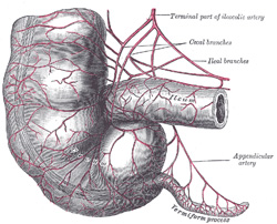
Terminal ileum, cecum,
and vermiform appendix
The [mesoappendix] is a triangular-shaped double-layered peritoneal membrane related to the vermiform appendix. One of the sides attaches to the vermiform appendix, the other is free, and the third one attaches to the ileum and the cecum. This last attachment varies in extension, giving the cecum varying degrees of mobility.
The mesoappendix contains the appendicular artery. This artery arises either from the ileocolic artery or the from the posterior ileocecal artery. The mesoappendix also contains the appendicular veins, lymphatics, lymphatic nodes, and fat.
In the female, there can be an extension of the mesoappendix that communicates with the broad ligament of the uterus. It is called the appendiculoovarian ligament, or Clado's ligament. This ligament may contain the appendiculoovarian artery, an anastomosis between the appendicular artery and the ovarian artery. The lymphatics contained in this appendiculoovarian ligament can also establish a lymphatic communication between the ovary and the vermiform appendix.
Sources:
1 "Tratado de Anatomia Humana" Testut et Latarjet 8 Ed. 1931 Salvat Editores, Spain
2. "Anatomy of the Human Body" Henry Gray 1918. Philadelphia: Lea & Febiger Image modified by CAA, Inc. Original image by Henry Vandyke Carter, MD., courtesy of bartleby.com
- Details
This article is part of the series "A Moment in History" where we honor those who have contributed to the growth of medical knowledge in the areas of anatomy, medicine, surgery, and medical research.
Alessandra Giliani (1307 – 1326). Italian prosector and anatomist. Alessandra Giliani is the first woman to be on record as being an anatomist and prossector. She was born on 1307 in the town of Persiceto in northern Italy.
She was admitted to the University of Bologna circa 1323. Most probably she studied philosophy and the foundations of anatomy and medicine. She studied under Mondino de Luzzi (c.1270 – 1326), one of the most famous teachers at Bologna.
Giliani was the prosector for the dissections performed at the Bolognese “studium” in the Bologna School of Anatomy. She developed a technique (now lost to history) to highlight the vascular tree in a cadaver using fluid dyes which would harden without destroying them. Giliani would later paint these structures using a small brush. This technique allowed the students to see even small veins.
Giliani died at the age of 19 on March 26, 1326, the same year that her teacher Mondino de Luzzi died. It is said that she was buried in front of the Madonna delle Lettere in the church of San Pietro e Marcellino at the Hospital of Santa Maria del Mareto in Florence by Otto Agenius Lustrulanus, another assistant to Modino de Luzzi.
Some ascribe to Agenius a love interest in Giliani because of the wording of the plaque that is translated as follows:
"In this urn enclosed are the ashes of the body of
Alessandra Giliani, a maiden of Persiceto.
Skillful with her brush in anatomical demonstrations
And a disciple equaled by few,
Of the most noted physician, Mondino de Luzzi,
She awaits the resurrection.
She lived 19 years: She died consumed by her labors
March 26, in the year of grace 1326.
Otto Agenius Lustrulanus, by her taking away
Deprived of his better part, inconsolable for his companion,
Choice and deservinging of the best from himself,
Has erected this plaque"
Sir William Osler says of Alessandra Giliani “She died, consumed by her labors, at the early age of nineteen, and her monument is still to be seen”
The teaching of anatomy in the times of Mondino de Luzzi and Alessandra Giliani required the professor to be seated on a high chair or “cathedra” from whence he would read an anatomy book by Galen or another respected author while a prosector or “ostensor” would demonstrate the structures to the student. The professor would not consider coming down from the cathedra to discuss the anatomy shown. This was changed by Andreas Vesalius.
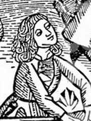
The image in this article is a close up of the title page of Mondino’s “Anothomia Corporis Humani” written in 1316, but published in 1478. Click on the image for a complete depiction of this title page. I would like to think that the individual doing the dissection looking up to the cathedra and Mondino de Luzzi is Alessandra Giliani… we will never know.
The life and death of Alessandra Giliani has been novelized in the fiction book “A Golden Web” by Barbara Quick.
Sources
1. “Books of the Body: Anatomical Ritual and Renaissance Learning” Carlino, A. U Chicago Press, 1999
2. “Encyclopedia of World Scientists” Oakes, EH. Infobase Publishing, 2002
3. “The Biographical Dictionary of Women in Science”Harvey, J; Ogilvie, M. Vol1. Routledge 2000
4. “The Evolution of Modern Medicine” Osler, W. Yale U Press 1921
5. “The Mondino Myth” Pilcher, LS. 1906
Original image courtesy of NLM


