![]()
Medical Terminology Daily (MTD) is a blog sponsored by Clinical Anatomy Associates, Inc. as a service to the medical community. We post anatomical, medical or surgical terms, their meaning and usage, as well as biographical notes on anatomists, surgeons, and researchers through the ages. Be warned that some of the images used depict human anatomical specimens.
You are welcome to submit questions and suggestions using our "Contact Us" form. The information on this blog follows the terms on our "Privacy and Security Statement" and cannot be construed as medical guidance or instructions for treatment.
We have 408 guests and no members online
")
Marcia Crocker Noyes
(1869 – 1946)
Further to my comment on old books and research that started with an interesting bookplate (Ex-Libris). I continued my research and found that the person in charge of the Osler library bookplate was a fascinating individual that today maybe a ghost in the MedChi library and building in Baltimore... This is certainly an article that can be called "A Moment in History"
Marcia Crocker Noyes was the librarian at The Maryland State Medical Society from 1896 to 1946 and was a founding member of the Medical Library Association.[1][2][3]
Sir William Osler, MD. a famous Johns Hopkins surgeon was a noted bibliophile and had a large personal collection of books on various topics. When he became the President of MedChi in 1896, he was dismayed at the condition of the library and knew that with the right person and some stewardship, it could become a significant collection. Sir William asked his friend, Dr. Bernard Steiner, a physician and President of the Enoch Pratt Free Library in Baltimore for suggestions of a librarian, and Dr. Steiner recommended Marcia Crocker Noyes. A native of New York, and a graduate of Hunter College, Marcia had moved to Baltimore for a lengthy visit with her sister, and took a “temporary” position at the Pratt Library, which turned into three years. Although she had no medical experience or background, she was enthusiastic, and most importantly, she was willing to move into the apartment provided for the librarian, who needed to be available 24 hours a day.
The image in this article is Ms. Noyes on her first year on the job. Marcia developed a book classification system for medical books, based on the Index Medicus, and called it the Classification for Medical Literature. The system uses the alphabet with capital letters for the major divisions of medicine and lower-case ones for the sub-sections. The system was used for many years, but it's now dated and the Faculty's original shelving scheme was never changed. The card catalogs still reflect her classification and many of the cards are written in Marcia's back-slanting handwriting.
Marcia knew enough to ask the Faculty's members about medical questions, terminology and literature. She gradually won over the predominantly male membership and they became her greatest allies; Sir William at the start, and then for nearly 40 years, Dr. John Ruhräh, a wealthy pediatrician with no immediate family of his own. She made a point of attending almost every Faculty function, and in 1904, under guidelines from the American Medical Association, Marcia was made the Faculty Secretary. For much of her first 10 years, she was the Faculty's only full-time employee, only being assisted by Mr. Caution, the Faculty's janitor. Later in life Marcia would say that she hired him because of his name!
Within ten years, the library had outgrown its space, and plans, spearheaded by Marcia and Sir William before his move to Oxford, were made to build a headquarters building, mainly to house the library's growing collection of medical books and journals.
Marcia was instrumental in the design and building of the new headquarters. She travelled to Philadelphia, New York and Boston to look at their medical society buildings, and eventually, the Philadelphia architectural firm, Ellicott & Emmart was selected to design and build the new Faculty building. Every detail of the building held her imprimatur, from the graceful staircase, to the light-filled reading room, and all of the myriad details of the millwork, marble tesserae, and most of all, the four-story cast iron stacks. She was on-site, climbing up unfinished staircases, checking out the progress of the building, which was built in less than one year at a cost of $90,000.
Among the features of the new building was a fourth-floor apartment for her. She referred to it as the "first penthouse in Baltimore" and it had a garden and rooftop terrace. The library collection eventually grew to more than 65,000 volumes from medical and specialty societies around the world. Journals were traded back and forth, and physicians eagerly anticipated the arrival of each new issue. At the same time, Marcia was involved in the Medical Library Association as one of eight founding members. The MLA promotes medical libraries and the exchange of information. One of the earliest mandates of the MLA was the Exchange, a distribution and trade service for those who had duplicates or little-used books in their collections. Initially, the Exchange was run out of the Philadelphia medical society, but in 1900 it was moved to Baltimore and Marcia oversaw it. Several hundred periodicals and journals were received and sent each month, a huge amount of work for a tiny staff. In 1904, the Faculty had run out of room to manage the Exchange, so it was moved to the Medical Society of the Kings County (Brooklyn). But without Marcia's excellent administrative skills, it floundered and in 1908, the MLA asked Marcia to take charge once again.
In 1909, when the new Faculty building opened, there was enough room to run the Exchange and with the help of MLA Treasurer, noted bibliophile and close friend, Dr. John Ruhräh, it once again became successful. Additionally, Marcia and Dr. Ruhräh combined forces to revive the MLA's bulletin, which had all but ceased publication in 1908, taking the Exchange with it. This duo maintained editorial control from 1911 until 1926. In 1934, around the time of Dr. Ruhräh's death, Marcia became the first “unmedicated” professional to head the MLA. During her tenure, the MLA incorporated, the first seal was adopted, and the annual meeting was held in Baltimore. Marcia wanted to write the history of the MLA once she retired from full-time work at the Faculty, but her health was beginning to fail. She had back problems and had suffered a serious burn on her shoulder as a young woman, possibly from her time running a summer camp, Camp Seyon, for young ladies in the Adirondack Mountains. In 1946, a celebration was planned to honor Marcia's 50 years at the Faculty. But she was adamant that the physicians wait until November, the actual date of her 50 years. However, they knew she was gravely ill, and might not make it until then, so a huge party was held in April. More than 250 physicians attended the celebration, but the ones she was closest to in the early years, were long gone. She was presented with a suitcase, a sum of money to use for travelling, and her favorite painting of Dr. John Philip Smith, a founder of the Medical College in Winchester, Virginia. It was painted by Edward Caledon Smith, a Virginia painter who had been a student of the painter Thomas Sully.[4] She adored this painting and vowed, jokingly, to take it with her wherever she went.
The painting was not to stay with her for very long, for she died in November 1946, and left it to the Faculty in her will. Her funeral was held in the Faculty's Osler Hall, named for her dear friend. More than 60 physicians served as her pallbearers, and she was buried at Baltimore's Green Mount Cemetery. In 1948, the MLA decided to establish an award in the name of Marcia Crocker Noyes. It was for outstanding achievement in medical library field and was to be awarded every two years, or when a truly worthy candidate was submitted. In 2014, the Faculty began giving a bouquet of flowers to the winner of the award in Marcia's name, and in honor of her work. Much evidence exists for this tradition, as we know that the physicians, especially Drs. Osler and Ruhräh, frequently gave her bouquets of flowers. Marcia also cultivated flower gardens at the Faculty and decorated the rooms with her work.
Today, the MedChi building is open for tours and if the rumors are to be believed Ms. Marcia Crocker Noyes is still at work in her beloved library as the "resident ghost" [1][5]

NOTE: This article has been modified from the original Wikipedia article on Marcia Crocker Noyes. The article itself is well-written with interesting images of the subject. I would encourage you to visit it. The second insert is from book 00736 in my personal library and shows in pencil, the incredibly small handwriting of Marsha C. Noyes.
Sources:
1. "Marcia, Marcia, Marcia" MedChi Archives blog.
2. "Marcia C. Noyes, Medical Librarian" (PDF). Bulletin of the Medical Library Association. 35 (1): 108–109. 1947. PMC 194645
3. Smith, Bernie Todd (1974). "Marcia Crocker Noyes, Medical Librarian: The Shaping of a Career" (PDF). Bulletin of the Medical Library Association. 62 (3): 314–324. PMC 198800Freely accessible. PMID 4619344.
4. Edward Caledon BRUCE (1825-1901)"
5. Behind the scenes tour MedChiBuilding
"Clinical Anatomy Associates, Inc., and the contributors of "Medical Terminology Daily" wish to thank all individuals who donate their bodies and tissues for the advancement of education and research”.
Click here for more information


- Details
This medical term is Greek and is composed of [γιατρός] (iatros) meaning "doctor", "physician", or "healer" and the suffix [-(o)genic], meaning "creation", "born of", or "beggining". An iatrogenic condition is that which is caused or created by the doctor or the hospital.
This is an expensive word, as iatrogenic conditions may lead to a lawsuit!
- Details
")
Click for a larger image
The word [leiomyoma] is of Greek origin with combined root terms. The term [-lei(o)-] arises from the Greek [λείος] meaning "smooth", the other root is [μυς] (mys) meaning "muscle". The suffix [-oma] means "tumor" or "mass". A [leiomyoma] is a "smooth muscle tumor". The medical plural form is [leiomyomata], or it can be [leiomyomas].
Since smooth muscle is involuntary muscle, leiomyomata are usually found in viscera. The most common leiomyomata are found in the uterus (see image), in the muscular or submucosal layer of the digestive system, mostly jejunum and ileum, and gallbladder, or in smooth muscle of the skin. The term itself does not imply that leiomyomata are cancerous, and most leiomyomata are not.
- Details
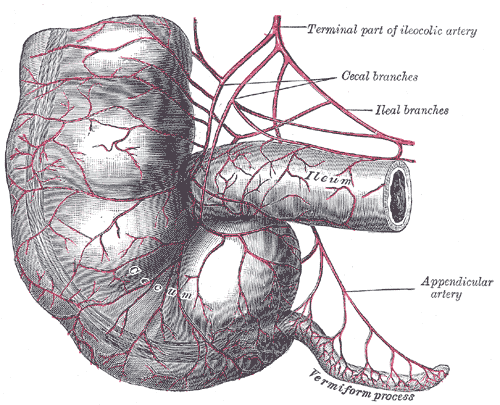
Terminal ileum, cecum,
and vermiform appendix
The [mesoappendix] is a triangular-shaped double-layered peritoneal membrane related to the vermiform appendix. One of the sides attaches to the vermiform appendix, the other is free, and the third one attaches to the ileum and the cecum. This last attachment varies in extension, giving the cecum varying degrees of mobility.
The mesoappendix contains the appendicular artery. This artery arises either from the ileocolic artery or the from the posterior ileocecal artery. The mesoappendix also contains the appendicular veins, lymphatics, lymphatic nodes, and fat.
In the female, there can be an extension of the mesoappendix that communicates with the broad ligament of the uterus. It is called the appendiculoovarian ligament, or Clado's ligament. This ligament may contain the appendiculoovarian artery, an anastomosis between the appendicular artery and the ovarian artery. The lymphatics contained in this appendiculoovarian ligament can also establish a lymphatic communication between the ovary and the vermiform appendix.
Sources:
1 "Tratado de Anatomia Humana" Testut et Latarjet 8 Ed. 1931 Salvat Editores, Spain
2. "Anatomy of the Human Body" Henry Gray 1918. Philadelphia: Lea & Febiger Image modified by CAA, Inc. Original image by Henry Vandyke Carter, MD., courtesy of bartleby.com
- Details
This article is part of the series "A Moment in History" where we honor those who have contributed to the growth of medical knowledge in the areas of anatomy, medicine, surgery, and medical research.

Click for a larger image
Alessandra Giliani (1307 – 1326). Italian prosector and anatomist. Alessandra Giliani is the first woman to be on record as being an anatomist and prossector. She was born on 1307 in the town of Persiceto in northern Italy.
She was admitted to the University of Bologna circa 1323. Most probably she studied philosophy and the foundations of anatomy and medicine. She studied under Mondino de Luzzi (c.1270 – 1326), one of the most famous teachers at Bologna.
Giliani was the prosector for the dissections performed at the Bolognese “studium” in the Bologna School of Anatomy. She developed a technique (now lost to history) to highlight the vascular tree in a cadaver using fluid dyes which would harden without destroying them. Giliani would later paint these structures using a small brush. This technique allowed the students to see even small veins.
Giliani died at the age of 19 on March 26, 1326, the same year that her teacher Mondino de Luzzi died. It is said that she was buried in front of the Madonna delle Lettere in the church of San Pietro e Marcellino at the Hospital of Santa Maria del Mareto in Florence by Otto Agenius Lustrulanus, another assistant to Modino de Luzzi.
Some ascribe to Agenius a love interest in Giliani because of the wording of the plaque that is translated as follows:
"In this urn enclosed are the ashes of the body of
Alessandra Giliani, a maiden of Persiceto.
Skillful with her brush in anatomical demonstrations
And a disciple equaled by few,
Of the most noted physician, Mondino de Luzzi,
She awaits the resurrection.
She lived 19 years: She died consumed by her labors
March 26, in the year of grace 1326.
Otto Agenius Lustrulanus, by her taking away
Deprived of his better part, inconsolable for his companion,
Choice and deservinging of the best from himself,
Has erected this plaque"
Sir William Osler says of Alessandra Giliani “She died, consumed by her labors, at the early age of nineteen, and her monument is still to be seen”
The teaching of anatomy in the times of Mondino de Luzzi and Alessandra Giliani required the professor to be seated on a high chair or “cathedra” from whence he would read an anatomy book by Galen or another respected author while a prosector or “ostensor” would demonstrate the structures to the student. The professor would not consider coming down from the cathedra to discuss the anatomy shown. This was changed by Andreas Vesalius.
The image in this article is a close up of the title page of Mondino’s “Anothomia Corporis Humani” written in 1316, but published in 1478. Click on the image for a complete depiction of this title page. I would like to think that the individual doing the dissection looking up to the cathedra and Mondino de Luzzi is Alessandra Giliani… we will never know.
The life and death of Alessandra Giliani has been novelized in the fiction book “A Golden Web” by Barbara Quick.
Sources
1. “Books of the Body: Anatomical Ritual and Renaissance Learning” Carlino, A. U Chicago Press, 1999
2. “Encyclopedia of World Scientists” Oakes, EH. Infobase Publishing, 2002
3. “The Biographical Dictionary of Women in Science”Harvey, J; Ogilvie, M. Vol1. Routledge 2000
4. “The Evolution of Modern Medicine” Osler, W. Yale U Press 1921
5. “The Mondino Myth” Pilcher, LS. 1906
Original image courtesy of NLM
- Details
")
Click for a larger image
The word [peritoneum] has a Greek origin [περίτόνοςαιον]. Loosely translated it has the prefix [peri-] meaning "around", the root [-ton-] from the Greek [tonos], meaning "to stretch", and the suffix [-eum] meaning "a membrane". It is "a membrane that is stretched around".
The peritoneum is a thin serosal membranous sac found in the abdominopelvic cavity. Histologically it is composed of a layer of mesothelium supported by a layer of connective tissue. Being a serosal sac, it contains in its interior a small amount of peritoneal fluid. The pathological accumulation of peritoneal fluid is called ascitis.
Although the peritoneum is one continuous membrane, and because of its relation to the organs and the abdominal wall, the peritoneum is described as formed by two components:
• Parietal peritoneum: The parietal peritoneum is that portion of the peritoneal sac related to or in contact with the walls of the abdomen and the pelvis.
• Visceral peritoneum: The visceral peritoneum is that portion of the peritoneal sac related to or in contact with the abdominopelvic viscera. I this case the peritoneum encases the viscera almost completely and is referred to as their serosa layer i,e: serosa layer of the ileum.
The double-layered portions of the peritoneal sac that stretch between organs or between organs and the abdominal wall are known by different names. They can be called an abdominopelvic [ligament], a [mesentery], a [meso..(something)], or an [omentum]. These structures are covered in separate articles. Some of these structures are:
- Falciform ligament: A sickle-shaped double fold of peritoneum related to the liver
- Ligament of Treitz: Also known as the "suspensory ligament of the duodenum"
- Infundibulopelvic ligament: A fold of peritoneum containing the ovarian arteries and veins
- Lesser omentum: The lesser omentum is one of the two double-folds of peritoneum related to the stomach
- Mesosigmoid: A double peritoneal membrane related to the sigmoid colon
- Transverse mesocolon: A double peritoneal membrane related to the transverse colon
- Mesoappendix: A double peritoneal membrane related to the vermiform appendix, etc.
Sources:
1. "Clinically Oriented Anatomy" Moore, KL. 3r Ed. Williams & Wilkins 1992
2. "The origin of Medical Terms" Skinner, AH, 1970
3. "Tratado de Anatomia Humana" Testut et Latarjet 8 Ed. 1931 Salvat Editores, Spain
Image modified from the original from Testut and Latajet, 1931. Public domain.
Thanks to Dr. Randall Wolf for suggesting this article
- Details

A derivate of the Latin root [cervix] or [cervicis] meaning "neck". The word [cervical] means "pertaining to the neck".
The term is used in many areas and structures of the human body:
• Cervical spine: refers to the spinal column region formed by the seven cervical vertebrae. See image
• Uterine cervix: The inferior region of the uterus which projects partially into the vagina.
• Cervical rib: An anatomic variation where one or more supernumerary ribs are found related to the lower cervical vertebrae. This anomaly can cause clinical symptoms.
Images property of: CAA.Inc. Artist: Dr. E. Miranda