![]()
Medical Terminology Daily (MTD) is a blog sponsored by Clinical Anatomy Associates, Inc. as a service to the medical community. We post anatomical, medical or surgical terms, their meaning and usage, as well as biographical notes on anatomists, surgeons, and researchers through the ages. Be warned that some of the images used depict human anatomical specimens.
You are welcome to submit questions and suggestions using our "Contact Us" form. The information on this blog follows the terms on our "Privacy and Security Statement" and cannot be construed as medical guidance or instructions for treatment.
We have 104 guests and no members online
")
Marcia Crocker Noyes
(1869 – 1946)
Further to my comment on old books and research that started with an interesting bookplate (Ex-Libris). I continued my research and found that the person in charge of the Osler library bookplate was a fascinating individual that today maybe a ghost in the MedChi library and building in Baltimore... This is certainly an article that can be called "A Moment in History"
Marcia Crocker Noyes was the librarian at The Maryland State Medical Society from 1896 to 1946 and was a founding member of the Medical Library Association.[1][2][3]
Sir William Osler, MD. a famous Johns Hopkins surgeon was a noted bibliophile and had a large personal collection of books on various topics. When he became the President of MedChi in 1896, he was dismayed at the condition of the library and knew that with the right person and some stewardship, it could become a significant collection. Sir William asked his friend, Dr. Bernard Steiner, a physician and President of the Enoch Pratt Free Library in Baltimore for suggestions of a librarian, and Dr. Steiner recommended Marcia Crocker Noyes. A native of New York, and a graduate of Hunter College, Marcia had moved to Baltimore for a lengthy visit with her sister, and took a “temporary” position at the Pratt Library, which turned into three years. Although she had no medical experience or background, she was enthusiastic, and most importantly, she was willing to move into the apartment provided for the librarian, who needed to be available 24 hours a day.
The image in this article is Ms. Noyes on her first year on the job. Marcia developed a book classification system for medical books, based on the Index Medicus, and called it the Classification for Medical Literature. The system uses the alphabet with capital letters for the major divisions of medicine and lower-case ones for the sub-sections. The system was used for many years, but it's now dated and the Faculty's original shelving scheme was never changed. The card catalogs still reflect her classification and many of the cards are written in Marcia's back-slanting handwriting.
Marcia knew enough to ask the Faculty's members about medical questions, terminology and literature. She gradually won over the predominantly male membership and they became her greatest allies; Sir William at the start, and then for nearly 40 years, Dr. John Ruhräh, a wealthy pediatrician with no immediate family of his own. She made a point of attending almost every Faculty function, and in 1904, under guidelines from the American Medical Association, Marcia was made the Faculty Secretary. For much of her first 10 years, she was the Faculty's only full-time employee, only being assisted by Mr. Caution, the Faculty's janitor. Later in life Marcia would say that she hired him because of his name!
Within ten years, the library had outgrown its space, and plans, spearheaded by Marcia and Sir William before his move to Oxford, were made to build a headquarters building, mainly to house the library's growing collection of medical books and journals.
Marcia was instrumental in the design and building of the new headquarters. She travelled to Philadelphia, New York and Boston to look at their medical society buildings, and eventually, the Philadelphia architectural firm, Ellicott & Emmart was selected to design and build the new Faculty building. Every detail of the building held her imprimatur, from the graceful staircase, to the light-filled reading room, and all of the myriad details of the millwork, marble tesserae, and most of all, the four-story cast iron stacks. She was on-site, climbing up unfinished staircases, checking out the progress of the building, which was built in less than one year at a cost of $90,000.
Among the features of the new building was a fourth-floor apartment for her. She referred to it as the "first penthouse in Baltimore" and it had a garden and rooftop terrace. The library collection eventually grew to more than 65,000 volumes from medical and specialty societies around the world. Journals were traded back and forth, and physicians eagerly anticipated the arrival of each new issue. At the same time, Marcia was involved in the Medical Library Association as one of eight founding members. The MLA promotes medical libraries and the exchange of information. One of the earliest mandates of the MLA was the Exchange, a distribution and trade service for those who had duplicates or little-used books in their collections. Initially, the Exchange was run out of the Philadelphia medical society, but in 1900 it was moved to Baltimore and Marcia oversaw it. Several hundred periodicals and journals were received and sent each month, a huge amount of work for a tiny staff. In 1904, the Faculty had run out of room to manage the Exchange, so it was moved to the Medical Society of the Kings County (Brooklyn). But without Marcia's excellent administrative skills, it floundered and in 1908, the MLA asked Marcia to take charge once again.
In 1909, when the new Faculty building opened, there was enough room to run the Exchange and with the help of MLA Treasurer, noted bibliophile and close friend, Dr. John Ruhräh, it once again became successful. Additionally, Marcia and Dr. Ruhräh combined forces to revive the MLA's bulletin, which had all but ceased publication in 1908, taking the Exchange with it. This duo maintained editorial control from 1911 until 1926. In 1934, around the time of Dr. Ruhräh's death, Marcia became the first “unmedicated” professional to head the MLA. During her tenure, the MLA incorporated, the first seal was adopted, and the annual meeting was held in Baltimore. Marcia wanted to write the history of the MLA once she retired from full-time work at the Faculty, but her health was beginning to fail. She had back problems and had suffered a serious burn on her shoulder as a young woman, possibly from her time running a summer camp, Camp Seyon, for young ladies in the Adirondack Mountains. In 1946, a celebration was planned to honor Marcia's 50 years at the Faculty. But she was adamant that the physicians wait until November, the actual date of her 50 years. However, they knew she was gravely ill, and might not make it until then, so a huge party was held in April. More than 250 physicians attended the celebration, but the ones she was closest to in the early years, were long gone. She was presented with a suitcase, a sum of money to use for travelling, and her favorite painting of Dr. John Philip Smith, a founder of the Medical College in Winchester, Virginia. It was painted by Edward Caledon Smith, a Virginia painter who had been a student of the painter Thomas Sully.[4] She adored this painting and vowed, jokingly, to take it with her wherever she went.
The painting was not to stay with her for very long, for she died in November 1946, and left it to the Faculty in her will. Her funeral was held in the Faculty's Osler Hall, named for her dear friend. More than 60 physicians served as her pallbearers, and she was buried at Baltimore's Green Mount Cemetery. In 1948, the MLA decided to establish an award in the name of Marcia Crocker Noyes. It was for outstanding achievement in medical library field and was to be awarded every two years, or when a truly worthy candidate was submitted. In 2014, the Faculty began giving a bouquet of flowers to the winner of the award in Marcia's name, and in honor of her work. Much evidence exists for this tradition, as we know that the physicians, especially Drs. Osler and Ruhräh, frequently gave her bouquets of flowers. Marcia also cultivated flower gardens at the Faculty and decorated the rooms with her work.
Today, the MedChi building is open for tours and if the rumors are to be believed Ms. Marcia Crocker Noyes is still at work in her beloved library as the "resident ghost" [1][5]

NOTE: This article has been modified from the original Wikipedia article on Marcia Crocker Noyes. The article itself is well-written with interesting images of the subject. I would encourage you to visit it. The second insert is from book 00736 in my personal library and shows in pencil, the incredibly small handwriting of Marsha C. Noyes.
Sources:
1. "Marcia, Marcia, Marcia" MedChi Archives blog.
2. "Marcia C. Noyes, Medical Librarian" (PDF). Bulletin of the Medical Library Association. 35 (1): 108–109. 1947. PMC 194645
3. Smith, Bernie Todd (1974). "Marcia Crocker Noyes, Medical Librarian: The Shaping of a Career" (PDF). Bulletin of the Medical Library Association. 62 (3): 314–324. PMC 198800Freely accessible. PMID 4619344.
4. Edward Caledon BRUCE (1825-1901)"
5. Behind the scenes tour MedChiBuilding
"Clinical Anatomy Associates, Inc., and the contributors of "Medical Terminology Daily" wish to thank all individuals who donate their bodies and tissues for the advancement of education and research”.
Click here for more information


- Details
Axial cross-section of the heart
This term is of Latin origin and means "nipple". The plural form of [papilla] is [papillae]. It was first used to describe the renal papillae.
The term [papillary] refers to a structure that resembles a nipple. Some of the uses of the term are:
- Papillary muscle: Muscles of the internal cardiac wall (see image)
- Duodenal papilla: A nipple-like projection in the internal wall of the descending duodenum caused by the hepatopancreatic ampulla (of Vater), also know as the major papilla
- Parotid papilla: An elevation of the buccal mucosa caused by the opening of duct of the parotid salivary gland.
The image shows a section of the heart along its long axis. For a larger view, click on the image
Source:
1. "Anatomy of the Human Body" Henry Gray 1918. Philadelphia: Lea & Febiger
2 "Tratado de Anatomia Humana" Testut et Latarjet 8 Ed. 1931 Salvat Editores, Spain
Original image in the public domain, by Henry VanDyke Carter, MD, courtesy of bartleby.com
- Details
The medical term [prognosis] is composed by the the Greek prefix [pro-] meaning "forward" and the root term [-gnos-], a derivative of the Greek [γνώση] which means "knowledge".
Prognosis is then "forward knowledge", an statement of outcome of the course of a pathology.
Words suggested by:Sara Mueller
- Details
The word [cava] is of Latin origin, arising from the word [cavus] meaning "hollow". The plural form for cava is cavae.
The term is used in human anatomy to name the superior vena cava and the inferior vena cava. Both vena cavae empty into the right atrium of the heart. It is incorrect to refer to both vena cavae as "vena cavas".
The derived root term fom [cavus] is [-cav-] meaning "hollow" or "cavern" and can be found in everyday terms such as "cavern","cavernous", and "excavate" (to hollow out). In human anatomy besides the vena cavae, there are the cavernous sinuses, the cavoatrial junction, etc.
- Details
This article is part of the series "A Moment in History" where we honor those who have contributed to the growth of medical knowledge in the areas of anatomy, medicine, surgery, and medical research.

Gabrielle Fallopius
Gabrielle Fallopius (1523 - 1563) also known as Gabrielle Falloppia, was born close to Modena, Italy. He was for some years a priest in the service of the Church, among others as a at Modena's cathedral, but soon turned to medicine. He received his medical degree in 1548 from the University of Ferrara when he was only 25 years old. A professor of human anatomy and surgery at the University of Padua, he was (as Vesalius) critical of the anatomy of Galen. He is known for his accurate description of the uterine tubes, salpinx, or oviducts, which carry his eponym, as the "Fallopian tubes".
His anatomical studies were focused on the anatomy on the head, where he added much to the knowledge of the internal ear and the ethmoid bone.
Less known is the accurate description he made of the inguinal ligament, later named after Francois Poupart. He published only one book during his lifetime, the "Observationes Anatomicae" in 1561. His collected works were published after his death.
Original image in the public domain, courtesy of Images from the History of Medicine
- Details
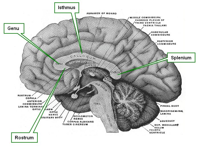
Median section of the brain
The term [corpus callosum] is Latin. The word [corpus] means "body", while the term [callosum] derives from [callosus] meaning "callous" or a structure with a hard consistency.
The corpus callosum is the largest of the nine interhemispheric commisures of the brain. It is formed by white matter consisting of axons that communicate both cerebral hemispheres. The corpus callosum is formed by several components:
• Rostrum: an anteroinferior region that resembles a bird's beak
• Genu: Latin for "knee", the genu is formed mostly by interfrontal fibers. These fibers form the anterior fornix
• Isthmus:also known as the "body" or "trunk", it is the main portion of the corpus callosum, allowing for interparietal and intertemporal communication
• Splenium: Latin for "bandage" the splenium allows for interhemispheric communication between the parietal, temporal, and occipital lobes. The fibers of the splenium form the posterior fornix
The image shown is a median section of the brain. Click on the image for a larger depiction. For a superior view of the corpus callosum click here.
Sources:
1 "Tratado de Anatomia Humana" Testut et Latarjet 8 Ed. 1931 Salvat Editores, Spain
2. "Anatomy of the Human Body" Henry Gray 1918. Philadelphia: Lea & Febiger
Image modified by CAA, Inc, Original image courtesy of bartleby.com
- Details

Click for a larger image
The term [vertebra prominens] is Latin and means the "prominent vertebra". It refers to the seventh cervical vertebra (C7) because of its long spinous process. Because of the anterior lordotic curvature of the cervical spine, their short spinous processes, and the presence of the ligamentum nuchae, the cervical vertebrae are usually not palpable with the exception of the vertebra prominens.
If you slide a finger down the nape of your neck in the midline, the first "bump" that is felt in the spine is the vertebra prominens, the next one down is T1, and so forth.
The vertebra prominens has foramina transversaria as do all cervical vertebrae. The only difference is that the vertebral artery usually is not found in the C7 foramina, opposite of the rest of the cervical vertebrae. The vertebral veins do pass through the foramina transversaria of the vertebra prominens.
Image property of: CAA.Inc. Photographer: David M. Klein