![]()
Medical Terminology Daily (MTD) is a blog sponsored by Clinical Anatomy Associates, Inc. as a service to the medical community. We post anatomical, medical or surgical terms, their meaning and usage, as well as biographical notes on anatomists, surgeons, and researchers through the ages. Be warned that some of the images used depict human anatomical specimens.
You are welcome to submit questions and suggestions using our "Contact Us" form. The information on this blog follows the terms on our "Privacy and Security Statement" and cannot be construed as medical guidance or instructions for treatment.
We have 789 guests and no members online
")
Marcia Crocker Noyes
(1869 – 1946)
Further to my comment on old books and research that started with an interesting bookplate (Ex-Libris). I continued my research and found that the person in charge of the Osler library bookplate was a fascinating individual that today maybe a ghost in the MedChi library and building in Baltimore... This is certainly an article that can be called "A Moment in History"
Marcia Crocker Noyes was the librarian at The Maryland State Medical Society from 1896 to 1946 and was a founding member of the Medical Library Association.[1][2][3]
Sir William Osler, MD. a famous Johns Hopkins surgeon was a noted bibliophile and had a large personal collection of books on various topics. When he became the President of MedChi in 1896, he was dismayed at the condition of the library and knew that with the right person and some stewardship, it could become a significant collection. Sir William asked his friend, Dr. Bernard Steiner, a physician and President of the Enoch Pratt Free Library in Baltimore for suggestions of a librarian, and Dr. Steiner recommended Marcia Crocker Noyes. A native of New York, and a graduate of Hunter College, Marcia had moved to Baltimore for a lengthy visit with her sister, and took a “temporary” position at the Pratt Library, which turned into three years. Although she had no medical experience or background, she was enthusiastic, and most importantly, she was willing to move into the apartment provided for the librarian, who needed to be available 24 hours a day.
The image in this article is Ms. Noyes on her first year on the job. Marcia developed a book classification system for medical books, based on the Index Medicus, and called it the Classification for Medical Literature. The system uses the alphabet with capital letters for the major divisions of medicine and lower-case ones for the sub-sections. The system was used for many years, but it's now dated and the Faculty's original shelving scheme was never changed. The card catalogs still reflect her classification and many of the cards are written in Marcia's back-slanting handwriting.
Marcia knew enough to ask the Faculty's members about medical questions, terminology and literature. She gradually won over the predominantly male membership and they became her greatest allies; Sir William at the start, and then for nearly 40 years, Dr. John Ruhräh, a wealthy pediatrician with no immediate family of his own. She made a point of attending almost every Faculty function, and in 1904, under guidelines from the American Medical Association, Marcia was made the Faculty Secretary. For much of her first 10 years, she was the Faculty's only full-time employee, only being assisted by Mr. Caution, the Faculty's janitor. Later in life Marcia would say that she hired him because of his name!
Within ten years, the library had outgrown its space, and plans, spearheaded by Marcia and Sir William before his move to Oxford, were made to build a headquarters building, mainly to house the library's growing collection of medical books and journals.
Marcia was instrumental in the design and building of the new headquarters. She travelled to Philadelphia, New York and Boston to look at their medical society buildings, and eventually, the Philadelphia architectural firm, Ellicott & Emmart was selected to design and build the new Faculty building. Every detail of the building held her imprimatur, from the graceful staircase, to the light-filled reading room, and all of the myriad details of the millwork, marble tesserae, and most of all, the four-story cast iron stacks. She was on-site, climbing up unfinished staircases, checking out the progress of the building, which was built in less than one year at a cost of $90,000.
Among the features of the new building was a fourth-floor apartment for her. She referred to it as the "first penthouse in Baltimore" and it had a garden and rooftop terrace. The library collection eventually grew to more than 65,000 volumes from medical and specialty societies around the world. Journals were traded back and forth, and physicians eagerly anticipated the arrival of each new issue. At the same time, Marcia was involved in the Medical Library Association as one of eight founding members. The MLA promotes medical libraries and the exchange of information. One of the earliest mandates of the MLA was the Exchange, a distribution and trade service for those who had duplicates or little-used books in their collections. Initially, the Exchange was run out of the Philadelphia medical society, but in 1900 it was moved to Baltimore and Marcia oversaw it. Several hundred periodicals and journals were received and sent each month, a huge amount of work for a tiny staff. In 1904, the Faculty had run out of room to manage the Exchange, so it was moved to the Medical Society of the Kings County (Brooklyn). But without Marcia's excellent administrative skills, it floundered and in 1908, the MLA asked Marcia to take charge once again.
In 1909, when the new Faculty building opened, there was enough room to run the Exchange and with the help of MLA Treasurer, noted bibliophile and close friend, Dr. John Ruhräh, it once again became successful. Additionally, Marcia and Dr. Ruhräh combined forces to revive the MLA's bulletin, which had all but ceased publication in 1908, taking the Exchange with it. This duo maintained editorial control from 1911 until 1926. In 1934, around the time of Dr. Ruhräh's death, Marcia became the first “unmedicated” professional to head the MLA. During her tenure, the MLA incorporated, the first seal was adopted, and the annual meeting was held in Baltimore. Marcia wanted to write the history of the MLA once she retired from full-time work at the Faculty, but her health was beginning to fail. She had back problems and had suffered a serious burn on her shoulder as a young woman, possibly from her time running a summer camp, Camp Seyon, for young ladies in the Adirondack Mountains. In 1946, a celebration was planned to honor Marcia's 50 years at the Faculty. But she was adamant that the physicians wait until November, the actual date of her 50 years. However, they knew she was gravely ill, and might not make it until then, so a huge party was held in April. More than 250 physicians attended the celebration, but the ones she was closest to in the early years, were long gone. She was presented with a suitcase, a sum of money to use for travelling, and her favorite painting of Dr. John Philip Smith, a founder of the Medical College in Winchester, Virginia. It was painted by Edward Caledon Smith, a Virginia painter who had been a student of the painter Thomas Sully.[4] She adored this painting and vowed, jokingly, to take it with her wherever she went.
The painting was not to stay with her for very long, for she died in November 1946, and left it to the Faculty in her will. Her funeral was held in the Faculty's Osler Hall, named for her dear friend. More than 60 physicians served as her pallbearers, and she was buried at Baltimore's Green Mount Cemetery. In 1948, the MLA decided to establish an award in the name of Marcia Crocker Noyes. It was for outstanding achievement in medical library field and was to be awarded every two years, or when a truly worthy candidate was submitted. In 2014, the Faculty began giving a bouquet of flowers to the winner of the award in Marcia's name, and in honor of her work. Much evidence exists for this tradition, as we know that the physicians, especially Drs. Osler and Ruhräh, frequently gave her bouquets of flowers. Marcia also cultivated flower gardens at the Faculty and decorated the rooms with her work.
Today, the MedChi building is open for tours and if the rumors are to be believed Ms. Marcia Crocker Noyes is still at work in her beloved library as the "resident ghost" [1][5]

NOTE: This article has been modified from the original Wikipedia article on Marcia Crocker Noyes. The article itself is well-written with interesting images of the subject. I would encourage you to visit it. The second insert is from book 00736 in my personal library and shows in pencil, the incredibly small handwriting of Marsha C. Noyes.
Sources:
1. "Marcia, Marcia, Marcia" MedChi Archives blog.
2. "Marcia C. Noyes, Medical Librarian" (PDF). Bulletin of the Medical Library Association. 35 (1): 108–109. 1947. PMC 194645
3. Smith, Bernie Todd (1974). "Marcia Crocker Noyes, Medical Librarian: The Shaping of a Career" (PDF). Bulletin of the Medical Library Association. 62 (3): 314–324. PMC 198800Freely accessible. PMID 4619344.
4. Edward Caledon BRUCE (1825-1901)"
5. Behind the scenes tour MedChiBuilding
"Clinical Anatomy Associates, Inc., and the contributors of "Medical Terminology Daily" wish to thank all individuals who donate their bodies and tissues for the advancement of education and research”.
Click here for more information


- Details
Since it's my birthday, I am taking the day off!
Not really, I am traveling to the "Vesalius and the Invention of the Modern Body" symposium in St. Louis. Looking forward to this meeting and the "Fabrica Vitae" exhibit by Pascale Pollier!!
I will post from the meeting and let you know what is going on!
See you tomorrow!
- Details
This word has three combined roots. [Chol-] or [chole-] meaning "bile", [-doch-] meaning "duct", and [-lith-], meaning " stone". The initial two combined roots [choledoch-] mean "bile duct." Many teach that [choledoch-] means "common bile duct'; although this is an accepted use of the term, it is not its true meaning.
The suffix [-iasis] means "condition". Therefore, the medical word [choledocholithiasis] means "condition of stones in the bile duct" , or more commonly understood as, "condition of stones in the common bile duct".
- Details
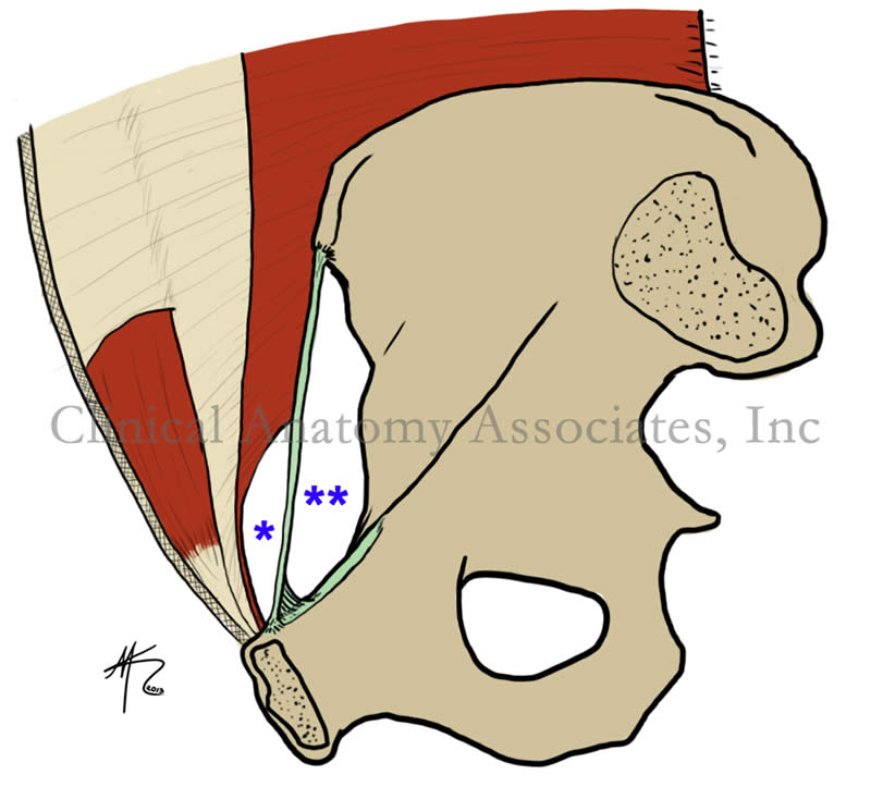
The term [myopectineal orifice] was coined originally by Dr. Henri Fruchaud, and refers to a "distinct area of weakness in the pelvic region". The term [myopectineal] arises from two root terms which are combined. The root term [-my-] means "muscle" and the term [-pect-] means "comb"or "pectinate". The word [pectineal] in this case refers to the pelvic bone area of origin of the pectinate muscle of the thigh.
Fruchaud postulated that the anterior abdominal wall has an area that is inherently weak, and that this area is genetically determined. As such, hernias are part of human nature, or as he stated, "a healthy man is, unknown to himself, a hernia bearer".
The myopectineal orifice, or MPO, is bound superiorly by the arching fibers of the transversus abdominis and internal oblique muscles, and inferiorly by the pectineal line.
The MPO is then composed by two regions separated by the inguinal ligament; the suprainguinal region, marked by one asterisk and site for direct and indirect inguinal hernias, and a small subsegment of the subinguinal region, (marked by two asterisks), site for femoral hernias.
In the accompanying sketch, the subinguinal region looks large, but this area is closed off by muscles, arteries, veins, and nerves, leaving only a small area of weakness (the femoral ring) where femoral hernias can arise.
Fruchaud advanced the separate concepts of inguinal hernias and femoral hernias and provided a new (for the time) concept of the repair of these hernias. Today, with laparoscopic herniorrhaphy, a surgeon attempts to repair the weak MPO instead of only the herniated locus.
Image property of:CAA.Inc.. Artist:David M. Klein
Source:
"Henri Fruchaud (1894–1960): A man of bravery, an anatomist a surgeon" Stoppa,R and Wantz,G. Hernia 1998,Vol 2,(1) 45 - 47
Clinical anatomy of the inguinofemoral hernias, as well as abdominal and perineal hernias are some of the lecture topics developed and delivered to the medical devices industry by Clinical Anatomy Associates, Inc. For more information Contact Us.
- Details

Click for a larger image
Also known as the [septomarginal trabecula], the [moderator band] is part of the conduction system of the heart.
The moderator band is found in the right ventricle of the heart and extends from the the interventricular septum to the base of the anterior papillary muscle. Although always present, it can have interesting anatomical variations, ranging from being double or triple, to being extremely thin or thick. In some cases, it can be confused as one of the trabeculae carnae.
The moderator band, usually clean on its superior aspect, may present with thin bands of tissue that extend towards the anterior wall of the right heart.
It was initially described by Leonardo Da Vinci, and English anatomists called it the "moderator band", because of its location. The initial belief was that the function of this structure was to prevent excessive ballooning of the right ventricle, "moderating" its dilation. Its descriptive name, the "septomarginal trabecula" was coined by Julius Tandler (1869 - 1936).
The moderator band as part of the conduction system of the heart, allows for some of the fibers of the right bundle branch to reach the wall of the right side of the heart. Its structure and function is similar to the atria internodal tracts, only in this case the conduction is not between the sinoatrial node and the interventricular node. The moderator band bypasses the apex of the heart and facilitates the passage of the electrical impulse from the interventricular septum to the right ventricular wall. is Click on the image for a larger version
Sources:
1. "Tratado de Anatomia Humana" Testut et Latarjet 8 Ed. 1931 Salvat Editores, Spain
2. "Gray's Anatomy"38th British Ed. Churchill Livingstone 1995
- Details

Click for a larger image
The word [trachelectomy] is composed of the root term [-trache-] which arises from the Greek word [τράχηλος] (trahelos), meaning “neck”, and the suffix [-ectomy] meaning “removal of”. The word [trachelectomy] then means “removal of the neck”.
Another way to refer to this procedure is [cervicectomy], where the root term [-cervic-] arises fron the Latin term [cervix], also meaning neck.
The procedure refers to the removal of the neck or cervix of the uterus. It can be performed as a procedure where the cervix is removed leaving the body of the uterus or [uterus proper] in place. This is done is younger females where there is a cervical cancer, but there is still the possibility of attaining pregnancy. The procedure carries a higher chance of miscarriage and the baby must be delivered via a Cesarean section.
A second reason to remove the cervix of the uterus is as a secondary procedure, after the uterus proper has been removed as part of a supracervical hysterectomy and the cervix, left behind, shows signs of cancer.
Medical terminology note: Trachelectomy and cervicectomy… why are there two terms for the same procedure? That is actually quite common in medical terminology, where you have words having the same meaning that arise from Latin and Greek. In fact, there are cases where there are more than two terms for the same organ or procedure, and let’s not count the vernacular terms on top! This makes medical terminology and interesting and fascinating topic. I would suggest that you click around the links for this article, you will find some interesting information. Dr. Miranda
- Details

Click for a larger image
UPDATED: The term [mesentery] is of Greek origin. The prefix [mes(o)-] arises from the Greek [μέσο] meaning "middle", the root term [-enter-] means "small intestine" or "intestine", and the suffix [-y] means "process" or "structure". Thus, the mesentery is "a structure in the middle".
The term [mesentery] can be used as a generic word to denote a double-layered peritoneal membrane that stretches between an abdominal viscus and the abdominal wall. A more precise use of the term is that of mesentery proper, which extends between the posterior abdominal wall and the jejunum and ileum. The superior mesenteric artery and veins are found at the root of the mesentery proper, along with a large accumulation of lymphatic nodes, and sympathetic and parasympathetic nerves.
Between the two layers of the mesentery proper, there are jejunal and ileal arteries and veins, a complex system of arterial and venous arches, as well as lymphatic vessels, autonomic nerves, and varying degrees of fat. Because of the presence of the mesentery proper, the jejunum and ileum are mobile or intraperitoneal, that is, they can slither, turn and twist with the movements of peristalsis. This movement is helped by the presence of a small amount of peritoneal fluid.

Click for a larger image
The fact that the mesentery is intraperitoneal is important in surgery. If the organ can already move around because of its mesentery, then it does not need to be "mobilized", it is already mobile!! If the organ (jejunum or ileum) have adhesions that limit their mobility within the abdominal cavity, the surgeon may have to perform and adhesiolysis to restore their mobility.
The first image shows an anatomical dissection where the greater omentum has been pulled anteriorly, exposing the small intestine and its mesentery, as well as the transverse mesocolon. Click on the image for a larger depiction. The second image (courtesy of Dr. Michiaki Akashi) is artwork depicting the surgical technique of transillumination, where the surgeon will shine a light through the mesentery to visualize the blood supply to the intestine prior to ligation and transection. The mesentery-like structure being transilluminated is the transverse mesocolon
WARNING: The first image is a photograph of a human dissection and can be considered descriptive.
NOTE: My personal thanks to Michiaki Akashi, M.D.for allowing us to use his artwork in this article. Dr. Akashi works as a surgeon and pathologist in the Saga Prefectural Hospital Koseikan in Saga, Japan.Dr. Miranda
First image property of: CAA.Inc. Photographer: David M. Klein