Medical Terminology Daily (MTD) is a blog sponsored by Clinical Anatomy Associates, Inc. as a service to the medical community. We post anatomical, medical or surgical terms, their meaning and usage, as well as biographical notes on anatomists, surgeons, and researchers through the ages. Be warned that some of the images used depict human anatomical specimens.
You are welcome to submit questions and suggestions using our "Contact Us" form. The information on this blog follows the terms on our "Privacy and Security Statement" and cannot be construed as medical guidance or instructions for treatment.
We have 488 guests online

Jean George Bachmann
(1877 – 1959)
French physician–physiologist whose experimental work in the early twentieth century provided the first clear functional description of a preferential interatrial conduction pathway. This structure, eponymically named “Bachmann’s bundle”, plays a central role in normal atrial activation and in the pathophysiology of interatrial block and atrial arrhythmias.
As a young man, Bachmann served as a merchant sailor, crossing the Atlantic multiple times. He emigrated to the United States in 1902 and earned his medical degree at the top of his class from Jefferson Medical College in Philadelphia in 1907. He stayed at this Medical College as a demonstrator and physiologist. In 1910, he joined Emory University in Atlanta. Between 1917 -1918 he served as a medical officer in the US Army. He retired from Emory in 1947 and continued his private medical practice until his death in 1959.
On the personal side, Bachmann was a man of many talents: a polyglot, he was fluent in German, French, Spanish and English. He was a chef in his own right and occasionally worked as a chef in international hotels. In fact, he paid his tuition at Jefferson Medical College, working both as a chef and as a language tutor.
The intrinsic cardiac conduction system was a major focus of cardiovascular research in the late nineteenth and early twentieth centuries. The atrioventricular (AV) node was discovered and described by Sunao Tawara and Karl Albert Aschoff in 1906, and the sinoatrial node by Arthur Keith and Martin Flack in 1907.
While the connections that distribute the electrical impulse from the AV node to the ventricles were known through the works of Wilhelm His Jr, in 1893 and Jan Evangelista Purkinje in 1839, the mechanism by which electrical impulses spread between the atria remained uncertain.
In 1916 Bachmann published a paper titled “The Inter-Auricular Time Interval” in the American Journal of Physiology. Bachmann measured activation times between the right and left atria and demonstrated that interruption of a distinct anterior interatrial muscular band resulted in delayed left atrial activation. He concluded that this band constituted the principal route for rapid interatrial conduction.
Subsequent anatomical and electrophysiological studies confirmed the importance of the structure described by Bachmann, which came to bear his name. Bachmann’s bundle is now recognized as a key determinant of atrial activation patterns, and its dysfunction is associated with interatrial block, atrial fibrillation, and abnormal P-wave morphology. His work remains foundational in both basic cardiac anatomy and clinical electrophysiology.
Sources and references
1. Bachmann G. “The inter-auricular time interval”. Am J Physiol. 1916;41:309–320.
2. Hurst JW. “Profiles in Cardiology: Jean George Bachmann (1877–1959)”. Clin Cardiol. 1987;10:185–187.
3. Lemery R, Guiraudon G, Veinot JP. “Anatomic description of Bachmann’s bundle and its relation to the atrial septum”. Am J Cardiol. 2003;91:148–152.
4. "Remembering the canonical discoverers of the core components of the mammalian cardiac conduction system: Keith and Flack, Aschoff and Tawara, His, and Purkinje" Icilio Cavero and Henry Holzgrefe Advances in Physiology Education 2022 46:4, 549-579.
5. Knol WG, de Vos CB, Crijns HJGM, et al. “The Bachmann bundle and interatrial conduction” Heart Rhythm. 2019;16:127–133.
6. “Iatrogenic biatrial flutter. The role of the Bachmann’s bundle” Constán E.; García F., Linde, A.. Complejo Hospitalario de Jaén, Jaén. Spain
7. Keith A, Flack M. The form and nature of the muscular connections between the primary divisions of the vertebrate heart. J Anat Physiol 41: 172–189, 1907.
"Clinical Anatomy Associates, Inc., and the contributors of "Medical Terminology Daily" wish to thank all individuals who donate their bodies and tissues for the advancement of education and research”.
Click here for more information
- Details
- Written by: Efrain A. Miranda, Ph.D.
Last week I attended this interdisciplinary symposium hosted by the Saint Louis University and Washington University. This three-day event was inspired by the landmark publication of Andrea Vesalius’s "De humani corporis fabrica, libri septem" (Basel, 1543 and 1555) and the new critical edition and translation of this work, the New Fabrica. Two of the keynote speakers were Daniel Garrison and Malcolm Hast, authors of the new Fabrica by Karger Publishers. Besides them there were several internationally-renowned speakers, art exhibits, presentation of academic papers of leading research, a public anatomy demonstration, rare books workshops, and a publishers’ exhibit hall.
Because the Fabrica represented a collaborative project involving a scientist (Vesalius), a humanist (Johannes Oporinus, the printer), and an artist (Jan van Kalkar), the goal of the conference was to encourage a network of scholars working in disparate fields to explore the potential for future interdisciplinary research. This objective was clearly attained, as I was able to speak and share with rare books curators, university librarians, artists, anatomist, physicians, poets, historians, etc., all of them brought together by the shared admiration for Andreas Vesalius, his work, his publications, and his legacy.
There were many highlights in this symposium and I will try to cover some of them in a series of articles. The first one was a presentation by Dr. Stephen N. Joffe, where he described the number of Vesalius' books still in existence in the US and estimates around the world. It was interesting to me that and estimated 600 first edition Fabricas were ever published, and that of those only a fraction exist today, most in university libraries!
Another highlight was the presentation by Pascale Pollier, a Belgian artist, of the Vesalius Continuum project, part of which are the Fabrica Vitae art exhibit that was available to the attendees and the public for the duration of the symposium. Another part of Vesalius Continuum was the meeting in Zakynthos, Greece in 2014. Pascale also presented the process of creation of a bust of Dr. Gunther Von Hagens, the inventor of the system of plastination.
Probably the most rewarding segments of this symposium were the question and answer sessions after each presentation.
- Details

Image courtesy of Dr. Randall Wolf
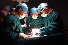
UPDATED: This Latin aphorism is at the core of Medicine and Surgery. It means "Above all, do no harm". Another translation would be "First, do no harm". The works of Hippocrates mentions the concept in his book Epidemics , and it has been presented in one way or another trough the ages. Thomas Sydenham (1624 - 1689), an English physician, is probably the first one to use it in an English publication, although his Latin phrase was "Primum est ut, non nocere". The first use of the modern phrasing "Primum Non Nocere" was by Lewis Atterbury Stimson (1844 - 1917), an American surgeon in 1879.
It seems almost counterintuitive that surgery would attempt to do no harm, but this is what moves innovation. From sharper needles that require less force to penetrate, and sharper scalpels that cause less trauma to tissues, to surgical staplers and minimally invasive techniques that attempt to reduce the size of the incision and the overall damage to the tissues.
The design of new surgical devices and new surgical techniques should always attempt to answer to this most important rule of surgery: "Primum Non Nocere".
As a point of interest, the Latin term [nocere] is the basis for the medical term [noxa] means "injury", "harm", or damage", this being the root for the term [noxious]
Sources
1. "Origin and Uses of Primum Non Nocere — Above All, Do No Harm!" Smith, CM J Clin Pharmacol 2005 45 (4): 371–377
2. "On abdominal drainage of adherent portions of ovarian cysts as a substitute for completed ovariotomy" Stimson LA Am J Med Sci 1879;78:88-100.
- Details
Since it's my birthday, I am taking the day off!
Not really, I am traveling to the "Vesalius and the Invention of the Modern Body" symposium in St. Louis. Looking forward to this meeting and the "Fabrica Vitae" exhibit by Pascale Pollier!!
I will post from the meeting and let you know what is going on!
See you tomorrow!
- Details
This word has three combined roots. [Chol-] or [chole-] meaning "bile", [-doch-] meaning "duct", and [-lith-], meaning " stone". The initial two combined roots [choledoch-] mean "bile duct." Many teach that [choledoch-] means "common bile duct'; although this is an accepted use of the term, it is not its true meaning.
The suffix [-iasis] means "condition". Therefore, the medical word [choledocholithiasis] means "condition of stones in the bile duct" , or more commonly understood as, "condition of stones in the common bile duct".
- Details
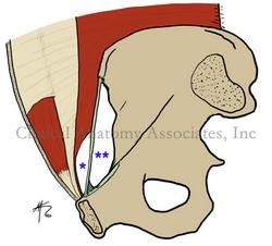
The term [myopectineal orifice] was coined originally by Dr. Henri Fruchaud, and refers to a "distinct area of weakness in the pelvic region". The term [myopectineal] arises from two root terms which are combined. The root term [-my-] means "muscle" and the term [-pect-] means "comb"or "pectinate". The word [pectineal] in this case refers to the pelvic bone area of origin of the pectinate muscle of the thigh.
Fruchaud postulated that the anterior abdominal wall has an area that is inherently weak, and that this area is genetically determined. As such, hernias are part of human nature, or as he stated, "a healthy man is, unknown to himself, a hernia bearer".
The myopectineal orifice, or MPO, is bound superiorly by the arching fibers of the transversus abdominis and internal oblique muscles, and inferiorly by the pectineal line.
The MPO is then composed by two regions separated by the inguinal ligament; the suprainguinal region, marked by one asterisk and site for direct and indirect inguinal hernias, and a small subsegment of the subinguinal region, (marked by two asterisks), site for femoral hernias.
In the accompanying sketch, the subinguinal region looks large, but this area is closed off by muscles, arteries, veins, and nerves, leaving only a small area of weakness (the femoral ring) where femoral hernias can arise.
Fruchaud advanced the separate concepts of inguinal hernias and femoral hernias and provided a new (for the time) concept of the repair of these hernias. Today, with laparoscopic herniorrhaphy, a surgeon attempts to repair the weak MPO instead of only the herniated locus.
Image property of:CAA.Inc.. Artist:David M. Klein
Source:
"Henri Fruchaud (1894–1960): A man of bravery, an anatomist a surgeon" Stoppa,R and Wantz,G. Hernia 1998,Vol 2,(1) 45 - 47
Clinical anatomy of the inguinofemoral hernias, as well as abdominal and perineal hernias are some of the lecture topics developed and delivered to the medical devices industry by Clinical Anatomy Associates, Inc. For more information Contact Us.
- Details
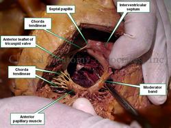
Also known as the [septomarginal trabecula], the [moderator band] is part of the conduction system of the heart.
The moderator band is found in the right ventricle of the heart and extends from the the interventricular septum to the base of the anterior papillary muscle. Although always present, it can have interesting anatomical variations, ranging from being double or triple, to being extremely thin or thick. In some cases, it can be confused as one of the trabeculae carnae.
The moderator band, usually clean on its superior aspect, may present with thin bands of tissue that extend towards the anterior wall of the right heart.
It was initially described by Leonardo Da Vinci, and English anatomists called it the "moderator band", because of its location. The initial belief was that the function of this structure was to prevent excessive ballooning of the right ventricle, "moderating" its dilation. Its descriptive name, the "septomarginal trabecula" was coined by Julius Tandler (1869 - 1936).
The moderator band as part of the conduction system of the heart, allows for some of the fibers of the right bundle branch to reach the wall of the right side of the heart. Click on the image for a larger version
Sources:
1. "Tratado de Anatomia Humana" Testut et Latarjet 8 Ed. 1931 Salvat Editores, Spain
2. "Gray's Anatomy"38th British Ed. Churchill Livingstone 1995


