![]()
Medical Terminology Daily (MTD) is a blog sponsored by Clinical Anatomy Associates, Inc. as a service to the medical community. We post anatomical, medical or surgical terms, their meaning and usage, as well as biographical notes on anatomists, surgeons, and researchers through the ages. Be warned that some of the images used depict human anatomical specimens.
You are welcome to submit questions and suggestions using our "Contact Us" form. The information on this blog follows the terms on our "Privacy and Security Statement" and cannot be construed as medical guidance or instructions for treatment.
We have 647 guests and no members online
")
Marcia Crocker Noyes
(1869 – 1946)
Further to my comment on old books and research that started with an interesting bookplate (Ex-Libris). I continued my research and found that the person in charge of the Osler library bookplate was a fascinating individual that today maybe a ghost in the MedChi library and building in Baltimore... This is certainly an article that can be called "A Moment in History"
Marcia Crocker Noyes was the librarian at The Maryland State Medical Society from 1896 to 1946 and was a founding member of the Medical Library Association.[1][2][3]
Sir William Osler, MD. a famous Johns Hopkins surgeon was a noted bibliophile and had a large personal collection of books on various topics. When he became the President of MedChi in 1896, he was dismayed at the condition of the library and knew that with the right person and some stewardship, it could become a significant collection. Sir William asked his friend, Dr. Bernard Steiner, a physician and President of the Enoch Pratt Free Library in Baltimore for suggestions of a librarian, and Dr. Steiner recommended Marcia Crocker Noyes. A native of New York, and a graduate of Hunter College, Marcia had moved to Baltimore for a lengthy visit with her sister, and took a “temporary” position at the Pratt Library, which turned into three years. Although she had no medical experience or background, she was enthusiastic, and most importantly, she was willing to move into the apartment provided for the librarian, who needed to be available 24 hours a day.
The image in this article is Ms. Noyes on her first year on the job. Marcia developed a book classification system for medical books, based on the Index Medicus, and called it the Classification for Medical Literature. The system uses the alphabet with capital letters for the major divisions of medicine and lower-case ones for the sub-sections. The system was used for many years, but it's now dated and the Faculty's original shelving scheme was never changed. The card catalogs still reflect her classification and many of the cards are written in Marcia's back-slanting handwriting.
Marcia knew enough to ask the Faculty's members about medical questions, terminology and literature. She gradually won over the predominantly male membership and they became her greatest allies; Sir William at the start, and then for nearly 40 years, Dr. John Ruhräh, a wealthy pediatrician with no immediate family of his own. She made a point of attending almost every Faculty function, and in 1904, under guidelines from the American Medical Association, Marcia was made the Faculty Secretary. For much of her first 10 years, she was the Faculty's only full-time employee, only being assisted by Mr. Caution, the Faculty's janitor. Later in life Marcia would say that she hired him because of his name!
Within ten years, the library had outgrown its space, and plans, spearheaded by Marcia and Sir William before his move to Oxford, were made to build a headquarters building, mainly to house the library's growing collection of medical books and journals.
Marcia was instrumental in the design and building of the new headquarters. She travelled to Philadelphia, New York and Boston to look at their medical society buildings, and eventually, the Philadelphia architectural firm, Ellicott & Emmart was selected to design and build the new Faculty building. Every detail of the building held her imprimatur, from the graceful staircase, to the light-filled reading room, and all of the myriad details of the millwork, marble tesserae, and most of all, the four-story cast iron stacks. She was on-site, climbing up unfinished staircases, checking out the progress of the building, which was built in less than one year at a cost of $90,000.
Among the features of the new building was a fourth-floor apartment for her. She referred to it as the "first penthouse in Baltimore" and it had a garden and rooftop terrace. The library collection eventually grew to more than 65,000 volumes from medical and specialty societies around the world. Journals were traded back and forth, and physicians eagerly anticipated the arrival of each new issue. At the same time, Marcia was involved in the Medical Library Association as one of eight founding members. The MLA promotes medical libraries and the exchange of information. One of the earliest mandates of the MLA was the Exchange, a distribution and trade service for those who had duplicates or little-used books in their collections. Initially, the Exchange was run out of the Philadelphia medical society, but in 1900 it was moved to Baltimore and Marcia oversaw it. Several hundred periodicals and journals were received and sent each month, a huge amount of work for a tiny staff. In 1904, the Faculty had run out of room to manage the Exchange, so it was moved to the Medical Society of the Kings County (Brooklyn). But without Marcia's excellent administrative skills, it floundered and in 1908, the MLA asked Marcia to take charge once again.
In 1909, when the new Faculty building opened, there was enough room to run the Exchange and with the help of MLA Treasurer, noted bibliophile and close friend, Dr. John Ruhräh, it once again became successful. Additionally, Marcia and Dr. Ruhräh combined forces to revive the MLA's bulletin, which had all but ceased publication in 1908, taking the Exchange with it. This duo maintained editorial control from 1911 until 1926. In 1934, around the time of Dr. Ruhräh's death, Marcia became the first “unmedicated” professional to head the MLA. During her tenure, the MLA incorporated, the first seal was adopted, and the annual meeting was held in Baltimore. Marcia wanted to write the history of the MLA once she retired from full-time work at the Faculty, but her health was beginning to fail. She had back problems and had suffered a serious burn on her shoulder as a young woman, possibly from her time running a summer camp, Camp Seyon, for young ladies in the Adirondack Mountains. In 1946, a celebration was planned to honor Marcia's 50 years at the Faculty. But she was adamant that the physicians wait until November, the actual date of her 50 years. However, they knew she was gravely ill, and might not make it until then, so a huge party was held in April. More than 250 physicians attended the celebration, but the ones she was closest to in the early years, were long gone. She was presented with a suitcase, a sum of money to use for travelling, and her favorite painting of Dr. John Philip Smith, a founder of the Medical College in Winchester, Virginia. It was painted by Edward Caledon Smith, a Virginia painter who had been a student of the painter Thomas Sully.[4] She adored this painting and vowed, jokingly, to take it with her wherever she went.
The painting was not to stay with her for very long, for she died in November 1946, and left it to the Faculty in her will. Her funeral was held in the Faculty's Osler Hall, named for her dear friend. More than 60 physicians served as her pallbearers, and she was buried at Baltimore's Green Mount Cemetery. In 1948, the MLA decided to establish an award in the name of Marcia Crocker Noyes. It was for outstanding achievement in medical library field and was to be awarded every two years, or when a truly worthy candidate was submitted. In 2014, the Faculty began giving a bouquet of flowers to the winner of the award in Marcia's name, and in honor of her work. Much evidence exists for this tradition, as we know that the physicians, especially Drs. Osler and Ruhräh, frequently gave her bouquets of flowers. Marcia also cultivated flower gardens at the Faculty and decorated the rooms with her work.
Today, the MedChi building is open for tours and if the rumors are to be believed Ms. Marcia Crocker Noyes is still at work in her beloved library as the "resident ghost" [1][5]

NOTE: This article has been modified from the original Wikipedia article on Marcia Crocker Noyes. The article itself is well-written with interesting images of the subject. I would encourage you to visit it. The second insert is from book 00736 in my personal library and shows in pencil, the incredibly small handwriting of Marsha C. Noyes.
Sources:
1. "Marcia, Marcia, Marcia" MedChi Archives blog.
2. "Marcia C. Noyes, Medical Librarian" (PDF). Bulletin of the Medical Library Association. 35 (1): 108–109. 1947. PMC 194645
3. Smith, Bernie Todd (1974). "Marcia Crocker Noyes, Medical Librarian: The Shaping of a Career" (PDF). Bulletin of the Medical Library Association. 62 (3): 314–324. PMC 198800Freely accessible. PMID 4619344.
4. Edward Caledon BRUCE (1825-1901)"
5. Behind the scenes tour MedChiBuilding
"Clinical Anatomy Associates, Inc., and the contributors of "Medical Terminology Daily" wish to thank all individuals who donate their bodies and tissues for the advancement of education and research”.
Click here for more information


- Details

Click for a larger image
UPDATED: The [common bile duct] also known as the [ductus choledocus]. is part of the hepatobiliary tree, taking bile from the gallbladder and liver to the second portion of the duodenum. The common bile duct begins at the junction of the common hepatic duct with the cystic duct, it continues inferiorly, usually to the right of the proper hepatic artery and anterior to the portal vein. It then passes posterior to the first portion of the duodenum, is surrounded by pancreatic tissue and ends at the hepatopancreatic ampulla of (Vater).
The junction of the common bile duct with the hepatopancreatic ampulla of (Vater) is the narrowest portion of the hepatobiliary tree, The lodging of a gallstone at this junction can be the cause for choledocholitiasis and jaundice.
In the accompanying image the common bile duct is elevated with a probe. The lesser omentum has been removed to show the common bile duct and vascular structures that are found between the two peritoneal layers that form the lesser omentum.
- Details
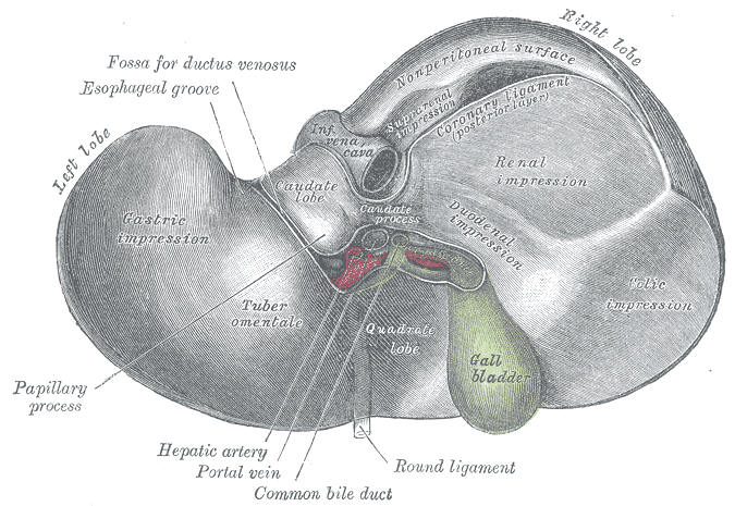
Inferior surface of the liver
The [gallbladder] is a bile transient storage organ, part of the hepatobiliary tree, situated in the anteroinferior aspect of the liver. The gallbladder is found in a depression on the inferior aspect of the right lobe of the liver, the gallbladder fossa or fossa vesicae felleae.
In the gallbladder we describe its dome-shaped fundus, the body of the organ, and the neck which is the area that opens into the cystic duct. Close to the neck, the gallbladder has a small pouch (Hartmann's pouch) which is important for surgeons during a laparoscopic cholecystectomy, as this is where they will lock one of the instruments that allows them to manipulate the gallbladder for dissection of the organ from the gallbladder fossa (the gallbladder bed). The other surgical grasper is placed at the gallbladder fundus.
The gallbladder is composed by three layers. From deep to superficial they are:
• Mucosa: Characterized by a columnar epithelium. Towards the neck of the gallbladder the mucosa creates spiral ridges that continue in to the cystic duct.
• Fibromuscular layer: This layer is composed by connective tissue and smooth muscle, mostly longitudinal
• Serosa: This is an incomplete layer and is formed by visceral peritoneum covering the area of the gallbladder not in contact with the liver. In an unusual anatomical variation, the serosa layer can be almost complete, forming a pseudomesentery that may contains some veins.
The gallbladder receives its blood supply by way of the cystic artery, a branch of the right hepatic artery. The venous return is by way of multiple small veins that empty into the liver venous system. In some cases, these veins may form large sinuses between the liver and the gallbladder causing potential troublesome bleeding during a cholecystectomy. For those who like medical history, Dr. Eric Muhe performed the first laparoscopic cholecystectomy on September 12, 1985! We are but a few days from the 30th anniversary!
For more information on terminology on "gall-", "bile", "chol", and "chole", click here.
Sources:
1 "Tratado de Anatomia Humana" Testut et Latarjet 8 Ed. 1931 Salvat Editores, Spain
2. "Anatomy of the Human Body" Henry Gray 1918. Philadelphia: Lea & Febiger
Image modified by CAA, Inc. Original image courtesy of bartleby.com
- Details

Last Friday April 17, I prepared and delivered a lecture on "Surgical Sutures, Needles, and Knots" which included a hands-on workshop on knots and wound closure on simulated tissue.
This was presented at the invitation of the Pre-Health club of the Mount Saint Joseph University in Cincinnati, OH. I am always glad to be invited to do these presentations as they allow me to maintain contact with the future generation of Health Care Professionals.
Of course this is a very short presentation compared to the longer course that Clinical Anatomy Associates, Inc. delivers for medical companies, but it shows these future professionals the complexity of the world of wound closure, healing, surgical sutures, needles, and knots.
We ended the lab with the challenge to do a two-layer closure of a simulated wound. Most of the attendees did a pretty good job. Congratulations!
My personal thanks to Dr. Eric Johnson who coordinated the meeting, and to the Pre-Health Club for their invitation. For more pictures of the meeting, see the Facebook album page of "Medical Terminology Daily"
- Details

Gallbladder and cystic duct
The cystic duct is a tubular structure that connects the neck of the gallbladder to the extrahepatic ductal system. It is 2-4 cm. in length and its lumen is about 2.6 +/- 0.7 mmm. The shape of the cystic duct varies, as it can be straight, angled, or acutely curved.
The mucosa of the cystic duct presents with 2-10 crescent-shaped folds that create a spiral-shaped inner structure referred to as the "Valve of Heister", first described by Lorenz Heister in 1732. These folds become smaller and scarcer towards the distal portion of the duct.
The cystic duct can present with several anatomical variations, from total absence where the neck of the gallbladder empties directly in to the common bile duct, to duplication, and even rare occasions where the cystic duct empties separately into the duodenal lumen.
The cystic duct is an important surgical landmark as it is one of the boundaries of the cystohepatic triangle or "Triangle of Calot", described by Jean-Francois Calot (1861 - 1944), which determines the location of the cystic artery, a critical structure that needs to be ligated and transected during a cholecystectomy.
Sources:
1 "Cystic Duct and Heister’s “Valves” Dasgupta,C, Stringer, MD, Clin Anat (2005) 18:81–87
2. "Tratado de Anatomia Humana" Testut et Latarjet 8 Ed. 1931 Salvat Editores, Spain
3. "Anatomy of the Human Body" Henry Gray 1918. Philadelphia: Lea & Febiger
Image modified by CAA, Inc. Original image courtesy of bartleby.com
- Details
The root term [-phor-] arises from the Greek word [φέρω] meaning “to bear”, “well”, and “healthy”. The meaning of the word today in medical terminology is “well-being”. The addition of the adjectival suffix [-ia] meaning “pertaining to” add to form [-phoria] meaning “pertaining to well-being” or “feeling well”. It is used in terms such as:
• Euphoria: The prefix [eu-] means “good”. The sensation of feeling good, of good well-being
• Dysphoria: The prefix [dys-] means “abnormal”. Abnormal feelings of well-being. In psychiatry these dysphoric moments can be seen in bipolar patients.
- Details
Click for a larger image
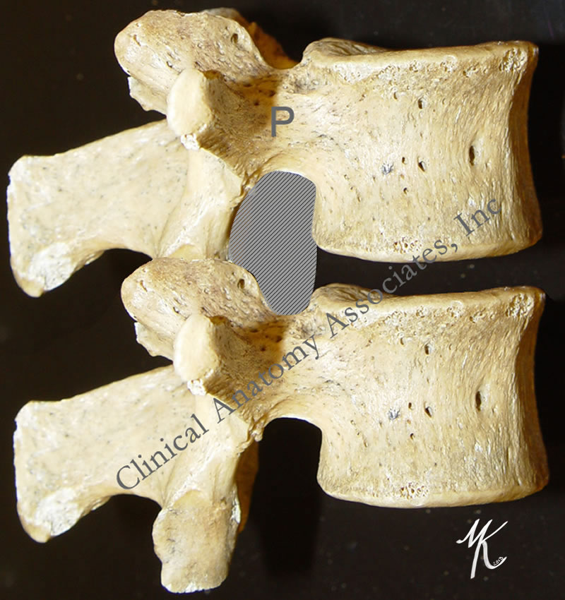
The term [intervertebral] means "between vertebrae", and [foramen] means "opening". The intervertebral foramina are bilateral openings between adjacent vertebrae. Each intervertebral foramen is found between adjacent pedicles ("P" in the large image), bound by the inferior vertebral notch and the superior vertebral notch of adjacent pedicles.
Although the term [intervertebral foramen] has been used for a long time, the concept has evolved to a more modern "intervertebral canal" or as some clinicians call it, the "lateral canal". The reason for this is that the intervertebral foramen is actually a tunnel whose length is determined by the width of the pedicles. This intervertebral canal has marked differences between the lateral, middle, and medial structures contained in the intervertebral canal.
Some of these structure are nerve roots, the dorsal root ganglion, the initial portion of the spinal nerve, dural sac, arteries, veins, recurrent nerves, fat, and a complex system of transforaminal and intraforaminal ligaments1. The structures contained in the intervertebral foramen can be compressed if the height of the intervertebral discs is compromised, or by a herniation of the intervertebral disc. The diameter of the intervertebral canal can also be reduced by bone and joint pathology.
If you hover over the image, the intervertebral foramen will be highlighted. For a larger version click on the image.
Images property of: CAA.Inc. Photographer: D.M. Klein
1 Thoracic and lumbar intraforaminal ligaments Akdemir, G.; J Neurosurg Spine 13:351-355, 2010