Medical Terminology Daily (MTD) is a blog sponsored by Clinical Anatomy Associates, Inc. as a service to the medical community. We post anatomical, medical or surgical terms, their meaning and usage, as well as biographical notes on anatomists, surgeons, and researchers through the ages. Be warned that some of the images used depict human anatomical specimens.
You are welcome to submit questions and suggestions using our "Contact Us" form. The information on this blog follows the terms on our "Privacy and Security Statement" and cannot be construed as medical guidance or instructions for treatment.
We have 808 guests online

Jean George Bachmann
(1877 – 1959)
French physician–physiologist whose experimental work in the early twentieth century provided the first clear functional description of a preferential interatrial conduction pathway. This structure, eponymically named “Bachmann’s bundle”, plays a central role in normal atrial activation and in the pathophysiology of interatrial block and atrial arrhythmias.
As a young man, Bachmann served as a merchant sailor, crossing the Atlantic multiple times. He emigrated to the United States in 1902 and earned his medical degree at the top of his class from Jefferson Medical College in Philadelphia in 1907. He stayed at this Medical College as a demonstrator and physiologist. In 1910, he joined Emory University in Atlanta. Between 1917 -1918 he served as a medical officer in the US Army. He retired from Emory in 1947 and continued his private medical practice until his death in 1959.
On the personal side, Bachmann was a man of many talents: a polyglot, he was fluent in German, French, Spanish and English. He was a chef in his own right and occasionally worked as a chef in international hotels. In fact, he paid his tuition at Jefferson Medical College, working both as a chef and as a language tutor.
The intrinsic cardiac conduction system was a major focus of cardiovascular research in the late nineteenth and early twentieth centuries. The atrioventricular (AV) node was discovered and described by Sunao Tawara and Karl Albert Aschoff in 1906, and the sinoatrial node by Arthur Keith and Martin Flack in 1907.
While the connections that distribute the electrical impulse from the AV node to the ventricles were known through the works of Wilhelm His Jr, in 1893 and Jan Evangelista Purkinje in 1839, the mechanism by which electrical impulses spread between the atria remained uncertain.
In 1916 Bachmann published a paper titled “The Inter-Auricular Time Interval” in the American Journal of Physiology. Bachmann measured activation times between the right and left atria and demonstrated that interruption of a distinct anterior interatrial muscular band resulted in delayed left atrial activation. He concluded that this band constituted the principal route for rapid interatrial conduction.
Subsequent anatomical and electrophysiological studies confirmed the importance of the structure described by Bachmann, which came to bear his name. Bachmann’s bundle is now recognized as a key determinant of atrial activation patterns, and its dysfunction is associated with interatrial block, atrial fibrillation, and abnormal P-wave morphology. His work remains foundational in both basic cardiac anatomy and clinical electrophysiology.
Sources and references
1. Bachmann G. “The inter-auricular time interval”. Am J Physiol. 1916;41:309–320.
2. Hurst JW. “Profiles in Cardiology: Jean George Bachmann (1877–1959)”. Clin Cardiol. 1987;10:185–187.
3. Lemery R, Guiraudon G, Veinot JP. “Anatomic description of Bachmann’s bundle and its relation to the atrial septum”. Am J Cardiol. 2003;91:148–152.
4. "Remembering the canonical discoverers of the core components of the mammalian cardiac conduction system: Keith and Flack, Aschoff and Tawara, His, and Purkinje" Icilio Cavero and Henry Holzgrefe Advances in Physiology Education 2022 46:4, 549-579.
5. Knol WG, de Vos CB, Crijns HJGM, et al. “The Bachmann bundle and interatrial conduction” Heart Rhythm. 2019;16:127–133.
6. “Iatrogenic biatrial flutter. The role of the Bachmann’s bundle” Constán E.; García F., Linde, A.. Complejo Hospitalario de Jaén, Jaén. Spain
7. Keith A, Flack M. The form and nature of the muscular connections between the primary divisions of the vertebrate heart. J Anat Physiol 41: 172–189, 1907.
"Clinical Anatomy Associates, Inc., and the contributors of "Medical Terminology Daily" wish to thank all individuals who donate their bodies and tissues for the advancement of education and research”.
Click here for more information
- Details
UPDATED: The term [fascia] means "band or bandage". It is derived from the Latin word [fascis] meaning "bundle", therefore a fascia is the bandage that ties a bundle. A bundle of sticks tied around an ax was the symbol of the lictors, the Roman imperial bodyguards. Many fasciae (Pl.) are eponymic, such as Camper's Fascia, Scarpa's fascia, Colle's fascia, etc.
In anatomy, the word [fascia] refers to a sheet of connective tissue. The term encompasses many types of fasciae (plural for fascia) which range from the thin deep or muscular fascia to the tendon-like thick fascia lata of the thigh. The image shows the anterior aspect of the thigh and its covering, the fascia lata. The opening shown in the fascia lata is the fossa ovalis of the thigh. Do not confuse with the fossa ovalis found in the interatrial wall of the heart. Click on the image for a larger depiction.
In the extremities, some fasciae may contain more than one muscle, creating fascial compartments. Trauma or vascular pathology in a fascial compartment may cause the muscles to swell to the point that the edema starts causing ischemia compromising the muscles to the point of necrosis. This is called "compartment syndrome" and may require a surgeon to cut open the fascia to relieve the pressure, a procedure called a "fasciotomy". For more information on [-otomy], click on this link.
Original images by Henry VanDyke Carter, MD and links courtesy ofbartleby.com
- Details
The term [urination] comes from the Greek [ούρα] meaning "urine" and refers to the "expulsion of urine". The term [micturition] has a Latin origin and refers to the "desire to empty the urinary bladder". Through use, these two terms have become synonymous.
Although the common use of [urination] is the act of bladder emptying, the proper use of the term describes the constant passage of urine from the ureters into the urinary bladder. The proper term for bladder emptying is [micturition].
The fact is that we are constantly urinating (right now as your are seated in front of your computer), but only micturate a few times a day.
Original images by Henry VanDyke Carter, MD and links courtesy ofbartleby.com
- Details
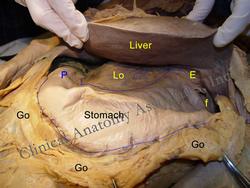
UPDATED: The word [pylorus] is Greek. It arises from the word [πύλη] (p?li)meaning [gate]. In ancient Greek [πυλωρός] (pylorus) meant "gatekeeper" or "gate guard", leaving us to assume that Greek physicians had an idea of the function of the pylorus.
The pylorus is a true anatomical sphincter and controls the emptying of the stomach. It is the most distal anatomical component of the stomach and can be see in the image marked with the letter "P". In history, it has been described by other names including "velut portanarium", and "pilorium" this last term was changed to "pylorus" by Andrea Vesalius.
Using surface anatomy the location of the pylorus can be found on or slightly superior to the transpyloric plane and slightly to the right of the midline.
Stenosis of the pylorus can lead to gastric emptying complications. A couple of surgical procedures to alleviate this condition are:
• Pyloromyotomy: From the root terms [pylor], meaning "pylorus", [-my-], meaning "muscle", and the suffix [-otomy], meaning "to open, or to cut"; therefore "opening of cutting the pyloric muscle"
• Pyloroplasty: From the root terms [pylor], meaning "pylorus", and the suffix[-oplasty], meaning "to reshape or reconstruct"; therefore "pyloric reshaping".
The image shows the anterior aspect of the stomach. The liver is retracted. Go= Greater omentum, Lo=Lesser omentum, E= Esophagus, f=Fundus, P= Pylorus.
Images property of:CAA.Inc.Photographer:D.M. Klein
- Details
A fistula is the abnormal communication or passageway between two hollow organs or between a hollow organ and the skin.
The term fistula is also used as a synonym for an anastomosis.
- Details
Andreas Vesalius opus magnus was the creation and the publication of his book “De Humani Corporis Fabrica, Libri Septem" (Seven books on the structure of the human body). This book was published on May 26th, 1543 by the printing press of Johannes Oporinus.
Much has been said and written about this book and the influence of Vesalius’ work on scientific thinking, the scientific method, and the displacement of dogmatic thinking based on the works of the ancient Greeks and Galen of Pergamon (129AD - 200AD) for a different view of the construction of the body based on direct and empirical observation.
Unfortunately, because of Vesalius’ following of Erasmus’ teachings on Latin, the book was written in a very difficult and circumvoluted language which made it difficult to understand. In addition, the book was very expensive for the times, with an estimated maximum printing of 600 copies.
Were it not for the images and the captions, as well as the many plagiarized versions of the Fabrica in different languages, Vesalius opus magnus would have been lost to history. Harvey Cushing wrote in his Vesalius bio-bibliography of 1943:”As a book, the Fabrica has been probably more admired and less read than any publication of equal significance in the history of science”.
Although several attempts have been done to translate the Fabrica, most of the works have been incomplete, or have tried to paraphrase or correct Vesalius’ words, leaving us with a watered-down image of the author and his intent.
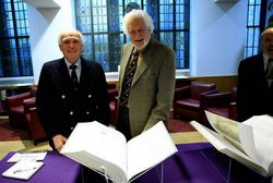
In 1993 Drs. Daniel H Garrison and Malcom H. Hast began a collaboration to translate the Fabrica of Vesalius. The 20- year story of how they obtained federal grants, discussed the translation, found a publisher, scanned and improved on the original images of the Fabrica, and how they even worked with Christian Mengelt to create a new typography for an annotated new Fabrica, was part of their presentation on the interdisciplinary symposium “Vesalius and the Invention of the Modern Body” hosted by the St. Louis University and the Washington University February 26-28, 2015.
This annotated new Fabrica is a translation of the 1543 first edition with comments on the 1555 second edition and it also includes passages and comments from a heavily edited 1555 second edition that has side margins comments and corrections now certified to be in Vesalius’ own handwriting. This book has been speculated to have been Vesalius’ personal copy and probably the basis of a potential third edition. This particular book is now known as "Vesalius' Annotated Fabrica"
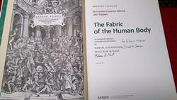
The "New Fabrica" was published in 2013 by Karger Publishing, a company based in Basel, Switzerland, the same city where the original Fabrica was published in 1543. The ISBN is 978-3-318-02246-9. Only 948 books were published and it has now been sold out. Because of the demand, an original is now considered a rare book.
Daniel H. Garrison received his degrees from Harvard (A.B. Classics, 1959) and Berkeley (PhD Comparative Literature, 1968). He was a member of the Classics Department at Northwestern University from 1966 until his retirement in 2010.
Malcolm H. Hast is Professor Emeritus of Otolaryngology – Head and Neck Surgery – and also past Professor of Cell and Molecular Biology (Anatomy) at Feinberg School of Medicine of Northwestern University. He is Fellow of the American Association for the Advancement of Science as well as Fellow of the Anatomical Society (UK) and a Chartered Biologist and Fellow of the Society of Biology (UK). He is also a recipient of The Gould International Award in Laryngology and a NATO Senior Fellowship in Science.
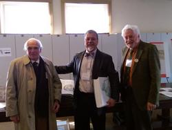
Personal note: I am honored to have met both Drs. Garrison and Hast at the symposium, shared some of the stories behind the new Fabrica and have them sign my own copy of this incredible book. Dr. Miranda
Sources:
1. "A Bio-blibliography of Andreas Vesalius" Cushung, H. 1943 Saunders
- Details
The terms “anatomy” and “dissection” are synonymous. In the days of Andreas Vesalius, the dissection of a corpse was a public event, where medical students would attend, as well as the paying public.
This event would go on for days as the dissector would explain the anatomy racing against time, as there were no means of body preservation.
Through the centuries after the public anatomies of the 1500’s, the dissection of donated bodies has been continued in the anatomy departments of medical schools helping medical students and surgeons prepare for the challenges of the practice of medicine and surgery.
A public anatomy was one of the events of the interdisciplinary symposium "Vesalius and the Invention of the Modern Body" hosted by the St. Louis University and the Washington University February 26-28, 2015. To my knowledge, a public anatomy has not been done in centuries (I may be wrong).
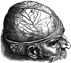
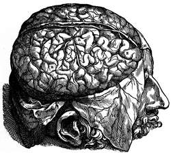
Some of the objectives were to demonstrate that Andrea Vesalius' description of the anatomy of the brain, its ventricular system, and the cranial nerves was logical, followed a process, and that the Fabrica, in its seventh book can be used as a dissector. The presentation was entitled “A Fabrica-guided Neo-Vesalian Public Dissection of the Brain Ventricular System 500 Years Later at St. Louis University” by Dr. Salomon Segal.
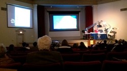
The dissection used excerpts and images from the Fabrica, as well as an advanced HD 3D camera, showing the brain and its structures with amazing clarity. The accompanying photo of the event is not well focused because of the light conditions, but shows the setup for the presentation.
This was an extremely professional presentation and although not completely “public” per se, the variety of attendees had a great feedback on the event. Proper attention to the care and respect towards the specimens and the anonymity of the donors was maintained at all times. I consider myself honored to have been a witness and a participant to this extraordinary event. Dr. Miranda.


