Medical Terminology Daily (MTD) is a blog sponsored by Clinical Anatomy Associates, Inc. as a service to the medical community. We post anatomical, medical or surgical terms, their meaning and usage, as well as biographical notes on anatomists, surgeons, and researchers through the ages. Be warned that some of the images used depict human anatomical specimens.
You are welcome to submit questions and suggestions using our "Contact Us" form. The information on this blog follows the terms on our "Privacy and Security Statement" and cannot be construed as medical guidance or instructions for treatment.
We have 799 guests online

Jean George Bachmann
(1877 – 1959)
French physician–physiologist whose experimental work in the early twentieth century provided the first clear functional description of a preferential interatrial conduction pathway. This structure, eponymically named “Bachmann’s bundle”, plays a central role in normal atrial activation and in the pathophysiology of interatrial block and atrial arrhythmias.
As a young man, Bachmann served as a merchant sailor, crossing the Atlantic multiple times. He emigrated to the United States in 1902 and earned his medical degree at the top of his class from Jefferson Medical College in Philadelphia in 1907. He stayed at this Medical College as a demonstrator and physiologist. In 1910, he joined Emory University in Atlanta. Between 1917 -1918 he served as a medical officer in the US Army. He retired from Emory in 1947 and continued his private medical practice until his death in 1959.
On the personal side, Bachmann was a man of many talents: a polyglot, he was fluent in German, French, Spanish and English. He was a chef in his own right and occasionally worked as a chef in international hotels. In fact, he paid his tuition at Jefferson Medical College, working both as a chef and as a language tutor.
The intrinsic cardiac conduction system was a major focus of cardiovascular research in the late nineteenth and early twentieth centuries. The atrioventricular (AV) node was discovered and described by Sunao Tawara and Karl Albert Aschoff in 1906, and the sinoatrial node by Arthur Keith and Martin Flack in 1907.
While the connections that distribute the electrical impulse from the AV node to the ventricles were known through the works of Wilhelm His Jr, in 1893 and Jan Evangelista Purkinje in 1839, the mechanism by which electrical impulses spread between the atria remained uncertain.
In 1916 Bachmann published a paper titled “The Inter-Auricular Time Interval” in the American Journal of Physiology. Bachmann measured activation times between the right and left atria and demonstrated that interruption of a distinct anterior interatrial muscular band resulted in delayed left atrial activation. He concluded that this band constituted the principal route for rapid interatrial conduction.
Subsequent anatomical and electrophysiological studies confirmed the importance of the structure described by Bachmann, which came to bear his name. Bachmann’s bundle is now recognized as a key determinant of atrial activation patterns, and its dysfunction is associated with interatrial block, atrial fibrillation, and abnormal P-wave morphology. His work remains foundational in both basic cardiac anatomy and clinical electrophysiology.
Sources and references
1. Bachmann G. “The inter-auricular time interval”. Am J Physiol. 1916;41:309–320.
2. Hurst JW. “Profiles in Cardiology: Jean George Bachmann (1877–1959)”. Clin Cardiol. 1987;10:185–187.
3. Lemery R, Guiraudon G, Veinot JP. “Anatomic description of Bachmann’s bundle and its relation to the atrial septum”. Am J Cardiol. 2003;91:148–152.
4. "Remembering the canonical discoverers of the core components of the mammalian cardiac conduction system: Keith and Flack, Aschoff and Tawara, His, and Purkinje" Icilio Cavero and Henry Holzgrefe Advances in Physiology Education 2022 46:4, 549-579.
5. Knol WG, de Vos CB, Crijns HJGM, et al. “The Bachmann bundle and interatrial conduction” Heart Rhythm. 2019;16:127–133.
6. “Iatrogenic biatrial flutter. The role of the Bachmann’s bundle” Constán E.; García F., Linde, A.. Complejo Hospitalario de Jaén, Jaén. Spain
7. Keith A, Flack M. The form and nature of the muscular connections between the primary divisions of the vertebrate heart. J Anat Physiol 41: 172–189, 1907.
"Clinical Anatomy Associates, Inc., and the contributors of "Medical Terminology Daily" wish to thank all individuals who donate their bodies and tissues for the advancement of education and research”.
Click here for more information
- Details
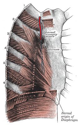
The [internal thoracic artery] is a bilateral artery of the thorax and is known to many as the [internal mammary artery], although this is not its proper anatomical name.
The internal thoracic artery is the first branch to arise off the subclavian artery. It descends inferiorly in a parasternal position, on the posterior aspect of the anterior thoracic cage. It is in contact with the ribs and as it descends it is covered posteriorly by the transversus thoracis muscle.
At the level of the sixth costal cartilage the internal thoracic artery gives off the musculophrenic artery and changes its name, continuing inferiorly as the superior epigastric artery. This artery will pass through one of the hiatuses of the respiratory diaphragm and in turn will become the inferior or deep epigastric artery at the level of the umbilicus. The deep epigastric artery (one of the boundaries of Hesselbach's triangle) will in turn open into the external iliac artery. The internal thoracic artery is part of a longitudinal collateral circulation arterial channel that parallels the aorta.
The internal thoracic artery will give off several branches. The first one is usually the pericardiacophrenic artery, a long artery that descends with the phrenic nerve alongside the parietal pericardium providing blood supply, as its name implies, to the pericardium and the phrenic nerve. Other branches are the anterior intercostal arteries, which communicate with the posterior intercostal arteries and the aorta.
The internal thoracic artery also gives arterial branches to the sternum and provides superficial, perforating branches to the medial side of the breast, hence its clinical name, the internal mammary artery.
The internal thoracic artery can be used to create a cardiac graft when performing a Coronary Artery Bypass Graft (CABG). Because of the two names used to denote this artery, surgeons will refer to the procedure either as a ITA (internal thoracic artery) or an IMA (internal mammary artery) CABG.
- Details
The word [ramus] is Latin and means “branch”. The plural form for [ramus] is [rami]. This word is used to denote “branches” or “divisions” in anatomical structures such as:
• Dorsal and ventral rami: the posterior (dorsal) and anterior (ventral) branches or divisions of a spinal nerve.
• Rami communicantes: small nerve connections between the spinal nerve and the sympathetic chain. These tiny nerve branches are part of the autonomic nervous system. Some are present at all levels of the spinal cord (gray rami communicans) and some only at levels T1 through L2 (white rami communicans)
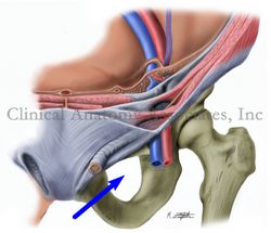
• Pubic rami: the superior and inferior lateral extensions of the pubic bone, which form the superior and inferior boundaries of the obturator foramen (see image)
Image property of:CAA.Inc.Artist:M. Zuptich
- Details
The word [proptosis] is composed of the prefix [pro-] meaning “forward”, the root term [-pt-] from the Greek [πτώση] (ptosi) which meaning “to fall”, and the suffix [-osis] meaning “condition”. Literal interpretation of the term would be “condition of falling forward”. Sometimes [–ptosis] can be considered a suffix, meaning “to sag” or "to droop".
Proptosis is used in medical terminology to describe a forward bulging to the eyes, similar to exophthalmos. The only difference is that the term exophthalmos is usually reserved to forward displacement of the eye related to or secondary to endocrinological dysfunction. All other eye protrusions are referred to as [proptosis
Proptosis can be caused by trauma, localized inflammation of the orbital tissues not related to thyroid dysfunction, tumors, etc.
- Details
The word [exophthalmos] is composed of the prefix [ex-] meaning “outer” or “outside”, the root term [-ophthalm-] which arises from the Greek word [οφθαλμός] (ophthalm?s) meaning “eye”, and the adjectival suffix [-os] meaning “pertaining to”. Literal interpretation of the term would be “pertaining to outside the eye”.
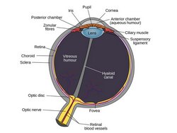
In reality the word [exophthalmos] is used to describe a condition where the eye is pushed anteriorly, protruding or bulging within the eye orbit. This condition can be unilateral or bilateral.
The term exophthalmos is usually reserved to forward displacement of the eye related to or secondary to endocrinological dysfunction. All other eye protrusions are referred to as [proptosis]. More on this word can be read on this article.
Thyroid dysfunction can lead to inflammation of the orbital tissues, including muscle and fatty tissues, causing edema and forward displacement of the eye.
- Details
The root term [-ophthalm-] arises from the Greek word [οφθαλμός] (ophthalm?s) meaning “eye” or "optic". It is used in several medical terms such as:
- Ophthalmology: The suffix [-ology] means “to study”. The study of the eye
- Ophthalmologist: A health care professional who specialized in eye diseases
- Ophthalmic: The adjectival suffix [-ic] means “pertaining to” and can be seen in anatomical terms such as the ophthalmic artery
- Ophthalmitis: The suffix [-itis] means “inflammation” or “infection”.
- Ophthalmoscope: An instrument to view the eye
- Anophthalmia: The prefix [an-] means "without" or "absence of". A congenital condition where one or both eyes do not develop
- Exophthalmos: The prefix [ex-] means "outer" or "outside". A protrusion of the eyes
Image by Rhcastilhos [Public domain], via Wikimedia Commons.
- Details
UPDATED: The root term [-mast-] arises from the Greek [μαστός] or [mastos] meaning "breast". A synonymous prefix is [mamm-] from the Latin [mamma], also meaning "breast". This prefix is used in medical terms such as:
- Mastectomy: The suffix [ectomy] means "removal of". Removal of the breast. This is an old operation, the first known records are from 180 A.D. The modern operation for radical mastectomy with careful removal of the related lymphatics was developed by William Halsted (1852 - 1922). The synonym [mammectomy] is correct, though rarely used. The word [masectomy] to refer to this procedure is incorrect and should not be used
- Mastoptosis: The suffix [-(o)ptosis] means "to fall", "to sag" or "go down". A falling or sagging of the breast. (Syn. mammoptosis)
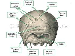
- Mastoid: The suffix [oid] means "similar to". Similar to a breast or with the shape of a breast, such as the mastoid process, a bony prominence of the temporal bone. See accompanying image
- Mastoplasty: The suffix [-(o)plasty] is used to mean "surgical reshaping". Surgical reshaping of the breast. Better known as a mammoplasty
- Mastopexy: A type of mastoplasty. The suffix "opexy" is used to mean "surgical fixation". Surgical fixation of the breast. In a mastopexy, the breast is "fixed" higher to reduce the mastoptosis. (Syn. mammopexy)
There is an interesting evolution of this word. The above applies only when the term [-mast-] is used as a root term, and combined with other word components. From the Greek, this term was passed on to Latin and thence evolved with the German term [m?sten] meaning "to feed" or "to fatten". This is why we have [mast cells] in histology. These are a type of mononuclear leukocyte described by Paul Ehrlich (1814 - 1915) in 1879, who named them [maztellen], which in German means "a well-fed cell".
For images of mastoptosis and mastopexy, CLICK HERE. Warning: images depict nude bodies.
Sources:
1. "The Origin of Medical Terms" Skinner, HA 1970 Hafner Publishing Co.
2. "Dorland's Illustrated Medical Dictionary" 28th Ed. W.B. Saunders. 1994
3. "Medical Terminology; Exercises in Etymology" Dunmore CW, Fleischer RM 2nd Ed. 1985
4. "Medical Meanings; A Glossary of Word Origins" Haubrich, WS. Am Coll Phys 1997


