Medical Terminology Daily (MTD) is a blog sponsored by Clinical Anatomy Associates, Inc. as a service to the medical community. We post anatomical, medical or surgical terms, their meaning and usage, as well as biographical notes on anatomists, surgeons, and researchers through the ages. Be warned that some of the images used depict human anatomical specimens.
You are welcome to submit questions and suggestions using our "Contact Us" form. The information on this blog follows the terms on our "Privacy and Security Statement" and cannot be construed as medical guidance or instructions for treatment.
We have 1531 guests online

Jean George Bachmann
(1877 – 1959)
French physician–physiologist whose experimental work in the early twentieth century provided the first clear functional description of a preferential interatrial conduction pathway. This structure, eponymically named “Bachmann’s bundle”, plays a central role in normal atrial activation and in the pathophysiology of interatrial block and atrial arrhythmias.
As a young man, Bachmann served as a merchant sailor, crossing the Atlantic multiple times. He emigrated to the United States in 1902 and earned his medical degree at the top of his class from Jefferson Medical College in Philadelphia in 1907. He stayed at this Medical College as a demonstrator and physiologist. In 1910, he joined Emory University in Atlanta. Between 1917 -1918 he served as a medical officer in the US Army. He retired from Emory in 1947 and continued his private medical practice until his death in 1959.
On the personal side, Bachmann was a man of many talents: a polyglot, he was fluent in German, French, Spanish and English. He was a chef in his own right and occasionally worked as a chef in international hotels. In fact, he paid his tuition at Jefferson Medical College, working both as a chef and as a language tutor.
The intrinsic cardiac conduction system was a major focus of cardiovascular research in the late nineteenth and early twentieth centuries. The atrioventricular (AV) node was discovered and described by Sunao Tawara and Karl Albert Aschoff in 1906, and the sinoatrial node by Arthur Keith and Martin Flack in 1907.
While the connections that distribute the electrical impulse from the AV node to the ventricles were known through the works of Wilhelm His Jr, in 1893 and Jan Evangelista Purkinje in 1839, the mechanism by which electrical impulses spread between the atria remained uncertain.
In 1916 Bachmann published a paper titled “The Inter-Auricular Time Interval” in the American Journal of Physiology. Bachmann measured activation times between the right and left atria and demonstrated that interruption of a distinct anterior interatrial muscular band resulted in delayed left atrial activation. He concluded that this band constituted the principal route for rapid interatrial conduction.
Subsequent anatomical and electrophysiological studies confirmed the importance of the structure described by Bachmann, which came to bear his name. Bachmann’s bundle is now recognized as a key determinant of atrial activation patterns, and its dysfunction is associated with interatrial block, atrial fibrillation, and abnormal P-wave morphology. His work remains foundational in both basic cardiac anatomy and clinical electrophysiology.
Sources and references
1. Bachmann G. “The inter-auricular time interval”. Am J Physiol. 1916;41:309–320.
2. Hurst JW. “Profiles in Cardiology: Jean George Bachmann (1877–1959)”. Clin Cardiol. 1987;10:185–187.
3. Lemery R, Guiraudon G, Veinot JP. “Anatomic description of Bachmann’s bundle and its relation to the atrial septum”. Am J Cardiol. 2003;91:148–152.
4. "Remembering the canonical discoverers of the core components of the mammalian cardiac conduction system: Keith and Flack, Aschoff and Tawara, His, and Purkinje" Icilio Cavero and Henry Holzgrefe Advances in Physiology Education 2022 46:4, 549-579.
5. Knol WG, de Vos CB, Crijns HJGM, et al. “The Bachmann bundle and interatrial conduction” Heart Rhythm. 2019;16:127–133.
6. “Iatrogenic biatrial flutter. The role of the Bachmann’s bundle” Constán E.; García F., Linde, A.. Complejo Hospitalario de Jaén, Jaén. Spain
7. Keith A, Flack M. The form and nature of the muscular connections between the primary divisions of the vertebrate heart. J Anat Physiol 41: 172–189, 1907.
"Clinical Anatomy Associates, Inc., and the contributors of "Medical Terminology Daily" wish to thank all individuals who donate their bodies and tissues for the advancement of education and research”.
Click here for more information
- Details
UPDATED: The term [ischemia] arises from the Greek word [ισχαιμία], meaning "to slow down the flow of blood". The root portion arises from the Greek [σφίγγω] meaning "to constrict" or "to stop". The uffix is [-emia] from the Greek [αίμα] (ema) meaning blood. Another potential origin is the Greek word [ischanein], meaning " to keep at bay" or 'to hold in check". It was Rudolf Virchow (1821-1902) who first used the term [ischemia] to denote a local reduction in the flow of blood.
Today the term ischemia means "localized reduction in the flow of blood to an organ or region of an organ". Ischemia occurs when there is a stenosis or stricture of an artery.
Note: The links to Google Translate include an icon that will allow you to hear the Greek or Latin pronunciation of the word.
- Details
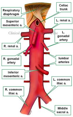
The bifurcation of the aorta is the point at which the abdominal aorta ends distally. At this point the aorta bifurcates giving origin to the right and left common iliac arteries. These arteries trend anterolaterally towards the pelvic brim.
The aortic bifurcation is usually found anterior to the inferior border of the 4th lumbar vertebra vertebra, slightly to the left of the midline. In surface anatomy, the bifurcation corresponds to a point slightly left to the midline and just about two fingerbreadths (two centimeters) inferior to the umbilicus.
This is an important landmark in surface anatomy in laparoscopic surgery. When placing the first periumbilical trocar the surgeon must angle the trocar posteroinferiorly towards the pelvic basin as to avoid perforating or lacerating the abdominal aorta. This situation has been studied in many journal articles.
Inferior to the aortic bifurcation is the confluence of both common iliac veins which give origin to the inferior vena cava.
The middle sacral artery arises from the lower portion of the abdominal aorta and appears inferior to the aortic bifurcation in the midline an continuing on its way to the anterior aspect of the sacrum.
The fact that the aorta bifurcates in front of the 4th lumbar vertebra leaves the L5-S1 intervertebral disc free of major arteries (with the exception of the middle sacral artery) allowing surgeons access to the intervertebral disc to perform laparoscopic removal of the disc with implantation of a device to allow intervertebral fusion in the case of intervertebral disc disease.
Sources:
1. “Major vascular injuries during laparoscopic procedures” Nordestgaard, AG et al Am J Surg (1995) 169,5: 543–545
2. “Evaluation of the direct trocar insertion technique at laparoscopy” Byron. JW et al Obst Gyn (1989) 74:3, 423-425
3. “Open versus closed establishment of pneumoperitoneum in laparoscopic surgery” Bonjer, HJ et al Br J Surg, 84: 599–602
4. “Serious Trocar Accidents in Laparoscopic Surgery: A French Survey of 103,852 Operations” Champault G et al Surg Lap Endosc (1996) 6(5):367-70
5. “Major vascular injuries during gynecologic laparoscopy” Chapron CM et al J Am Coll Surg (1997) 185:5 461-465
Image property of:CAA.Inc.Artist: Victoria G. Ratcliffe
- Details
Adjectival medical term that means “pertaining to a hospital”. The word is a derivate of the Greek word [νοσοκομείο] (nosokomio) meaning “hospital”. This term is itself composed by two Greek terms: [νόσος] (nosos), meaning “disease” or “injury” and [κομέω], meaning “to take care of”, so the Greek term [νοσοκομείο] means “to take care of a sick person” and the place where you do that is logically, a “hospital”.
This term was later adopted by Roman doctors, giving rise to the Latin term “nosocomium”, from which we derive our English “nosocomial”.
Although we use the term “hospital-acquired infection”, a proper way of saying this is “nosocomial infection”. A synonym for [nosocomial] is [iatrogenic].
Interestingly, the Latin root for “injury”, or [noxa] gave us the Golden Rule of Surgery” “Primum Non Nocere”
Thanks to Sharon L. Mueller, RN for suggesting this article.
Note: The links to Google Translate include an icon that will allow you to hear the Greek or Latin pronunciation of the word.
- Details
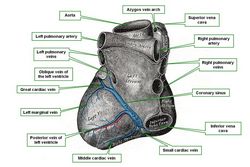
The middle cardiac vein is a vein that runs alongside or parallel to the posterior interventricular artery, also known as the posterior descending artery (PDA).
The middle cardiac vein appears close to the cardiac apex and ascends in the posterior interventricular sulcus (groove) to empty into the coronary sinus. It is responsible for venous drainage of the posterior aspect of the right and left ventricular wall as well as the posterior aspect of the interventricular septum.
Sources:
1 "Tratado de Anatomia Humana" Testut et Latarjet 8 Ed. 1931 Salvat Editores, Spain
2. "Anatomy of the Human Body" Henry Gray 1918. Philadelphia: Lea & Febiger
Original image modified. Image courtesy of bartleby.com
- Details
This is a Facebook post. If you cannot see it, click on the following link: https://www.facebook.com/CAAInc/posts/1000784059938724
- Details
- Hits: 14824
UPDATED: In both these words the suffix [-osis] means "condition". The root term [-kyph-] is Greek and means "bent or bowed" without an indication of the direction of bending, thus the term was originally used for any abnormal spinal curvature. It was Hippocrates who first used this term to denote "hunchback". Since then the term [kyphosis] denotes a curvature of the spine towards posterior, or better described, a spinal curvature in the median plane with a posterior convexity.
Hippocrated also used the Greek term [lordosis] to denote a curvature opposite to kyphosis. Lordosis is then a spinal curvature in the median plane with an posterior concavity.
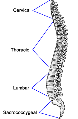
In the human spine, as viewed from the lateral aspect (see image), there are four normal curvatures. The cervical and lumbar curvatures are lordotic, while the thoracic and sacrococcygeal curvatures are kyphotic. Based on this description kyphosis and lordosis are normal conditions of the human spine.
A pathological, excessive, or exacerbated curvature should be denoted with the terms [hyperkyphosis] and [hyperlordosis] respectively; the prefix [hyper-] meaning "excessive". Through use, the terms [kyphosis] and [lordosis] are also used to denote pathological conditions. Hyperkyphosis has mostly a thoracic presentation, while hyperlordosis has mostly a lumbar presentation.
In vernacular terms, an individual with hyperkyphosis is known as a "hunchback", while an individual with hyperlordosis is known as a "swayback".
Image property of: CAA.Inc. Artist:D.M. Klein


